RSC Analytical Chemistry journals Emerging Investigator Series

Our Analytical Chemistry journals Analyst, Analytical Methods and Lab on a Chip are committed to early career researchers in the analytical chemistry and engineering fields. Our Emerging Investigator Series provide a platform for early career researchers to showcase their best work to a broad audience.

If you have an independent career and are within 10 years of obtaining your PhD or within 5 years of your first independent position you may be eligible for our Analyst, Analytical Methods or Lab on a Chip Emerging Investigator Series.

Analyst Emerging Investigator Series

Series Editors: Ryan Bailey, Laura Lechuga and Jaebum Choo Find out more

Analytical Methods Emerging Investigator Series

Series Editors: Fiona Regan and Juan Garcia-Reyes Find out more

Lab on a Chip Emerging Investigator Series

Series Editors: Dino Di Carlo, Yoon-Kyoung Cho and Piotr Garstecki Find out more 

Appropriate consideration will be given to career breaks and alternative career paths.


Digg This
Reddit This
Stumble Now!
Share on Facebook
Bookmark this on Delicious
Share on LinkedIn
Bookmark this on Technorati
Post on Twitter
Google Buzz (aka. Google Reader)

Organ-on-a-Chip systems-translating concept into practice Thematic Collection

We are pleased to announce a new Thematic Collection on Organ-on-a-Chip systems, translating concept into practice!

The first collection of papers on “Organ-body-and disease-on-a-Chip” collection has proved to be popular with the community. The collection has given this emerging field an identity and an effective venue for others to learn of the breadth, depth, and importance of this emerging area. We are delighted to announce that Michael Shuler (Cornell University, USA) will be acting as Thought Leader this follow-up collection.

We believe that a second collection highlighting efforts to translate this concept into practice would be valuable. While proof-of-concept papers for potential devices remains important, there has been significant progress in the last two years towards addressing the practical issues of translating these concepts into workable systems that will be adopted by industry and approved by regulators. While pharmaceuticals remain the primary target, it is clear that these devices will play important roles in the cosmetic, food, and chemical industries.

For regulatory approval and industrial adoption these devices need to be simple (easy to run by a technician), largely self-contained, low cost, reliable, incorporate advanced analytical techniques, and have efficient software to convert measurements into predictions of human response. Some of the initial proof-of-concept devices are too complicated and hence costly to be implemented industrially.  For an academic paper a lab can afford to have a high failure rate of systems as long as sufficient systems function to provide a robust data set.  For an industrial setting a high success rate will be necessary for adoption.  Automation of devices and efficient data collection and interpretation will be necessary for systems to have a broad impact and reduce labour costs.  Although much of the industrial data are proprietary, it should be possible to take historical cases where a drug candidate was approved and then withdrawn from the market due to toxicity and determine if the failure of the drug could have been anticipated from studies with a microphysiological (MPS) system.  Such examples could provide a compelling rationale for inclusion of MPS systems particularly in the later stages of the preclinical drug development process.

A series of papers that address aspects of the issues involved in moving from “proof-of-principle” devices to systems that can be routinely incorporated into testing of drugs, cosmetics, food ingredients, and chemicals would be valuable to the development of the field of microphysiological systems. We seek contributions that will help us fulfill this goal.

Lab on a Chip publishes the best work on significant and original work related to minia-turisation, at the micro- and nano-scale, of interest to a multidisciplinary readership. The journal seeks to publish work at the interface between physical technological advancements and high impact applications that are of direct interest to a broad audience.

Extraordinarily novel organ-on-a-chip systems that demonstrate unique new functions are also welcome.

Interested in submitting to the collection? 

We welcome submissions of original research articles and reviews to this collection and the collection is open for submissions.

Articles will be published as they are accepted and added to this online collection. They will receive extensive promotion throughout the submission period and as a complete collection.

If you are interested in submitting to the series, please get in touch with the Lab on a Chip Editorial Office at loc-rsc@rsc.org.

Digg This
Reddit This
Stumble Now!
Share on Facebook
Bookmark this on Delicious
Share on LinkedIn
Bookmark this on Technorati
Post on Twitter
Google Buzz (aka. Google Reader)

MicroTAS 2018 Highlights

The 22nd International Conference on Miniaturized Systems for Chemistry and Life Sciences (aka MicroTAS) was held last year in Kaohsiung, Taiwan. Welcoming more than 1000 participants, MicroTAS 2018 conference brought together several disciplines including microfluidics, microfabrication, nanotechnology, integration, materials& surfaces, analysis & synthesis, and detection technologies for life sciences & chemistry. Besides the exciting scientific program and great presentations, social/networking events (welcome reception, student mixer, women night out, and conference banquet) have made MicroTAS 2018 conference an unforgettable one. In this article, we would like to share some of the conference highlights with our Lab on a Chip blog readers.


Unraveling endothelial cell phenotypic regulation by spatial hemodynamic flows with microfluidics 

Sarvesh Varma, Guillermo Garcia-Cardena, & Joel Voldman

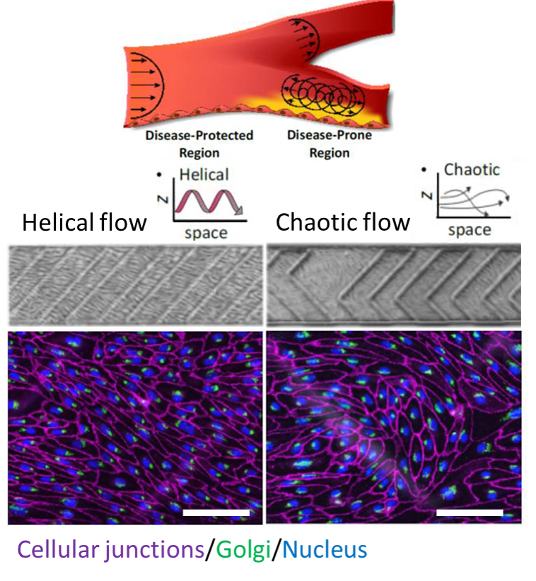
Did you know that artery bifurcations are prone to atherosclerosis? Blood flow profiles in vessels can help us to gain insights towards atherosclerosis. In this work, the authors fabricated a soft microdevice to study the effects of helical and chaotic flows on endothelial cells located in vein walls. They hypothesized and demonstrated that a helical (uniform) flow profile results in endothelial cells aligning upstream to flow and gain atheroprotective properties, while the chaotic flow results in misalignment of cells that give rise to atherosclerosis.

The figure shows the morphological adaptations of cells in response to distinct spatial flows, scale bars are 0.1 mm.


glass-like polymer


3D printing of microfluidic glass reactors 

Patrick Risch, Frederik Kotz, Dorothea Helmer & Bastian Rapp

Microfluidic devices are mostly made from PDMS, although this material is not always well-suited for thermal, optical, mechanical and chemical changes. In this work, the authors present a new resin formulation to inspire the 3D printing of glass, which is more durable than PDMS. The resin was fabricated using stereolithography printer and this technique is useful for rapid prototyping of microfluidic devices made from glass for optical detection or chemical reaction applications.

A 3D gradient generator is shown in this figure, scale bar is 2 mm.


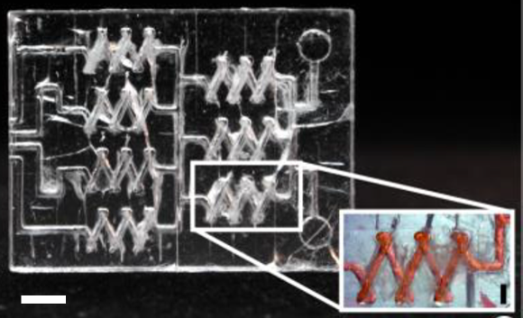
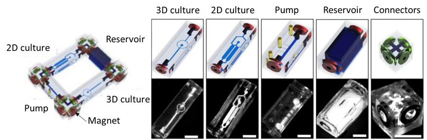
A Tetris-like modular microfluidic platform for mimicking multi-organ interactions 

Louis Ong Jun Ye, Terry Chng, Chong Lor Huai, Seep Li Huan & Toh Yi-Chin

Modularization is undoubtedly on the rise in microfluidics and this work demonstrates an interesting approach. The authors focused on solving ‘limited compatibility with existing devices‘ problem. To achieve that, ring magnets were utilized to connect different parts of PDMS building blocks that were previously fabricated using micro molds. A modular platform assembled using this approach was shown to culture cells as a proof-of-concept study. The platform is expected to allow facile configuration of complex experimental set-ups involving multiple tissues.

The image shows a modular device (left), and its parts (right) connected each other via magnets, scale bars are 1 cm.


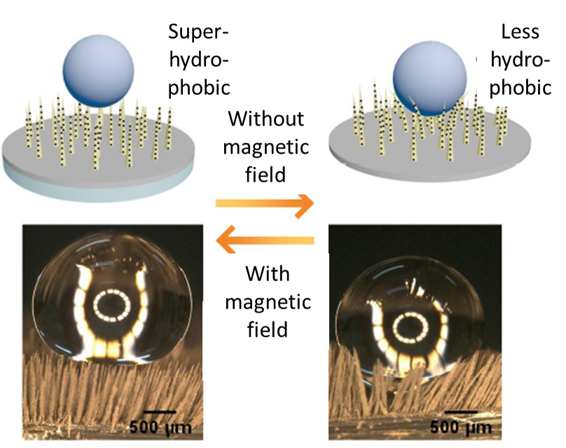
A magneto-switchable superhydrophobic surface for droplet manipulation

Chao Yang & Gang Li

Surface hydrophobicity is an important feature when it comes to bio and chemical applications. In this work, magneto switchable micro-pillars were made from PDMS and carbonyl iron particles. The pillars erect under influence of a magnetic field, resulting in subsequent switching of the wettability and adhesion of the surface between the water-repellent and water-adhesive states. The surface becomes superhydrophobic (water-repellent) when the magnetic field is applied. The authors demonstrated droplet lifting and transportation on a surface using this approach.

The image depicts the effect of an external magnetic field on the stiffness of micro-pillars.


About the Web writer

Burcu Gumuscu is a postdoctoral fellow in Herr Lab at UC Berkeley in the United States. Her research interests include the development of microfluidic devices for quantitative analysis of proteins from single-cells, next-generation sequencing, compartmentalized organ-on-chip studies, and desalination of water on the microscale.

Digg This
Reddit This
Stumble Now!
Share on Facebook
Bookmark this on Delicious
Share on LinkedIn
Bookmark this on Technorati
Post on Twitter
Google Buzz (aka. Google Reader)

Screening lipid libraries with microfluidics

Written by Darius Rackus

The plasma membrane is a key component of living organisms and is essential to all life. It separates the inside of the cell from the outside, compartmentalizes reactions, and selectively allows transport across it. While a lot of research has gone into how different proteins and surface molecules control the functions of the plasma membrane, less is known about how lipid composition gives rise to specific properties. For instance, transmembrane proteins, which are also a large class of drug targets, may have different requirements for the lipid environment or may have their function modified depending on local lipid composition. Recently, researchers from the David Weitz Lab at Harvard, have developed a microfluidic chip which they used to screen the largest lipid library to date, in order to identify which lipid compositions have specificity for certain protein transmembrane domains. This allows researchers to investigate the effect of local lipid concentration on transmembrane proteins.

The plasma membrane is often described as a ‘simple barrier’. But if that’s the case, then “why does nature go through the trouble of making so many different types of lipids?” explained Roy Ziblat, the lead author on the paper. Ziblat believes that the lipid membrane role is far bigger than a mere barrier and it serves as a substrate for accelerating bio-reactions. The role of the lipids composing the membrane is to control which biomolecules participate in these reactions, by their selectivity to membrane proteins. Having limited success with existing techniques, Ziblat turned to microfluidics to try to answer this question.

The microfluidic chip comprises an array of 108 wells in PDMS where lipid films can be deposited and dried before sealing the chip with another layer of PDMS. Liposomes are generated within the wells by swelling in aqueous buffer. These liposomes are then tested to see whether or not transmembrane domain peptides will insert into them. However, because the transmembrane domain peptides are insoluble, they can’t simply be added into the chip. To get around this, Ziblat et al. turned to cell-free protein synthesis. By loading the chip with DNA for the transmembrane domain peptide and PURExpress (a commercial cocktail of ribosomes, enzymes, and nucleotides for transcription and translation), the peptides can be synthesized in close proximity to the liposomes, thus minimizing precipitation and increasing the chance of insertion. The paper by Ziblat et al., which was featured on the cover of the 7th December issue of Lab on a Chip, also includes a helpful video description of these methods. Ziblat said he first made the video to better communicate his methods with his supervisor and colleagues, but it really helps the reader understand a very technical methodology.

Going forward, Ziblat hopes to use the device to study other membrane interactions, such as virus-cell binding. There’s also hope that this new device and method can be used to identify what the authors call “druggable lipids”—peptides that interact with specific lipids and thus better direct drugs toward specific cells or even organelles.


To download the full article, click the link below:

Determining the lipid specificity of insoluble protein transmembrane domains

R. Ziblat, J. C. Weaver, L. R. Arriaga, S. Chong and D. A. Weitz

Lab Chip, 2018, 18, 3561

DOI: 10.1039/c8lc00311d


About the webwriter

Darius Rackus (right) is a postdoctoral researcher in the Dittrich Bionalytics Group at ETH Zürich. His research interests are in developing integrated microfluidic tools for healthcare and bioanalysis

Digg This
Reddit This
Stumble Now!
Share on Facebook
Bookmark this on Delicious
Share on LinkedIn
Bookmark this on Technorati
Post on Twitter
Google Buzz (aka. Google Reader)

Emerging Investigator Series – Jerome Charmet

Jérôme Charmet received the Diplôme d’Ingénieur in Microtechnology Engineering from HES-SO Arc in Switzerland in 1998, the M.Sc. degree in Biomedical Engineering from the University of Bern, Switzerland, in 2010, and the PhD degree from the University of Cambridge in 2015. Overall, he worked for more than 10 years in both industrial and academic positions, including Intel Corporation, the National Centre for Sensor Research of Dublin City University in Ireland, the Microtechnology Institute of HES-SO Arc in Switzerland and the Centre for Misfolding Diseases of the University of Cambridge, UK. He joined the University of Warwick as an Assistant Professor in 2016 where he is developing integrated microfluidic platforms to study complex fluids and biological environments with applications in diagnosis, monitoring and drug screening/discovery. Read more about his group research here.

Read his Emerging Investigator article “Resolving protein mixtures using microfluidic diffusional sizing combined with synchrotron radiation circular dichroism” and read about him in the interview below:

Your recent Emerging Investigator Series paper focuses on protein mixtures using microfluidic diffusional sizing combined with synchrotron radiation circular dichroism. How has your research evolved from your first article to this most recent article?

It has evolved quite a lot, in fact it was not even directly related to microfluidics!

What aspect of your work are you most excited about at the moment?

I have started to explore organs-on-a-chip platforms with some colleagues biologists and I find it quite fascinating. But to be honest, every aspects of my work is exciting. I have a great team and collaborators I really enjoy to work with on a daily basis!

In your opinion, what applications can your current approach be used for?

In the manuscript we have used diffusional sizing to resolve the secondary structure of a complex mixture of proteins using synchrotron radiation circular dichroism, but the approach can be applied to other biomolecules with other bulk measurement techniques. We are taking advantage of laminar flow to separate the mixture into “controllable” fractions. By measuring the mixture and the different fractions, we can retrieve information about each component in the mixture.

What do you find most challenging about your research?

It’s multidisciplinary nature.. but it is also one of the most rewarding (when it works).

In which upcoming conferences or events may our readers meet you?

I’m just back from MicroTAS 2018 in Kaohsiung (Taiwan). Next, I will be attending the 8th Annual UK and Ireland Early Career Blood Brain Barrier Symposium 2018 in Oxford.

How do you spend your spare time?

Hiking, running … and these days spending as much time as possible with my 1 year old son.

Which profession would you choose if you were not a scientist?

I got to work with art conservator-restorers (…for some reason) and it is something I would definitely enjoy.

Can you share one piece of career-related advice or wisdom with other early career scientists?

It will sound a bit cheesy, but I will say “believe in your own ideas and importantly, find the right environment to develop them”.

Digg This
Reddit This
Stumble Now!
Share on Facebook
Bookmark this on Delicious
Share on LinkedIn
Bookmark this on Technorati
Post on Twitter
Google Buzz (aka. Google Reader)

Lab on a Chip Thematic Collections

We’ve brought together all of our latest Lab on a Chip Article Collections, Themed Issues, and Editor’s Choice collections to enable you to easily navigate to content most relevant to you. We hope you enjoy reading the papers in these collections!

Ongoing Collections

Thematic Collections

Digg This
Reddit This
Stumble Now!
Share on Facebook
Bookmark this on Delicious
Share on LinkedIn
Bookmark this on Technorati
Post on Twitter
Google Buzz (aka. Google Reader)

Lab on a Chip presents prestigious prizes at MicroTAS 2018

The µTAS 2018 Conference was held during 11th-15th November in Kaohsiung, Taiwan.  Simon Neil, Executive Editor of Lab on a Chip, attended the conference and announced the prestigious Lab on a Chip awards which include the Pioneers of Miniaturization Lectureship (in partnership with Dolomite Microfluidics), the Widmer Young Researcher Poster Prize and the Art in Science competition (in partnership with NIST). All three competitions received many fantastic submissions and we are delighted to present the winners, below:

“Pioneers of Miniaturization” Lectureship

Professor Sunghoon Kwon (Seoul National University) won the 13th “Pioneers of Miniaturization” Lectureship, sponsored by Dolomite and Lab on a Chip. The “Pioneers of Miniaturization” Lectureship rewards early to mid-career scientists who have made extraordinary or outstanding contributions to the understanding or development of miniaturised systems. Professor Sunghoon Kwon received a certificate, a monetary award and delivered a short lecture titled “Miniaturization for Personalised Medicine” at the conference.


Left to right: Simon Neil (Lab on a Chip), Sunghoon Kwon (winner) and Mark Gilligan (Dolomite)


Art in Science Competition

Lab on a Chip Executive Editor Simon Neil and Greg Cooksey from the National Institute of Standards Technology (NIST) presented the Art in Science award and a cake featuring the winning image at the Royal Society of Chemistry booth to Nam-Trung Nguyen for his entry “The Green Planet”. This award aims to highlight the aesthetic value in scientific illustrations while still conveying scientific merit.


Left to right: Simon Neil (Lab on a Chip), Greg Cooksey (NIST) and winner, Nam-Trung Nguyen with the personalised cake, and the winning image ‘The Green Planet’: an image of a floating liquid marble, decorated with green fluorescent beads. The image was taken with a colour USB camera. The liquid marble is made of a water droplet containing green fluorescent beads and coated with Teflon powder.


Widmer Young Researcher Poster Prize

The Widmer Young Researcher Poster Prize was awarded to Richard Cheng from the University of Toronto for his poster on “In Situ Delivery And Patterning Of Skin Cell Containing Biomaterial Sheet Using A Microfluidic Bioprinter”.


Simon Neil (left) with Richard Cheng (winner)


Congratulations to all the winners at the conference, we look forward to seeing you at µTAS 2019 in Basel, Switzerland! 


Digg This
Reddit This
Stumble Now!
Share on Facebook
Bookmark this on Delicious
Share on LinkedIn
Bookmark this on Technorati
Post on Twitter
Google Buzz (aka. Google Reader)

Art in Science Competition Winner Announced at MicroTAS 2018

Lab on a Chip and the National Institute of Standards Technology (NIST) presented the Art in Science award at the µTAS 2018 Conference on 14 November 2018 at the Royal Society of Chemistry booth. The award highlights the aesthetic value in scientific illustrations while still conveying scientific merit. The competition received many fantastic submissions this year which were judged by Simon Neil, Lab on a Chip Executive Editor, Greg Cooksey, NIST representative and Manabu TokeshiLab on a Chip Associate Editor .

Simon Neil and Greg Cooksey announced the winner of the competition was Nam-Trung Nguyen with his entry “The Green Planet” and presented Dr Nguyen with his award and certificate and a cake featuring the winning image.

The Green Planet 

Nam-Trung Nguyen, Griffith University, Australia

The Green Planet


The runners up are:


Magnetic Artificial Cilia with a Brush-shaped Cap 

Shuaizhong Zhang, Eindhoven University of Technology, Netherlands

magnetic artificial cilia with a brush-shaped cap


Embracing Chaos

Samantha Byrnes, Intellectual Ventures Laboratory, USA


A big thank you to all the contributors this year!

Digg This
Reddit This
Stumble Now!
Share on Facebook
Bookmark this on Delicious
Share on LinkedIn
Bookmark this on Technorati
Post on Twitter
Google Buzz (aka. Google Reader)

Emerging Investigator Series – Weian Zhao

Dr. Weian Zhao is an Associate Professor at the Sue and Bill Gross Stem Cell Research Center, Chao Family Comprehensive Cancer Center, Department of Biomedical Engineering, and Department of Pharmaceutical Sciences at University of California, Irvine. Dr. Zhao is also the co-founder of Velox Biosystems Inc, Baylx Inc, and Amberstone Biosciences LLC, start-up companies that aim to develop technologies for rapid diagnosis, stem cell therapy, and drug discovery, respectively. Dr. Zhao’s research aims to 1) elucidate and eventually control the fate of transplanted stem cells and immune cells to treat cancer and autoimmune diseases, and 2) develop novel miniaturized devices for early diagnosis and monitoring for conditions including sepsis, antibiotic resistance and cancer. Dr. Zhao has received several awards including the MIT’s Technology Review TR35 Award: the world’s top 35 innovators under the age of 35 and NIH Director’s New Innovator Award. Dr. Zhao completed his BSc and MSc degrees in Chemistry at Shandong University and then obtained his PhD in Chemistry at McMaster University in 2008. During 2008-2011, Dr. Zhao was a Human Frontier Science Program (HFSP) Postdoctoral Fellow at Harvard Medical School, Brigham and Women’s Hospital and MIT.

Read Dr Zhao’s Emerging Investigator article “Functional TCR T cell screening using single-cell droplet microfluidics” and read more about him in the interview below:

Your recent Emerging Investigator Series paper focuses on functional TCR T cell screening using single-cell droplet microfluidics. How has your research evolved from your first article to this most recent article?

I was trained as a colloidal and surface chemist. My first paper back when I was a PhD student was about building nanostructures using DNA as a template. Over the years, my research has evolved towards addressing immediate, major unmet need in biology and medicine; this is achieved by developing new tools and technologies, often using an interdisciplinary and collaborative approach, as exemplified by this new paper.

What aspect of your work are you most excited about at the moment?

It is the perspective that what we do in research can potentially solve real-world problems and help the patients in a short term. Like this new paper illustrated, our single-cell system holds great potential to accelerate development of cancer immunotherapeutics. There is a great and urgent demand in the clinic for many more of this type of promising drugs that can benefit patients who do not have lots of time to wait.

In your opinion, how can droplet microfluidic technologies contribute to immune-screening and immuno-therapy and personalised medicine in general?

Droplet microfluidic technologies and other exciting single-cell systems being developed in our field can contribute to immune-screening, immuno-therapy and personalised medicine by significantly reducing the time needed for the discovery phase. For example, conventional screening platforms for immunologic agents such as antibodies or T cells, usually take months to years to obtain a therapeutic candidate whereas the new single-cell platforms can potentially do this in the matter of days to weeks. In this new paper, this ability is enabled by directly interrogating the “functions” of individual cell clones in greater depth by using single-cell analysis. As biological samples are heterogeneous, this key information is often lost in conventional, population based studies.

What do you find most challenging about your research?

It is always the people. I take time to identify and build the right team that, once in place, can almost conquer any challenges in research itself. I also wish I could spend more time to do research rather than writing grants.

In which upcoming conferences or events may our readers meet you?

IEEE EMBS Micro & Nanotechnology in Medicine 2018, Physical Science of Cancer (GRS) 2019, and PEGS 2019

How do you spend your spare time?

Mostly with the family, and watching Manchester United games.

Which profession would you choose if you were not a scientist?

Probably try to become a football manager in the English Premier League. There is a lot of similarity in running a research laboratory and running a football club, isn’t there?

Can you share one piece of career-related advice or wisdom with other early career scientists?

Ask for help and mentorship from people who have succeeded in what you are trying to do. Try to accelerate your progress through collaboration and partnership.

Digg This
Reddit This
Stumble Now!
Share on Facebook
Bookmark this on Delicious
Share on LinkedIn
Bookmark this on Technorati
Post on Twitter
Google Buzz (aka. Google Reader)

Simple microfluidic cell sorter device to replace manual tissue dissociation protocols

Even a tiny group of cells has the ability to populate a tumor in tissues. Determining cellular diversity and identifying these small cell groups gains importance when it comes to selection of treatment strategies. Tissue samples taken from patients are required to be dissociated into single-cell suspensions, therefore identification can be efficiently done at single-cell level using a powerful suite of technologies including flow cytometry, mass cytometry, and single cell RNA sequencing. However, breaking a tissue down to a single-cell suspension is not an easy task. The old-school way is to cut the tissue sample into small pieces with a blade and mechanically dissociated by vigorous shaking after the application of proteolytic enzymes. Large aggregates are removed by filtering the suspension through a strainer. This technique significantly increases the sample loss, drops the speed of the process and is not ideal for immediate downstream analysis.

In this month’s Lab on a Chip HOT article series, a group of researchers led by Dr Jered Haun at University of California Irvine presented a novel and simple approach that improves the quality of single-cell suspensions obtained from tissue samples using microfluidics. Jeremy Lombardo, a co-author of this article, explains that “the goal of this work was to fully replace manual intensive tissue dissociation protocols by using microfluidic devices.” The developed tool is a microdevice consisting of two nylon membranes, one with 25-50 µm mesh, and the other with 10-15 µm mesh, attached to micron-sized pores and microchannels. The device is made of laser-micromachined hard plastic (PET, aka. polyethylene terephthalate), which enables operation at high flow rates (>10 mL/min) when compared to PDMS (a silicon-based organic material). Also, the chip has multiple layers for connecting nylon mesh membranes at different levels (Figure 1).

cell sorter

Figure 1. Microfluidic cell sorter device for tissue  samples. The sketch shows the inner layers, consisting of two membranes for operating the device in direct or tangential filtration modes. Membrane mesh size can be adjusted to the cell size. Micrographs on the right show lattice network with several pore sizes used in this work. Pore sizes are (top to bottom) 50, 25, 15, 10 µm diameter.

Working principle of the cell sorter device

The inlet of the device connects to a microporous membrane to introduce tissue samples. The Sample passing through the membrane exits through the effluent outlet. It is also possible to direct a portion of the sample along the surface of the membrane that is connected to the cross-flow outlet. The device is either operated in a direct filtration regime to maximize sample recovery and processing speed, or in a tangential filtration regime to sweep larger tissue fragments and cell aggregates away to prevent clogging.

While the researchers initially hypothesized that under pressure-driven flow, cell and tissue aggregates might disaggregate as they pass through the membranes of the device, they were pleasantly surprised by the drastic level of single cell increases seen in the initial testing of these devices, says Jeremy Lombardo and adds “The hardest part in developing and testing this device was to find a combination of membrane pore sizes that could best dissociate cell aggregates and tissue without compromising cell viability. Thorough testing of various pore sizes and combinations were ultimately carried out with both cell line and murine tissue models before we settled on the final 50 and 15 μm pore sizes.”

Advantages, challenges and the future

The authors summarized the advantages of this platform for Lab on a Chip blog readers: “The device is extremely simple to operate as well as inexpensive to fabricate. It can easily be incorporated into many tissue dissociation applications for improved single cell yields as a standalone device but could also be easily integrated with other downstream microfluidic operations (cell sorting, detection etc.).” According to the authors, “in the current format of cell sorter device, cells that are very large in size would likely be difficult to process, as they would likely span multiple pores of the filters and be traditionally filtered away instead of dissociated.” Although seeming like a challenge, this can easily be addressed by adjusting the filter membrane pore sizes to accommodate these larger cell types.

For the future of the device, the authors indicate, “We are also currently working on integrating this device with upstream, larger scale tissue dissociation devices that we have developed previously to create a fully automatable microfluidic tissue dissociation platform.


To download the full article for free* click the link below:

Microfluidic filter device with nylon mesh membranes efficiently dissociates cell aggregates and digested tissue into single cells
Xiaolong Qiu, Jeremy A. Lombardo, Trisha M. Westerhof, Marissa Pennell, Anita Ng, Hamad Alshetaiwi, Brian M. Luna, Edward L. Nelson, Kai Kessenbrock, Elliot E. Hui, and Jered B. Haun
Lab Chip, 2018, Lab on a Chip Recent Hot Articles
DOI: 10.1039/c8lc00507a

About the Webwriter
Burcu Gumuscu is a postdoctoral fellow in Herr Lab at UC Berkeley in the United States. Her research interests include development of microfluidic devices for quantitative analysis of proteins from single-cells, next generation sequencing, compartmentalized organ-on-chip studies, and desalination of water on the microscale.

Digg This
Reddit This
Stumble Now!
Share on Facebook
Bookmark this on Delicious
Share on LinkedIn
Bookmark this on Technorati
Post on Twitter
Google Buzz (aka. Google Reader)