Lab on a Chip Issue 9 now online

Another great issue of LOC is now available online, including Paul Vulto et al.‘s HOT article describing their new phaseguide technology for filling and emptying of microfluidic structures, independent of the chamber and channel geometry, which is highlighted on the outside front cover.

Phaseguides: a paradigm shift in microfluidic priming and emptying
Paul Vulto, Susann Podszun, Philipp Meyer, Carsten Hermann, Andreas Manz and Gerald A. Urban
Lab Chip, 2011, 11, 1596-1602

On the inside front cover we have another HOT article from James P. Landers et al. describing a PMMA microfluidic DNA purification device with embedded microfabricated posts, which have been functionalized to allow for pH-induced DNA binding.

Solid phase extraction of DNA from biological samples in a post-based, high surface area poly(methyl methacrylate) (PMMA) microdevice
Carmen R. Reedy, Carol W. Price, Jeff Sniegowski, Jerome P. Ferrance, Matthew Begley and James P. Landers
Lab Chip, 2011, 11, 1603-1611

The issue also features a Focus article from Holger Becker on his wish-list of unsolved technological issues — which is part of our series of Focus articles on the commercialization of microfluidics.

All I want for Christmas…
Holger Becker
Lab Chip, 2011, 11, 1571-1573

View the full issue here

Digg This
Reddit This
Stumble Now!
Share on Facebook
Bookmark this on Delicious
Share on LinkedIn
Bookmark this on Technorati
Post on Twitter
Google Buzz (aka. Google Reader)

HOT article: phaseguides – primed and ready to revolutionise microfluidics

So you’ve designed this fantastic little lab-on-a-chip device that will revolutionise disease/virus/cancer detection or discover fantastic new drugs, but you can’t get your sample into all the complicated channels or bubbles are ruining your flow.  Sound familiar?

Well now Paul Vulto (Albert-Ludwigs-Universität Freiburg) and colleagues have developed a novel technique for priming microfluidic devices that could solve all these problems.  By using patterned ridges in their devices they are able to guide the liquid–air interfaceto fill complicated geometries – by forcing it to align with the ridge.     This allows them to fill complex microfluidic chambers and channels, independant of their shape, simply by the carefully patterning of these ridges.  The team have aptly coined the term  ‘phaseguides‘ to descibe their invention and hope that it ‘will prove a leap forward towards more simple, flexible and reliable microfluidic systems’.

This figure demonstrates the impressive use of phaseguides by the filling of a butterfly shaped chamber.  Inserts (a and b) demonstrate that when filling without a phaseguide pattern the liquid spreads radially leaving the structure largely un-filled.   Inserts (c–f) demonstrate that with phaseguides (visualized by dashed lines in (c)), the structure can be completely filled with liquid.

This paper comes highly recommended by our expert reviewers and is free to access for 6 weeks – why not download it today!

Phaseguides: a paradigm shift in microfluidic priming and emptying
Paul Vulto, Susann Podszun, Philipp Meyer, Carsten Hermann, Andreas Manz and Gerald A. Urban
Lab Chip, 2011, 11, 1596-1602
DOI: 10.1039/C0LC00643B

Digg This
Reddit This
Stumble Now!
Share on Facebook
Bookmark this on Delicious
Share on LinkedIn
Bookmark this on Technorati
Post on Twitter
Google Buzz (aka. Google Reader)

Free Lab on a Chip articles in Nature technology feature

Work published in Lab on a Chip features heavily in Nature’s latest Special Technology Feature ‘Tissue models: A living system on a chip‘.

The article, summarising the current state-of-the-art in creating living tissue models on chips, references work from LOC Editorial Board member Donald Ingber (Harvard Medical School) and LOC publications from Michael Shuler (Cornell),  John March (Cornell), Linda Griffith (MIT) and Axel Günther (University of Toronto).

We’ve made these great articles free to access for 2 weeks – why not take a look!

A microfluidic device for a pharmacokinetic–pharmacodynamic (PK–PD) model on a chip
Jong Hwan Sung, Carrie Kam and Michael L. Shuler
Lab Chip, 2010, 10, 446-455

Microscale 3-D hydrogel scaffold for biomimetic gastrointestinal (GI) tract model
Jong Hwan Sung, Jiajie Yu, Dan Luo, Michael L. Shuler and John C. March
Lab Chip, 2010, 11, 389-392

Perfused multiwell plate for 3D liver tissue engineering
Karel Domansky, Walker Inman, James Serdy, Ajit Dash, Matthew H. M. Lim and Linda G. Griffith
Lab Chip, 2010, 10, 51-58

A microfluidic platform for probing small artery structure and function

Axel Günther, Sanjesh Yasotharan, Andrei Vagaon, Conrad Lochovsky, Sascha Pinto, Jingli Yang, Calvin Lau, Julia Voigtlaender-Bolz and Steffen-Sebastian Bolz
Lab Chip, 2010, 10, 2341-2349
From our 2010 Emerging Investigators themed issue

Digg This
Reddit This
Stumble Now!
Share on Facebook
Bookmark this on Delicious
Share on LinkedIn
Bookmark this on Technorati
Post on Twitter
Google Buzz (aka. Google Reader)

Lab on a Chip issue 8 now available online!

This month’s issue features another great selection of articles, including a Focus article from Mathias Uhlen and Helene Andersson Svahn on affinity reagents for lab on a chip applications.

Featured on the outside front cover is the HOT article from William Rodriguez and Rashid Bashir et al., demonstrating a microfabricated biochip to determine the CD4+ T lymphocyte count in HIV patients.

A microfabricated electrical differential counter for the selective enumeration of CD4+ T lymphocytes
Nicholas N. Watkins, Supriya Sridhar, Xuanhong Cheng, Grace D. Chen, Mehmet Toner, William Rodriguez and Rashid Bashir

On the inside front cover we have Rodolphe Marie’s micro device that can isolate centimetre-long portions of human DNA to help study the genetic make-up of diseased cells.  This paper was recently featured in Chemistry World – you can read the story here.

A device for extraction, manipulation and stretching of DNA from single human chromosomes
Kristian H. Rasmussen, Rodolphe Marie, Jacob M. Lange, Winnie E. Svendsen, Anders Kristensen and Kalim U. Mir

Finally, the back cover highlights work from  Gang Logan Liu, using a standard CD drive to count microparticles and cells with a digital microfluidic compact disc.  This article was also recently highlighted in Chemistry World.

Microparticle and cell counting with digital microfluidic compact disc using standard CD drive
Syed M. Imaad, Nathan Lord, Gulsim Kulsharova and Gang Logan Liu

Other HOT articles in this issue include:

Integrated photocatalytic micropillar nanoreactor electrospray ionization chip for mimicking phase I metabolic reactions
Teemu Nissilä, Lauri Sainiemi, Mika-Matti Karikko, Marianna Kemell, Mikko Ritala, Sami Franssila, Risto Kostiainen and Raimo A. Ketola

Capillary-driven automatic packaging
Yuzhe Ding, Lingfei Hong, Baoqing Nie, Kit S. Lam and Tingrui Pan

A magnetic bead-based assay for the rapid detection of methicillin-resistant Staphylococcus aureus by using a microfluidic system with integrated loop-mediated isothermal amplification
Chih-Hung Wang, Kang-Yi Lien, Jiunn-Jong Wu and Gwo-Bin Lee


Digg This
Reddit This
Stumble Now!
Share on Facebook
Bookmark this on Delicious
Share on LinkedIn
Bookmark this on Technorati
Post on Twitter
Google Buzz (aka. Google Reader)

HOT article: packed to precision – capillary-driven automatic packaging for microfluidic devices

The fabrication  of 3D microstructures for microfluidic devices continues to be a challenge as traditional microfabrication techniques are not suitable for the construction of these devices.  For example, PDMS (polydimethylsiloxane) is one of the most common substrate materials for microfluidic devices, but standard packaging techniques are not able to bond multiple substrate layers with the high precision required.

Tingrui Pan (University of California, Davis) et al. have now come up with an easy and robust technique which they hope will make this problem a thing of the past.  Their new technique, CAP (capillary-driven automatic packaging) uses the interactions between a liquid capillary bridge and the top and bottom substrates to align multiple substrate layers with high precision, and has a bonding strength comparable to standard oxygen plasma processes.  The technique is also transferable to other materials, requires no thermal or mechanical treatment, nor any specialist equipment.

Illustration of the CAP-enabled microdevice fabrication process, including (a) micropatterning of a shadow mask made of dry film, (b) PDMS replica molding, (c) selective oxygen plasma treatment through the shadow mask, (d) DI water loading in the defined hydrophilic regions, and finally (e) self-alignment and self-engagement steps between two chips with identical capillary alignment patterns.

Pan et al. believe that this technique has the ability to be employed in microdevices for point-of-care diagnosis, controlled drug delivery, and combinatorial biological screening – why not take a look and see for yourself – the article’s free to access for four weeks!

Capillary-driven automatic packaging
Yuzhe Ding, Lingfei Hong, Baoqing Nie, Kit S. Lam and Tingrui Pan
Lab Chip, 2011, 11, 1464-1469
DOI: 10.1039/C0LC00710B

Digg This
Reddit This
Stumble Now!
Share on Facebook
Bookmark this on Delicious
Share on LinkedIn
Bookmark this on Technorati
Post on Twitter
Google Buzz (aka. Google Reader)

HOT article: magnetic bead-based assay for MRSA rapid detection – what an attractive idea!

In recent years methicillin-resistant Staphylococcus aureus (MRSA) has become one of the most prevalent antibiotic resistant pathogens in hospitals and as such there is an urgent need for a way of rapidly detecting it to limit the spread of the infection.

Current methods of detection, involving the use of PCR, require quite bulky, power-intensive and quite expensive apparatus. Gwo-Bin Lee et al. have developed a portable hand-held system where the entire diagnostic protocol, from bio-sample pre-treatment to optical detection, can be automatically completed within an hour and with a limit of detection 1000- fold higher than conventional bench-top PCR systems.

The entire process can be accomplished automatically via magnetic bead-based hybridization of the target DNA purified from clinical samples, a loop-mediated isothermal amplification process for the amplification of the target genes, and then spectrophotometric analysis of the amplified target genes.

To find out more take a look at this HOT article which is FREE to view for 4 weeks!

A magnetic bead-based assay for the rapid detection of methicillin-resistant Staphylococcus aureus by using a microfluidic system with integrated loop-mediated isothermal amplification
Chih-Hung Wang, Kang-Yi Lien, Jiunn-Jong Wu and Gwo-Bin Lee
Lab Chip, 2011, 11, 1521-1531
DOI: 10.1039/C0LC00430H

Digg This
Reddit This
Stumble Now!
Share on Facebook
Bookmark this on Delicious
Share on LinkedIn
Bookmark this on Technorati
Post on Twitter
Google Buzz (aka. Google Reader)

HOT article: no need to shed light on this T cell counter!

Antiretroviral therapy (ART) increases the longevity and quality of life for HIV patients.  The lack of objective diagnostic tests to determine when to start ART and to monitor its successes hinders the effective use of treatment.  An important diagnostic procedure is to obtain a patient’s CD4+ lymphocyte count, which is traditionally carried out using optical instrumentation. However, the cost and technical requirements of such equipment make them infeasible as analytical methods in poorer countries.

William Rodriguez (Daktari Diagnostics) and Rashid Bashir (University of Illinois) present a solution to this problem with their novel microfabricated biochip to enumerate CD4+ T lymphocytes from healthy human subject blood samples.

Their biochip incorporates electrical impedance sensing coupled with immunoaffinity chromatography to electrically differentiate CD4+ cells from other leukocytes with accuracy comparable to current optical diagnostic methods.  This negates any requirement for labelling or optical detection, while its microfabricated nature suggests it may be an inexpensive, simple and portable alternative to current flow cytometric practises.

Learn more about this device by reading this HOT article, which is free to access for the next 4 weeks!

A microfabricated electrical differential counter for the selective enumeration of CD4+ T lymphocytes
Nicholas N. Watkins, Supriya Sridhar, Xuanhong Cheng, Grace D. Chen, Mehmet Toner, William Rodriguez and Rashid Bashir
Lab Chip, 2011, 11, 1437-1447
DOI: 10.1039/C0LC00556H

Digg This
Reddit This
Stumble Now!
Share on Facebook
Bookmark this on Delicious
Share on LinkedIn
Bookmark this on Technorati
Post on Twitter
Google Buzz (aka. Google Reader)

Top ten most accessed articles in February

This month sees the following articles in Lab on a Chip that are in the top ten most accessed:-

Microfluidics and complex fluids 
Ph Nghe, E. Terriac, M. Schneider, Z. Z. Li, M. Cloitre, B. Abecassis and P. Tabeling 
Lab Chip, 2011, 11, 788-794, DOI: 10.1039/C0LC00192A, Tutorial Review 

Rapid prototyping of microstructures in polydimethylsiloxane (PDMS) by direct UV-lithography 
Tim Scharnweber, Roman Truckenmüller, Andrea M. Schneider, Alexander Welle, Martina Reinhardt and Stefan Giselbrecht 
Lab Chip, 2011, 11, 1368-1371, DOI: 10.1039/C0LC00567C, Technical Note 

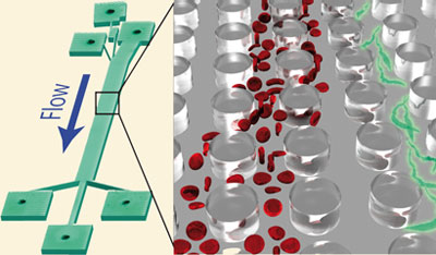
Continuous separation of breast cancer cells from blood samples using multi-orifice flow fractionation (MOFF) and dielectrophoresis (DEP) 
Hui-Sung Moon, Kiho Kwon, Seung-Il Kim, Hyunju Han, Joohyuk Sohn, Soohyeon Lee and Hyo-Il Jung 
Lab Chip, 2011, 11, 1118-1125, DOI: 10.1039/C0LC00345J, Paper 

nanoLAB: An ultraportable, handheld diagnostic laboratory for global health 
Richard S. Gaster, Drew A. Hall and Shan X. Wang 
Lab Chip, 2011, 11, 950-956, DOI: 10.1039/C0LC00534G, Paper 

Programmed trapping of individual bacteria using micrometre-size sieves 
Min-Cheol Kim, Brett C. Isenberg, Jason Sutin, Amit Meller, Joyce Y. Wong and Catherine M. Klapperich 
Lab Chip, 2011, 11, 1089-1095, DOI: 10.1039/C0LC00362J, Paper 

Lab-on-a-chip based immunosensor principles and technologies for the detection of cardiac biomarkers: a review 
Mazher-Iqbal Mohammed and Marc P. Y. Desmulliez 
Lab Chip, 2011, 11, 569-595, DOI: 10.1039/C0LC00204F, Tutorial Review 

Simple room temperature bonding of thermoplastics and poly(dimethylsiloxane) 
Vijaya Sunkara, Dong-Kyu Park, Hyundoo Hwang, Rattikan Chantiwas, Steven A. Soper and Yoon-Kyoung Cho 
Lab Chip, 2011, 11, 962-965, DOI: 10.1039/C0LC00272K, Technical Note 

Stand-alone self-powered integrated microfluidic blood analysis system (SIMBAS) 
Ivan K. Dimov, Lourdes Basabe-Desmonts, Jose L. Garcia-Cordero, Benjamin M. Ross, Antonio J. Ricco and Luke P. Lee 
Lab Chip, 2011, 11, 845-850, DOI: 10.1039/C0LC00403K, Paper 

Integrated systems for rapid point of care (PoC) blood cell analysis 
Cees van Berkel, James D. Gwyer, Steve Deane, Nicolas Green, Judith Holloway, Veronica Hollis and Hywel Morgan 
Lab Chip, 2011, 11, 1249-1255, DOI: 10.1039/C0LC00587H, Paper 

Affinity reagents for lab on chips 
Mathias Uhlen and Helene Andersson Svahn 
Lab Chip, 2011, Advance Article, DOI: 10.1039/C1LC90005F, Focus 

Why not take a look at the articles today and blog your thoughts and comments below.

Fancy submitting an article to Lab on a Chip? Then why not submit to us today or alternatively email us your suggestions.

Digg This
Reddit This
Stumble Now!
Share on Facebook
Bookmark this on Delicious
Share on LinkedIn
Bookmark this on Technorati
Post on Twitter
Google Buzz (aka. Google Reader)

Microfluidics to diagnose sleeping sickness

Another Lab on a Chip article has been highlighted in Chemistry World!

Parasites (green) are separated from red blood cells in the device according to shape, not size

Human African trypanosomiasis, or sleeping sickness, is caused by parasites in the blood called trypanosomes. The disease is transmitted by tsetse flies and is fatal is left untreated. Standard diagnosis is done by looking for the parasites in blood samples using a microscope. However, the concentration of parasites is often very low, so they need to be separated from the red blood cells before analysis. Many separation methods have been developed, but they are expensive and too complex to use in remote areas where the disease is common.

Jonas Tegenfeldt from the University of Lund, and his colleagues, have developed a microfluidic device that separates the parasites from the blood cells using their shape, because parasites and red blood cells are very difficult to separate by size.

Read Amaya Camara-Campos’ full story online here or go straight to the LOC article:

Separation of parasites from human blood using deterministic lateral displacement
Stefan H. Holm, Jason P. Beech, Michael P. Barrett and Jonas O. Tegenfeldt
Lab Chip, 2011, 11, 1326
DOI: 10.1039/c0lc00560f

Digg This
Reddit This
Stumble Now!
Share on Facebook
Bookmark this on Delicious
Share on LinkedIn
Bookmark this on Technorati
Post on Twitter
Google Buzz (aka. Google Reader)

Lab on a Chip Issue 7 now online – focus on UK research

As part of our 10th anniversary series we focus this month on UK research, with an introductory editorial from  Andrew deMello and Hywel Morgan.

Highlighted on the front cover is work by John deMello and colleagues, applying enhanced temperatures for micro continuous-flow synthesis of nanoparticles with excellent reaction control:

A stable droplet reactor for high temperature nanocrystal synthesis
A. M. Nightingale, S. H. Krishnadasan, D. Berhanu, X. Niu, C. Drury, R. McIntyre, E. Valsami-Jones and J. C. deMello

On the inside front cover we have work from Miles Padgett and Roberto Di Leonardo. In their critical review they provide a comprehensive overview of optical tweezers and holographic optical tweezers for lab-on-a-chip applications:

Holographic optical tweezers and their relevance to lab on chip devices
Miles Padgett and Roberto Di Leonardo

The issue also includes HOT articles on observations of the positional dependence of particles in microfludic impedance cytometry, a great new method for fast, cheap PDMS patterning using direct UV lithography and on the back cover we highlight a microfluidic paper-based chemiluminescence biosensor for simultaneous glucose and uric acid developed by Jinghua Yu et al.

Digg This
Reddit This
Stumble Now!
Share on Facebook
Bookmark this on Delicious
Share on LinkedIn
Bookmark this on Technorati
Post on Twitter
Google Buzz (aka. Google Reader)