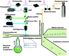
Focus on circulating tumor cells in microfluidics

This important Focus article looks at the most up to date research on the isolation and characterisation of circulating tumor cells (CTCs) using microfluidic techniques.

CTCs are produced when tumor cells break off and enter the circulatory system. This makes them important targets for diagnosis and monitoring. However they are only present in extremely small quantities in the blood, meaning plenty of technical difficulties arise when trying to accurately characterise and assess them in a blood sample.

This Focus highlights: 

  1. Considerations in design of CTC isolation, separation and detection systems
  2. Disadvantages of macroscale systems
  3. Magnetic-based microfluidic systems
  4. Affinity chromatography-based microfluidic systems
  5. Size and deformability-based microdevices
  6. Dielectrophoretic-based microdevices

Tony Jun Huang et al. based at The Pennsylvania State University, Massachusetts Institute of Technology (MIT) and Harvard Medical School, USA, finish by discussing the possible combination of the above systems and the future of such technology in terms of clinical relevance for different cancers and technological development.

We’ve made this article free to access for the next 4 weeks*, read it now by clicking the link below:

Probing circulating tumor cells in microfluidics
Peng Li, Zackary S. Stratton, Ming Dao, Jerome Ritz and Tony Jun Huang
DOI: 10.1039/C2LC90148J

 *Free access to individuals is provided through an RSC Publishing personal account. Registration is quick, free and simple

Digg This
Reddit This
Stumble Now!
Share on Facebook
Bookmark this on Delicious
Share on LinkedIn
Bookmark this on Technorati
Post on Twitter
Google Buzz (aka. Google Reader)

First HOT article of the week – a simpler method for measuring ultralow interfacial tension

Surface tension is an important property of all liquids and influences many of their characteristics, such as droplet formation. Surface tension at the interface of two different liquids is interfacial tension. An example of this is the separation of oil and water in solution.

There are other solutions of dissimilar liquids that have extremely small interfacial tensions, 1000 times less than that of oil and water. Measuring this accurately by traditional techniques, such as force tensiometry methods, is a problem. Although many more methods, have been proposed to improve upon this, a simple and accurate technique is yet to be found.

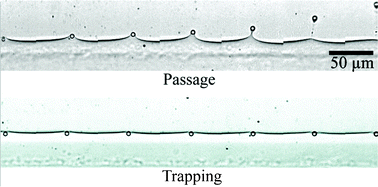
Howard Stone has now led a team at Princeton University and Harvard University, USA, in producing the first microfluidic tensiometer with the capability of measuring ultralow interfacial tension. Their development uses only a small amount of reagents and magnetic particles followed by application of a mathematical model to calculate the interfacial tension.

The measurement is taken by observing the distance between the center of the magnetic particle and the flowing interface. The paramagnetic beads may pass through the interface or be trapped by it due to the balance of magnetic and interfacial forcesonly when this ratio passes a threshold value do the particles pass through.  The method uses these threshold values to then calculate the ultralow interfacial tension.

This simple, miniaturised tensiometer has application in many different systems, with advantages for the applications of ultralow interfacial tension including enhanced oil recovery and microemulsions.

Microfluidic ultralow interfacial tensiometry with magnetic particles
Scott S. H. Tsai, Jason S. Wexler, Jiandi Wan and Howard A. Stone
DOI: 10.1039/C2LC40797C

*Free access to individuals is provided through an RSC Publishing personal account. Registration is quick, free and simple

Digg This
Reddit This
Stumble Now!
Share on Facebook
Bookmark this on Delicious
Share on LinkedIn
Bookmark this on Technorati
Post on Twitter
Google Buzz (aka. Google Reader)

Point-of-care diagnostics using capillary forces – a HOT article!

Point-of-care diagnostics is the aim for much medical research. Often the problem is the ease of preparation of the biological sample, which usually requires multiple steps before the diagnostic test can even begin. Automation of sample preparation has been explored and another option is pressure-driven microfluidics. However, this requires complex external equipment to run the chip only suitable for use in the laboratory away from the patient.

To get true point-of-care diagnostics, sample preparation has to be fast, simple and cheap.

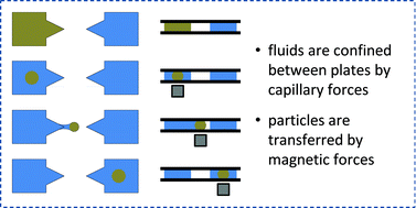
Stationary microfluidics is now being explored.  Liquid is present in fixed positions on the chip and it is magnetic particles that move between the points holding liquid. This often involves separating the liquids using an oil barrier, but this has disadvantages of over-complicating the device by having more than one liquid (the test solution), meaning an additional very careful pipetting step. Not all test solutions or all diagnostic tests would be compatible with this oil barrier, for example, purification of protein. Oil-free versions are now being designed, usually “open droplet” devices, but this again needs accurate pipetting and risks contamination of the sample. Using a large tube limits the miniaturisation capability.

Menno Prins has led a team based at Philips Research and Eindhoven University of Technology, The Netherlands, to solve this problem. They demonstrate technology called Magneto–Capillary Valve (MCV) technology. There is no need for an oil phase.  The confinement of the liquid occurs by capillary forces and they can be separated by a gas. The device operation is a balancing act between magnetic and capillary forces.

In this HOT article, they give numerous device architectures and clearly demonstrate the advantages of this technique in purifying and enriching nucleic acids and proteins. They look in detail at how the device works and its applicability for a wide range of problems in point-of-care-diagnostics.

This research is best read in detail, and as this HOT article is free to access for the next 4 weeks*, you can give it a read now by clicking on the link below:

Magneto-capillary valve for integrated purification and enrichment of nucleic acids and proteins
Remco C. den Dulk, Kristiane A. Schmidt, Gwénola Sabatté, Susana Liébana and Menno W. J. Prins
DOI: 10.1039/C2LC40929A

*Free access to individuals is provided through an RSC Publishing personal account. Registration is quick, free and simple

Digg This
Reddit This
Stumble Now!
Share on Facebook
Bookmark this on Delicious
Share on LinkedIn
Bookmark this on Technorati
Post on Twitter
Google Buzz (aka. Google Reader)

Microdevices in cell biology: not one, but three HOT articles!

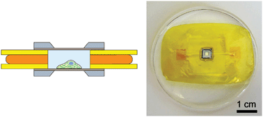
microfluidic x-ray cellsBritta Weinhausen and Sarah Köster study hydrated eukaryotic cells device using X-rays in a new microfluidic device in their Technical Innovation, free to access for 4 weeks*.

The team at Georg-August-Universität Göttinghen, Germany, overcome some of the limitations of previous work to create a new type of device suitable for studying hydrated cells, with the possibility of applying it to living cells.

They use UV-curable glue (NOA 81) allowing sophisticated channel structures to be defined, two thin Kapton foils, with advantageous properties for X-ray studies, and a Si3N4 membrane window, upon which cells can be grown and fixed before the device is closed with a second membrane. Despite the small difference in electron density between the cellular material and water, they successfully produce X-ray dark-field images:

Microfluidic devices for X-ray studies on hydrated cells
Britta Weinhausen and Sarah Köster
DOI: 10.1039/C2LC41014A


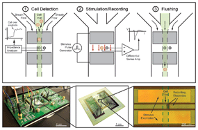
cytometryAs mentioned in the LOC issue 2 blog, Luke Lee et al. at the University of California, Berkeley and Stanford University, featured on the front cover with their work on sorting stem cells using a cell cytometer based on the electrophysiological response to stimulus. As a HOT article that was featured on the cover, this article has been free to access* for 6 weeks from mid-December so get your skates on to read it in full in the next week:

Label-free electrophysiological cytometry for stem cell-derived cardiomyocyte clusters
Frank B. Myers, Christopher K. Zarins, Oscar J. Abilez and Luke P. Lee
DOI: 10.1039/C2LC40905D


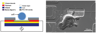
electroporationIn this Technical Innovation, Rashid Bashir led a team of researchers from the USA, Sweden and Singapore in using silicon nanowires for rapid lysis of single cells by ultra-localized electroporation to release cell components for analysis. They integrate label-free magnetic manipulation to correctly position cells with the use of field effect transistors to apply an electric field for cell lysis.

Their method eliminates the use of microfluidic trapping techniques, transparent substrates for optical tweezing and high voltages, amongst other requirements for previous devices.

This work is featured on the inside front cover of Issue 3, so it’s currently free to access for 6 weeks*. Do have a read of the article here:

Ultra-localized single cell electroporation using silicon nanowires
Nima Jokilaakso, Eric Salm, Aaron Chen, Larry Millet, Carlos Duarte Guevara, Brian Dorvel, Bobby Reddy, Amelie Eriksson Karlstrom, Yu Chen, Hongmiao Ji, Yu Chen, Ratnasingham Sooryakumar and Rashid Bashir
DOI: 10.1039/C2LC40837F

*Free access to individuals is provided through an RSC Publishing personal account. Registration is quick, free and simple

Digg This
Reddit This
Stumble Now!
Share on Facebook
Bookmark this on Delicious
Share on LinkedIn
Bookmark this on Technorati
Post on Twitter
Google Buzz (aka. Google Reader)

HOT article: A hydrodynamic study on streamer formation in biofilms

Researchers led by Aloke Kumar at the Oak Ridge National Laboratory, USA, and Thomas Thundat at the University of Alberta, Canada, used a porous microfluidic device to study biofilm streamer development in porous media.

The microfluidic method allowed close observation and measurement of streamers developing between microposts embedded in a microchannel. The formation of streamers in the device forming a web connecting different microposts was strongly influenced by the hydrodynamics.

Higher flow rates resulted in thicker structures and higher numbers of streamers as they grew much faster. At very high flow rate, their formation was only transient as they were destabilized after forming. The fluid flow seems to be the cause of both the formation and destabilization of the streamers. Interestingly, streamers formed parallel to the flow direction in regions of high flow rate and transverse to it in regions of low flow rate. In contrast with a previous report, they observe streamers distributed throughout the height of the device, attributed to secondary flow in the corners of the device. Carrying out numerical simulations, Kumar and Thundat et al. showed that secondary flows in the z-direction do not have a large role to play.

Development of streamers is complex and a great deal of detail remains to be elucidated, however, this Lab on a Chip article indicates that streamer formation might lead to mature microbial structures under the right conditions.

This HOT article is free to access* on our site for the next four weeks, so why not download the paper here:

A web of streamers: biofilm formation in a porous microfluidic device
Amin Valiei ,  Aloke Kumar ,  Partha P. Mukherjee ,  Yang Liu and Thomas Thundat
DOI: 10.1039/C2LC40815E

*Free access to individuals is provided through an RSC Publishing personal account. Registration is quick, free and simple

Digg This
Reddit This
Stumble Now!
Share on Facebook
Bookmark this on Delicious
Share on LinkedIn
Bookmark this on Technorati
Post on Twitter
Google Buzz (aka. Google Reader)

LOC issue 2 – sorting stem cell-derived cardiomyocyte clusters, cross talk between cancer and immune cells and Genome Sequence Scanning

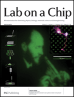
Welcome to Issue 2 of Lab on a Chip! This issue features the winner of the Lab on a Chip Pioneers of Miniaturisation Lectureship, Andrew deMello, on the back cover. Read more about this award and others given at MicroTAS here.

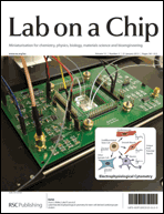
On the front cover of issue 2 is featured work from the group of Luke Lee at the University of California, Berkeley, in conjunction with colleagues at Stanford University.

In the paper, the authors use a ‘non-genetic, label-free cell cytometry method based on electrophysiological response to stimulus’ to sort undifferentiated stem cells from heterogeneous stem cell progeny.

The cell cytometer can identify human induced pluripotent stem cell-derived cardiomyocyte clusters from their extracellular field potential signals – these stem cells can then be used for various stem cell therapies.

Label-free electrophysiological cytometry for stem cell-derived cardiomyocyte clusters
Frank B. Myers, Christopher K. Zarins, Oscar J. Abilez and Luke P. Lee
DOI: 10.1039/C2LC40905D

Work from the Italian National Research Council – Institute for Photonics and Nanotechnologies and the Istituto Superiore di Sanità is featured on the inside front cover.

The groups, led by Fabrizio Mattei and Luca Businaro, have used an on-chip co-culture system to investigate interactions between cancer cells and a host’s immune system.

Cross talk between cancer and immune cells: exploring complex dynamics in a microfluidic environment
Luca Businaro, Adele De Ninno, Giovanna Schiavoni, Valeria Lucarini, Gabriele Ciasca, Annamaria Gerardino, Filippo Belardelli, Lucia Gabriele and Fabrizio Mattei
DOI: 10.1039/C2LC40887B

On the inside back cover, work from Robert Meltzer and co-workers at Pathogenetix, Inc. is featured.

In their paper, they present a novel compound funnel design for use with Genome Sequence Scanning (GSS) technology, which improves molecule throughput.

High-throughput genome scanning in constant tension fluidic funnels
Joshua W. Griffis, Ekaterina Protozanova, Douglas B. Cameron and Robert H. Meltzer
DOI: 10.1039/C2LC40943G

As with all our cover articles these are free to access for 6 weeks (following a simple registration for an RSC Publishing account).

Other HOT articles featured in the issue include:

Benchtop fabrication of microfluidic systems based on curable polymers with improved solvent compatibility
Michinao Hashimoto, Robert Langer and Daniel S. Kohane
DOI: 10.1039/C2LC40888K

Microfluidic assisted self-assembly of chitosan based nanoparticles as drug delivery agents
Fatemeh Sadat Majedi, Mohammad Mahdi Hasani-Sadrabadi, Shahriar Hojjati Emami, Mohammad Ali Shokrgozar, Jules John VanDersarl, Erfan Dashtimoghadam, Arnaud Bertsch and Philippe Renaud
DOI: 10.1039/C2LC41045A

Microfluidic devices for X-ray studies on hydrated cells
Britta Weinhausen and Sarah Köster
DOI: 10.1039/C2LC41014A

For even more exciting microfluidics research, read the full issue here.

Digg This
Reddit This
Stumble Now!
Share on Facebook
Bookmark this on Delicious
Share on LinkedIn
Bookmark this on Technorati
Post on Twitter
Google Buzz (aka. Google Reader)

Worried about food allergens? There’s an app for that

Building on their work on a mobile phone – or cell phone – app to detect bacteria, US scientists have now adapted the technology to detect allergens in foods.

The iTube platform uses colorimetric assays and a smartphone to reveal the presence of allergens in food samples

The iTube platform uses colorimetric assays and a smartphone to reveal the presence of allergens in food samples

The device, developed by Aydogan Ozcan and his colleagues at the University of California, Los Angeles, is a compact and lightweight attachment for a mobile phone’s camera unit, which is used to image tubes containing food samples illuminated by light emitting diodes. ‘If there is an allergen in the sample, the transmitted light intensity changes,’ says Ozcan. ‘By quantifying the transmitted light intensity using a smart application on the phone, we can quantify the amount of allergen in the sample in parts per million.’

 To read the full article, visit Chemistry World.

A personalized food allergen testing platform on a cellphone
Ahmet Coskun, Justin Wong, Delaram Khodadadi, Richie Nagi, Andrew Tey and Aydogan Ozcan
Lab Chip, 2012, Accepted Manuscript
DOI: 10.1039/C2LC41152K

Digg This
Reddit This
Stumble Now!
Share on Facebook
Bookmark this on Delicious
Share on LinkedIn
Bookmark this on Technorati
Post on Twitter
Google Buzz (aka. Google Reader)

Top ten most accessed articles in August 2012

The following articles were in the Lab on a Chip top ten most accessed for the month of August:

Microfluidic designs and techniques using lab-on-a-chip devices for pathogen detection for point-of-care diagnostics
Amir M. Foudeh, Tohid Fatanat Didar, Teodor Veres and Maryam Tabrizian
Lab Chip, 2012, 12, 3249-3266
DOI: 10.1039/C2LC40630F

Configurable 3D-Printed millifluidic and microfluidic ‘lab on a chip’ reactionware devices
Philip J. Kitson, Mali H. Rosnes, Victor Sans, Vincenza Dragone and Leroy Cronin
Lab Chip, 2012, 12, 3267-3271
DOI: 10.1039/C2LC40761B

Electrokinetics with “paper-and-pencil” devices
Pratiti Mandal, Ranabir Dey and Suman Chakraborty
Lab Chip, 2012, 12, 4026-4028
DOI: 10.1039/C2LC40681K

Microfluidic evaporator for on-chip sample concentration
Xavier Casadevall i Solvas, Vladimir Turek, Themistoklis Prodromakis and Joshua B. Edel
Lab Chip, 2012, 12, 4049-4054
DOI: 10.1039/C2LC40746A

Joining plasmonics with microfluidics: from convenience to inevitability
Jaeyoun Kim
Lab Chip, 2012, 12, 3611-3623
DOI: 10.1039/C2LC40498B

A novel microfluidic microplate as the next generation assay platform for enzyme linked immunoassays (ELISA)
Junhai Kai, Aniruddha Puntambekar, Nelson Santiago, Se Hwan Lee, David W. Sehy, Victor Moore, Jungyoup Han and Chong H. Ahn
Lab Chip, 2012, 12, 4257-4262
DOI: 10.1039/C2LC40585G

Optical imaging techniques in microfluidics and their applications
Jigang Wu, Guoan Zheng and Lap Man Lee
Lab Chip, 2012, 12, 3566-3575
DOI: 10.1039/C2LC40517B

Concentration gradient generation of multiple chemicals using spatially controlled self-assembly of particles in microchannels
Eunpyo Choi, Hyung-kwan Chang, Chae Young Lim, Taesung Kim and Jungyul Park
Lab Chip, 2012, 12, 3968-3975
DOI: 10.1039/C2LC40450H

Frontiers of optofluidics in synthetic biology
Cheemeng Tan, Shih-Jie Lo, Philip R. LeDuc and Chao-Min Cheng
Lab Chip, 2012, 12, 3654-3665
DOI: 10.1039/C2LC40828G

Electrode-free picoinjection of microfluidic drops
Brian O’Donovan, Dennis J. Eastburn and Adam R. Abate
Lab Chip, 2012, 12, 4029-4032
DOI: 10.1039/C2LC40693D

Why not take a look at the articles today and blog your thoughts and comments below.

Fancy submitting an article to Lab on a Chip? Then why not submit to us today or alternatively email us your suggestions.

Digg This
Reddit This
Stumble Now!
Share on Facebook
Bookmark this on Delicious
Share on LinkedIn
Bookmark this on Technorati
Post on Twitter
Google Buzz (aka. Google Reader)

Lab on a Chip gives prestigious awards at µTAS 2012

This year’s MicroTAS conference was held in October, at the Okinawa Convention Center in Okinawa, Japan.

As in previous years, Harp Minhas, Editor of Lab on a Chip, was in attendance at the conference to announce the prestigious Lab on a Chip awards, which include the Pioneers of Miniaturisation Lectureship (supported by Corning Inc), the Widmer Young Researcher Poster Prize, and the Art in Science Award (co-sponsored by NIST).

The Widmer Young Researcher Poster Prize

This year’s winner of the Widmer Young Researcher Poster Prize was Klaus Eyer from Professor Petra Dittrich’s lab at ETH Zürich, with his poster entitled ‘Single Cell ELISA’.

Left to right: David Juncker (Poster award chair), Klaus Eyer (winner), Harp Minhas (Lab on a Chip)

Art in Science Award

The Art in Science Award is given each year “to draw attention to the aesthetic value in scientific illustrations while still conveying scientific merit.” This year’s award was presented to Yi Zhang, a PhD student from the Department of Biomedical Engineering, Johns Hopkins University School of Medicine, USA.

Left to right: Michael Gaitan (NIST), Yi Zhang (winner), Harp Minhas (Lab on a Chip)

The image, entitled ‘Stretching the Rainbow‘, shows a droplet with multiple rainbow stripes being stretched by the magnetic particle on a surface energy traps (SETs)-enabled magnetic digital microfluidic platform. In this particular scenario, the droplet is immobilized by the SET while the magnetic particles are trying to split from the droplet. The rainbow is the natural colour resulting from the diffraction pattern caused by a DVD disc, on which the droplet sits.

Stretching the Rainbow

Pioneers of Miniaturisation Lectureship

The Pioneers of Miniaturisation Lectureship recognises outstanding achievements and significant contributions to the understanding and advancement of micro- and nano-scale science. This year, the Lectureship was awarded to Professor Andrew deMello at ETH Zurich, Switzerland. Among his many achievements and awards, Andrew first demonstrated combinatorial chemistry and controlled nanoparticle synthesis in continuous flow microfluidic devices; co-authored the first demonstration of continuous flow PCR, which has over 750 citations to date; pioneered the application of high-contract fluorescence lifetime imaging to microfluidic environments; founded Molecular Vision Ltd, an in vitro diagnostic company, providing point of care tests for cardiovascular and kidney disease; and has published over 40 articles on droplet-based microfluidics since 2007.

Left to right: Harp Minhas (Lab on a Chip), Andrew deMello (winner), Po Ki Yuen (Corning Inc)

Please join us at Lab on a Chip in congratulating all of our prize winners!

Digg This
Reddit This
Stumble Now!
Share on Facebook
Bookmark this on Delicious
Share on LinkedIn
Bookmark this on Technorati
Post on Twitter
Google Buzz (aka. Google Reader)

Idea featured in Lab on a Chip article to benefit from the RSC’s support of Marblar

The RSC has teamed up with an Oxford University spin-out to sponsor six challenges on Marblar – a radical new online platform for finding applications for unused scientific discoveries.

More than 95% of technologies developed in universities never make it to market, leaving publicly or philanthropy-funded research collecting dust on the shelf.  Often, given that academic research can be so ahead of its time, the commercial relevance of these technologies isn’t immediately obvious.

Marblar aims to remove this bottleneck by crowdsourcing ideas for real-world applications from the global science and technology community, with the ultimate goal to create new products and new companies that will drive job creation around these innovative discoveries.

An idea from one Lab on a Chip article – SlipChip – has been chosen as one of the RSC sponsored challenges.

SlipChip is a low-cost microfluidic device that uses only two pieces of plastic or glass to enable the user to perform multiple small-scale chemical reactions simply and precisely. By simply ‘slipping’ the glass or plastic ‘chips’ across each other, a number of reactions can be carried out in nanoscale volumes in parallel.

SlipChip was developed at the University of Chicago by Professor Rustem Ismagilov and his then graduate student, Feng Shen. They’ve since created a spinout company called SlipChip based on this technology and Ismagilov has continued his work at the California Institute of Technology. Given the technology’s ability to precisely manipulate reactions in a programmable way, they see applications in multiple fields. Through Marblar, they hope to find ideas beyond their discipline that can exploit SlipChip’s ability to ‘count molecules’, as well as new capabilities for the technology.

Visit the Marblar website to get involved, or read the Lab on a Chip article below:

Digital PCR on a SlipChip
Feng Shen, Wenbin Du, Jason E. Kreutz, Alice Fok and Rustem F. Ismagilov
DOI: 10.1039/C004521G

Digg This
Reddit This
Stumble Now!
Share on Facebook
Bookmark this on Delicious
Share on LinkedIn
Bookmark this on Technorati
Post on Twitter
Google Buzz (aka. Google Reader)