Innovative cell culture and analysis microdevices

Here we have two HOT articles for you to read, both of which are free to access for the next 4 weeks*:

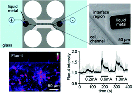
Researchers at North Carolina State University, USA, integrate microelectrodes into a neuron culture platform creating a single device with which cells can be electrically stimulated and their behaviour recorded simultaneously for scientific experiments. They do this using gallium and its alloys, which are liquid at room temperature and flow into microchannels to create the microelectrodes. This integrated biocompatible device avoids the complication of manually aligning the electrodes and provides subcellular accuracy.

Integration of pre-aligned liquid metal electrodes for neural stimulation within a user-friendly microfluidic platform
Nicholas Hallfors, Asif Khan, Michael D. Dickey and Anne Marion
DOI: 10.1039/C2LC40954B

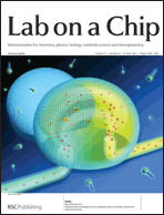
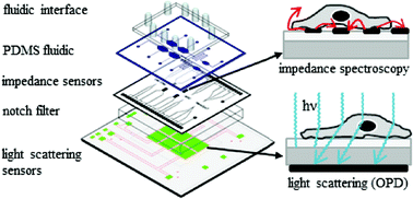
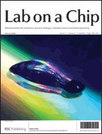
As featured on the outside front cover of Issue 5, HOT article number two is from a team led by Peter Ertl at the Austrian Institute of Technology, Austria, and Siemens AG, Germany. They combine two methods of cell analysis into one miniaturised device for dual-parameter analysis of cell cultures. The first technique is electrical impedance spectroscopy, normally used in analysing adherent cell cultures. The second is optical light scattering, normally used in flow cytometry. The combination of the two to detect light scattering from adherent cells enables rapid identification of cell number variations during culture, assessment of cell adhesion and interactions in physiological conditions. Applications are envisaged in cell–drug interaction assays and cytotoxicity screening. It also identifies intracellular granularity, which indicates apoptosis.

Standardization of microfluidic cell cultures using integrated organic photodiodes and electrode arrays
Verena Charwat, Michaela Purtscher, Sandro F. Tedde, Oliver Hayden and Peter Ertl
DOI: 10.1039/C2LC40965H

*Free access to individuals is provided through an RSC Publishing personal account. Registration is quick, free and simple

Digg This
Reddit This
Stumble Now!
Share on Facebook
Bookmark this on Delicious
Share on LinkedIn
Bookmark this on Technorati
Post on Twitter
Google Buzz (aka. Google Reader)

Video of microfluidic origami – digital microfluidics with integrated mass spectrometry to monitor microreactions in real time

Lab on a Chip editorial board member Aaron Wheeler and Andrea Kirby at the University of Toronto enable analysis on-chip with a new device format for digital microfluidics (DMF) based chemical synthesis that they are calling “microfluidic origami”.

The setup allows DMF to be integrated with mass spectrometry for in-line analysis. Offline analysis on the benchtop is particularly troublesome for mass spectrometry as it requires further handling steps and takes further time. There are three devices that have coupled DMF with electrospray ionisation before. However, these either have challenges associated with device fabrication or need external equipment and tricky alignment of device and ESI emitter.

This video from the Wheeler laboratory clearly shows how the device is folded, like origami, and explains how it works.

http://www.youtube.com/watch?v=-lpQhPBlu7M 

The “origami” aspect is down to a folded nanoelectrospray ionization (nanoESI) emitter on a single flexible polyimide film. This work combines previously known concept of folded ESI emitters and that of making DMF devices with flexible substrates to move droplets on non-planar surfaces to get an integrated device on one flexible substrate. They include a two-plate-to-one-plate DMF interface to give ease of droplet delivery from the mixing region to the nanoESI emitter.

They validate the device with a Morita-Baylis-Hilman reaction, monitoring it in-line in real time. This is the first time a digital microfluidic device has been used to monitor chemical synthesis in real time with in-line mass spectrometry.

This HOT article has been made free to access for the next 4 weeks*, so after watching the video you can read the scientific detail of how they fabricated and validated the device:

Microfluidic origami: a new device format for in-line reaction monitoring by nanoelectrospray ionization mass spectrometry
Andrea E. Kirby and Aaron R. Wheeler
DOI: 10.1039/C3LC41431K

Digg This
Reddit This
Stumble Now!
Share on Facebook
Bookmark this on Delicious
Share on LinkedIn
Bookmark this on Technorati
Post on Twitter
Google Buzz (aka. Google Reader)

Webinar: The power of modern HPTLC

Join Chemistry World and Advion for a webcast on the latest developments in HPTLC technology.

WHAT: Professor Morlock from the University of Giessen, Germany, will give an overview of current HPTLC methodology, explore some examples of HPTLC-MS coupling and review other current hyphenations in HPTLC. By the end of this free webinar, you will be able to:
– Recognise the power of modern HPTLC
– Learn about current hyphenations in HPTLC
– Understand the principle of elution-based HPTLC-MS
– Recognise how HPTLC hyphenations efficiently support analyses

WHEN: Wednesday, 20 March 2013 – 15:00 GMT

HOW: Click here to register (free)

Register today, even if you can’t make it on 20th March, and we’ll send you a link to the recorded webinar.

Digg This
Reddit This
Stumble Now!
Share on Facebook
Bookmark this on Delicious
Share on LinkedIn
Bookmark this on Technorati
Post on Twitter
Google Buzz (aka. Google Reader)

HOT article: Nodes in pore channel create signature to count even the smallest particles in a heterogeneous population

Counting of cells and even smaller particles, such as single molecules of DNA and viruses, can be carried out using resistive-pulse sensing (RPS). The particles move through a pore set up with a flow of current across it. When a particle goes through, it changes the resistance, which can be measured and used to count the particles and even size them. The difficulty is that small molecules in a heterogeneous mixture get lost amongst the large molecules and remain undetected as the pore must be large enough for the largest particle. The signal-to-noise ratio is the limiting factor on sensitivity.

Researchers at the University of California, Berkeley, USA, led by Lydia Sohn present solutions to these limitations in this HOT article with a method they title node-pore sensing (NPS). Instead of one uniform pore width, the microfluidic device has a pore with a sequence of nodes down the length of the pore channel. The nodes in their device are larger than the rest of the channel.  The presence of these nodes means that the device sensitivity is three orders above that of other RPS sensors. The simple method enables the spacing, number and size of these nodes to be changed.

The number and spacing of the nodes changes the normal pulse signal output accordingly, producing a very identifiable corresponding rise and fall pattern. Using this signature, the particle can be detected even with usually problematic signal-to-noise ratios.  By changing the flow rate and specifically designing the node arrangement, this signature pattern can be controlled to distinguish even very small particles through a large pore. The size range of particles that can be measure with the one pore is therefore considerable.

This article is well worth a read, offering numerous advantages over current devices and offering the potential for point-of-care analysis. It’s free to access for the next four weeks*, read it by clicking the link below:

Node-pore sensing: a robust, high-dynamic range method for detecting biological species
Karthik Balakrishnan, George Anwar, Matthew R. Chapman, Trongtuong Nguyen,  Anand Kesavarajud and Lydia L. Sohn
DOI:10.1039/C3LC41286E

*Free access to individuals is provided through an RSC Publishing personal account. Registration is quick, free and simple

Digg This
Reddit This
Stumble Now!
Share on Facebook
Bookmark this on Delicious
Share on LinkedIn
Bookmark this on Technorati
Post on Twitter
Google Buzz (aka. Google Reader)

Issue 7 online! The latest HOT miniaturisation research and critical reviews on microfabricated organ systems, nucleic acid amplification and microfluidics in IVF

As highlighted on the outside front cover, physicists, engineers and chemists at the University of Illinois , USA, describe a new laser-based label-free resonant optical biosensor with high resolution and high sensitivity. The difference with this sensor is that they use a photonic crystal resonant reflector surface to introduce optical gain to the sensor from an external source via simulated emission. All of our cover articles are free to access for 6 weeks,* so you can read the full work now:

External cavity laser biosensor
Chun Ge, Meng Lu, Sherine George, Timothy A. Flood, Clark Wagner, Jie Zheng, Anusha Pokhriyal, J. Gary Eden, Paul J. Hergenrother and Brian T. Cunningham
DOI: 10.1039/C3LC41330F

On the inside front cover, researchers at California Institute of Technology, USA, and LeukoDx Inc., Israel, present a point-of-care test for leukocyte counting using a microflow cytometer and fluorescent dye. This method eliminated excessive dilution and sheath flow, giving a minimal needed sample volume.

Four-part leukocyte differential count based on sheathless microflow cytometer and fluorescent dye assay
Wendian Shi, Luke Guo, Harvey Kasdan and Yu-Chong Tai
DOI: 10.1039/C3LC41059E

Work from Tino Frank and Savaş Tay at ETH Zurich is featured on the inside back cover. To improve studies of cell signalling in vitro, this article introduces a simple cell culture platform that can produce programmable diffusion-based gradients using microfluidics based on modifying flow over time. Read it here:

Flow-switching allows independently programmable, extremely stable, high-throughput diffusion-based gradients
Tino Frank and Savaş Tay
DOI: 10.1039/C3LC41076E

The outside back cover highlights the work of Sung Gap Im at KAIST, South Korea. This article is about a doubly cross-linked nano-adhesive system (DCNA) and the team demonstrate fabrication of microfluidic devices with flexible and rigid substrates with high strength and stability. The flexible devices could be manipulated without delamination occurring.

A doubly cross-linked nano-adhesive for the reliable sealing of flexible microfluidic devices
Jae Bem You, Kyoung-Ik Min, Bora Lee, Dong-Pyo Kim and Sung Gap Im
DOI: 10.1039/C2LC41266G


In addition to the primary microfluidics research and high number of HOT articles in issue 7, there are also three critical reviews:

Microfabricated mammalian organ systems and their integration into models of whole animals and humans
Jong H. Sung, Mandy B. Esch, Jean-Matthieu Prot, Christopher J. Long, Alec Smith, James J. Hickman and Michael L. Shuler
DOI: 10.1039/C3LC41017J

Thinking big by thinking small: application of microfluidic technology to improve ART
J. E. Swain, D. Lai, S. Takayama and G. D. Smith
DOI: 10.1039/C3LC41290C

Nucleic acid amplification using microfluidic systems
Chen-Min Chang, Wen-Hsin Chang, Chih-Hung Wang, Jung-Hao Wang, John D. Mai and Gwo-Bin Lee
DOI: 10.1039/C3LC41097H

View the whole issue here

*Free access to individuals is provided through an RSC Publishing personal account. Registration is quick, free and simple


Digg This
Reddit This
Stumble Now!
Share on Facebook
Bookmark this on Delicious
Share on LinkedIn
Bookmark this on Technorati
Post on Twitter
Google Buzz (aka. Google Reader)

How to tickle a cell: on-chip microrobotic mechanical sensor

In the past researchers developed many different microrobots for the mechanical studies of single cells. Some of them require complicated micromanipulators; others can be controlled with optical tweezers, but are limited in the force that they exert and so are inapplicable to cells with stiff cell walls.

A team from institutes in the USA and Japan, including MIT and RIKEN, describes in a recent Lab Chip article a new type of microrobot that can be installed in a microfluidic chip and used for mechanical stimulation of single cells. In this device a robotic arm with a mechanical probe at its end is built from a magnetic material and can be moved within the structure of the microchip by applying magnetic force. This design means that there are no complex mechanical assemblies necessary to move the arm. The probe itself consists of a microneedle placed on a flexible base. The force applied with the needle is measured by the extent of base deformation.

Tomohiro Kawahara and colleagues built a microchip with the microrobotic probe in its main chamber and use it to study the reaction of small single-cellular organisms, diatoms, to mechanical stimulus. They are able to adjust the applied force so that the cell itself isn’t damaged. Upon stimulation they observe agglomeration of chloroplasts into two separate groups. They also quantify the stimulus required for chloroplast assembly to occur.

Although the experiment described by Kawahara and colleagues is a simple proof-of-concept, it does mean that a wealth of phenomena can now be examined in more detail than ever before. Another example where mechanical stimulus elicits a response is in the small organism dinoflagellate, which bioluminesces when mechanically probed. The response of many cells to this sort of stimulus mimics their response to attack by pathogens and Miyawaki’s microrobot now opens the way to studying such responses.

This HOT article is free to access for the next 4 weeks*, just click on the link below:

On-chip microrobot for investigating the response of aquatic microorganisms to mechanical stimulation
Tomohiro Kawahara, Masakuni Sugita, Masaya Hagiwara, Fumihito Arai, Hiroyuki Kawano, Ikuko Shihira-Ishikawa and Atsushi Miyawaki
DOI: 10.1039/C2LC41190C

 *Free access to individuals is provided through an RSC Publishing personal account. Registration is quick, free and simple

Published on behalf of Rafal Marszalek, Molecular BioSystems web science writer. Rafal is an Assistant Editor of Genome Biology at BioMed Central


Digg This
Reddit This
Stumble Now!
Share on Facebook
Bookmark this on Delicious
Share on LinkedIn
Bookmark this on Technorati
Post on Twitter
Google Buzz (aka. Google Reader)

Two post-doctoral research positions in microfluidics

For those of you reading this blog who are active researchers in the field of microfluidics, here are two job opportunities that have been brought to our attention, which may well be of interest:

Research Associate (MEMPHIS project) at UCL Department of Chemical Engineering
Full time, funded for three years in the first instance, available immediately, closing date 09/03/2013
Salary: £32,375 – £39,132 per annum

Applications are invited for a research associate position in the Department of Chemical Engineering at UCL. The main role will be to carry out experimental investigations of multiphase flows. This post will be funded by a £5M EPSRC Programme Grant that will harness the synergy between world-leading scientists from four prestigious institutions: Imperial College, Birmingham, Nottingham and UCL, to create the next generation modelling tools for complex multiphase flows. This will require a programme of focussed, multi-scale experiments on multiphase flows to validate and update numerical codes.

The experimental work will involve application of optical diagnostic methods on small and large scale geometries, with an additional aim of developing routes to create novel multiphase structures which can be exploited by our industrial partners. It will be expected that the post holder will interact closely with the numerical component of the Programme, carried out at all the participating universities, in order to validate, and update the numerical tools, and to provide input into the design of experiments. Experience in PIV preferred.

For key requirements and further details please download the full job advert by clicking on MEMPHIS_Advert_Feb13.

For queries about the application process contact Miss Agata Blaszczyk at a.blaszcuyk@ucl.ac.uk; for informal inquiries please contact Dr P. Angeli at p.angeli@ucl.ac.uk or visit http://www.ucl.ac.uk/chemeng/vacancies for further information.


Post-Doctoral Research Fellow (Boiling in Microchannels) at Brunel University School of Engineering and Design
Three years beginning 1st May 2013, closing date 21/03/2013
Salary £32,590 – £38,464 per annum

Applications are sought for a Post-Doctoral Research Fellow to join a very active research team in the area of two-phase flow in microchannels. The research will work on a project entitled “Boiling in Microchannels – Integrated design of closed-loop cooling system for devices operating at high heat fluxes”.  The project is in collaboration with Edinburgh University and is funded by EPSRC. It is also sponsored by four UK based companies. The project aims to study fundamental and practical aspects of flow boiling in micro single and multichannel arrangements that will elucidate physical phenomenal and lead to actual prototype designs for use in high flux devices. The work at Brunel will be experimental.

For key requirements and further details please download the full job advert by clicking on DEA0559-1;

If you have any queries please contact Professor Karayiannis at tassos.karayiannis@brunel.ac.uk or visit https://jobs.brunel.ac.uk/WRL

Digg This
Reddit This
Stumble Now!
Share on Facebook
Bookmark this on Delicious
Share on LinkedIn
Bookmark this on Technorati
Post on Twitter
Google Buzz (aka. Google Reader)

HOT review: Forming the wiring of the nervous system – discovering the mechanisms of axon guidance through new technologies

Today’s HOT article is a review article that was featured on the front cover of our Neuroengineering themed issue.

In this review, Santiago Costantino et al. at the University of Montreal and McGill University, Canada, critically discuss the most recent insights into axon guidance that have been enabled by developments in methods and technologies for engineered cell substrates.

This review includes:

  1. Extracellular cues and growth cones
  2. Historical background
  3. The stripe assay in discovery of guidance cues and signalling mechanisms
  4. Microcontact printing – development from the stripe assay
  5. Insights via microfluidics
  6. Studies using laser-assisted printing and patterning 3D hydrogels

The focus is on the biological insights into how axons recognise and interpret external signals in order to reach their synaptic target that have come out of using these technologies, rather than the development of the technology itself. This cover article is still free to access for another couple of weeks*, read it now by clicking the link below:

Engineered cell culture substrates for axon guidance studies: moving beyond proof of concept
Joannie Roy, Timothy E. Kennedy and Santiago Costantino
DOI: 10.1039/C2LC41002H

*Free access to individuals is provided through an RSC Publishing personal account. Registration is quick, free and simple

Digg This
Reddit This
Stumble Now!
Share on Facebook
Bookmark this on Delicious
Share on LinkedIn
Bookmark this on Technorati
Post on Twitter
Google Buzz (aka. Google Reader)

Welcome to Lab on a Chip Issue 6! A microrobot, adaptive coatings, protein detection, flow cytometry, a critical review and the final Acoustofluidics paper

On the outside front cover is a Technical Innovation from Oliver Hayden, Michael Helou and co-workers in Germany describing a magnetic time-of-flight flow cytometry workflow for point-of-care tests, integrating sample preparation with the use of superparamagnetic labels:

Time-of-flight magnetic flow cytometry in whole blood with integrated sample preparation
Michael Helou, Mathias Reisbeck, Sandro F. Tedde, Lukas Richter, Ludwig Bär, Jacobus J. Bosch, Roland H. Stauber, Eckhard Quandt and Oliver Hayden
DOI: 10.1039/C3LC41310A


The inside front cover highlights work by Martin Gijs’s team at EPFL, Switzerland, on a microfluidic method for ultra-sensitive protein detection in serum using magnetic beads:

Attomolar protein detection using a magnetic bead surface coverage assay
H. Cumhur Tekin, Matteo Cornaglia and Martin A. M. Gijs
DOI: 10.1039/C3LC41285G


Mengsu Yang and co-workers at the City University of Hong Kong are advertised on the back cover, which illustrates their research on monitoring of intracellular calcium signals after mechanical stimulation using a single integrated microfluidic device:

Microfluidics study of intracellular calcium response to mechanical stimulation on single suspension cells
Tao Xu, Wanqing Yue, Cheuk-Wing Li, Xinsheng Yao and Mengsu Yang
DOI: 10.1039/C3LC40880A


Remember, all of our cover articles are free to access for 6 weeks!*

*Free access to individuals is provided through an RSC Publishing personal account. Registration is quick, free and simple


The final paper in our highly interesting Acoustofluidics series of tutorial articles is published in Issue 6. It points out the ability to combine acoustic forces with others for microfluidic applications:

Acoustofluidics 23: acoustic manipulation combined with other force fields
Peter Glynne-Jones and Martyn Hill
DOI: 10.1039/C3LC41369A

Issue 6 also includes a Critical Review on sample preparation for cell analysis technology from Dino Di Carlo et al. at the University of California, USA:

Microfluidic sample preparation for diagnostic cytopathology
Albert J. Mach, Oladunni B. Adeyiga and Dino Di Carlo
DOI: 10.1039/C2LC41104K

HOT articles in Issue 6 include:

On-chip microrobot for investigating the response of aquatic microorganisms to mechanical stimulation
Tomohiro Kawahara, Masakuni Sugita, Masaya Hagiwara, Fumihito Arai, Hiroyuki Kawano, Ikuko Shihira-Ishikawa and Atsushi Miyawaki
DOI: 10.1039/C2LC41190C

Dynamic pH mapping in microfluidic devices by integrating adaptive coatings based on polyaniline with colorimetric imaging techniques
Larisa Florea, Cormac Fay, Emer Lahiff, Thomas Phelan, Noel E. O’Connor, Brian Corcoran, Dermot Diamond and Fernando Benito-Lopez
DOI: 10.1039/C2LC41065F

For more fascinating Lab on a Chip content, read the full issue here

Digg This
Reddit This
Stumble Now!
Share on Facebook
Bookmark this on Delicious
Share on LinkedIn
Bookmark this on Technorati
Post on Twitter
Google Buzz (aka. Google Reader)

Constant stretching of DNA in a microfluidic device for higher throughput Genome Sequence Scanning

This HOT article by Robert Meltzer et al. at Pathogenetix Inc., USA, describes new geometries for the stretching funnel in Genome Sequence Scanning that increase DNA throughput by 30 times.

Genome Sequence Scanning (GSS) detects sequence-specific fluorescent tags on DNA fragments. It is used to identify bacteria by first lysing open the bacteria cell to release the DNA, then using a restriction endonuclease enzyme to digest the DNA into smaller fragments. Fluorescent tags are added that recognise specific repeated elements present in all bacterial genomes. GSS characterises the bacterial genome by the spatial distribution of the tags.

genome sequence scanning, GSSThe detection is done in a continuous flow microfluidic device with confocal microscopy. In order to carry out the spatial recognition of the fluorescent tags along the length of the DNA fragment, it needs to be stretched out into a linear form using a funnel. High molecule throughput is important as the detection confidence of this technology relies on observing as many tags as possible in the specified experimental period.

The team looks at the relationship between the funnel taper shape and related parameters, such as fluid velocity and fragment length, to improve the current designs and increase throughput. Their new geometries are able to keep the tension applied to the DNA constant during the detection process. Because DNA fragments come in various lengths, a very important goal is to maximise the range of lengths that can be stretched effectively with the funnel. The influence of channel etch depth on fluid flow, and therefore throughput, is also considered.

You can learn more about the work of Pathogenetix on Genome Sequence Scanning by visiting their website. This HOT article, which was featured on the cover of Issue 2, is free to access for the next 4 weeks* and you can read it by clicking the link below:

High-throughput genome scanning in constant tension fluidic funnels
Joshua W. Griffis, Ekaterina Protozanova, Douglas B. Cameron and Robert H. Meltzer
DOI: 10.1039/C2LC40943G#

*Free access to individuals is provided through an RSC Publishing personal account. Registration is quick, free and simple


Keep up to date with the latest Lab on a Chip news and blogs by following us on Twitter! @LabonaChip

Digg This
Reddit This
Stumble Now!
Share on Facebook
Bookmark this on Delicious
Share on LinkedIn
Bookmark this on Technorati
Post on Twitter
Google Buzz (aka. Google Reader)