Game on!

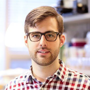
Written by Darius Rackus, PhD researcher at University of Toronto

Researchers at Standford University develop multi-level programming language for biotic games using swarms of microorganisms

Computer games are a ubiquitous pastime and a great example of how a single programming language give rise to a myriad of games. But what about biotic games? How could you program biological systems to function in an interactive way? Biotic games are interactive applications that interface biology and computer science for the promotion of science. The Riedel-Kruse Lab at Standford specialize in developing biotic games that use light to control swarms of Euglena gracilis—a phototaxic microorganism that avoids—and can direct, capture, and move whole swarms or individual organisms.

But programming swarms of microorganisms is no easy task. Swarms exhibit collective behaviour and therefore need to be controlled through local context rather than at the individual level. In their recent publication, the Riedel-Kruse Lab developed a set of hierarchical programming abstractions that allows swarms of Euglena within a biological processing unit (BPU; i.e., chip, microscope, and light stimuli) to be programmed in a single and efficient language at the stimulus, swarm, and system levels. At the lowest level, stimulus space programming (which the authors analogize to machine code) allows the programmer to have direct control over the various stimuli (e.g., turn left light on for 3 s), independent of the Euglena. Higher level programming at the swarm and system levels are more general and commands are given in terms of what the user wants the Euglena or system to do. For instance, swarm space commands direct the swarm in different operations such as move, split, and combine. System space commands incorporate conditional statements that can be used to confine a specific number of Euglena to a certain region or to clear Euglena from the field of view, for example.


While Lam et al. used this new language to program a biotic game, this new language and approach to swarm programming could be generalized for any type of swarm and stimuli. One application could be to program swarms to construct complex structures on the microscale. In future, by increasing access to BPUs through cloud computing and releasing this new programming language it will be possible for hobbyists and researchers alike to write new programs and applications. And maybe this is just the beginning of a revolution like the one ushered in by the release of the personal microcomputer.


To download the full article for free* click the link below:

Device and programming abstractions for spatiotemporal control of active micro-particle swarms

Amy T. Lam, Karina G. Samuel-Gama, Jonathan Griffin, Matthew Loeun, Lukas C. Gerber, Zahid Hossain, Nate J. Cira, Seung Ah Lee and Ingmar H. Riedel-Kruse

Lab Chip, 2017,17, 1442-1451

DOI: 10.1039/C7LC00131B


*Free to access until 24th May 2017.


About the Webwriter

Darius Rackus is finishing his Ph.D. at the University of Toronto working in the Wheeler Lab. His research interests are in combining sensors with digital microfluidics for healthcare applications.

Digg This
Reddit This
Stumble Now!
Share on Facebook
Bookmark this on Delicious
Share on LinkedIn
Bookmark this on Technorati
Post on Twitter
Google Buzz (aka. Google Reader)

Microfluidics, Physics & Chemistry of – Gordon Research Conference

Lab on a Chip is delighted to be a sponsor of: 

Microfluidics, Physics & Chemistry of  – Gordon Research Conference.

The event will take place on 4th – 9th June 2017 in Barga, Italy.

The conference brings together scientists, engineers and clinicians to discuss and advance cutting edge knowledge of microfluidics. Microfluidics are small scale systems that could be used to diagnose disease, enable unique physical and biological experiments and create new materials.

The goal of the 2017 meeting is to bridge the gap between scientists and engineers focused on fundamentals and those translating fundamental work into new applications. The conference is sure to spark animated discussion, new interactions and fruitful collaborations!

The conference will consist of topical sessions and active poster sessions. Topics include:

  • Point of Care Technologies
  • Nucleic Acid Analysis and Next Generation Sequencing
  • Fields, Forces, and Flows
  • Field-Structure Interactions
  • Wearable and Implantable Systems
  • Cell Mechanics
  • Living Systems
  • 3D Printing

The event is being chaired by Dino Di Carlo (UCLA, Lab on a Chip Editorial Board member) and Catherine M. Klapperich (Boston University) and vice chaired by Hang Lu (Georgia Institute of Technology, Lab on a Chip Associate Editor) and Cullen R. Buie (MIT).

Invited speakers include Lab on a Chip Advisory Board members Yanyi Huang (Peking University), Tony Huang (Duke University) and Albert Folch (University of Washington).

Apply by 7th May to secure your place!

Digg This
Reddit This
Stumble Now!
Share on Facebook
Bookmark this on Delicious
Share on LinkedIn
Bookmark this on Technorati
Post on Twitter
Google Buzz (aka. Google Reader)

Optical DNA maps

Just like Google maps, DNA maps can tell us the distance between two genes, and allow us to zoom in on the region of interest. DNA mapping started with human genome project, where DNA sequencing techniques opened a way to unveil the genetic information. However, determining the unique places and repetitions of four “chemical letters” found in our DNA—together known as the genes—is a difficult mission due to temperature, pH, and pressure sensitivity of the molecule.  DNA mapping technology allows for easy identification of large structural variations in DNA and therefore provides long-range information of the genome and can more.

Optical DNA mapping has emerged in the past decade as a powerful alternative to other DNA sequencing techniques since it can easily be applied with reduced risk of DNA damage. Over 100000 basepairs of DNA molecules, which are quite difficult to handle with other techniques, are labeled, stretched, and rendered in a single image. The stretching part is done using nanochannels (and therefore lab-on-a-chip technology), while the labeling part can be done by either enzymatic or affinity-based techniques (Figure 1). The concept and applications of optical DNA mapping has recently been very well explained in a tutorial review written by Vilhelm Müller and Fredrik Westerlund from Chalmers University of Technology in Sweden.

In enzymatic labelling nucleotides at particular regions on a single DNA strand are replaced by new ones using a DNA polymerase. The replacement nucleotides are then utilized to incorporate fluorophores into the DNA strand and allow for visualization. Nicking enzymes and methyl-transferases present two different approaches to employ enzymatic labelling process. While the use of differently colored fluorophores extends the applicability of this technique, the final resolution depends on the degree of stretching and the density of fluorophores on the region.

Affinity-based labelling is based on non-covalent interactions which can be enabled by either denaturation mapping or competitive binding. In denaturation mapping, DNA is heated to discriminate between the bases by their different bond energies. While G-C-basepairs still hold both strands of DNA—due to 3 hydrogen bonds holding them—, A-T-basepairs will melt—due to 2 hydrogen bonds holding them—. At this stage, an intercalating fluorescent dye can be linked to G-C-basepairs, allowing for imaging. Competitive binding relies on the usage of a fluorescent intercalating dye and a molecule selective for either A-T or G-C regions. Therefore, fluorescent dye cannot bind where the selective molecules have already bound. An optical map of DNA molecules can be obtained in this way. Affinity-based labelling is also highly dependent on the degree of stretching.


Optical DNA mapping techniques are useful tools for a wide range of applications from assembly of complex genomes to bacterial plasmid epidemiology. The concept opens up exciting research directions as it allows for automation of whole analysis using lab-on-a-chip systems and observation of the results using smartphones.

optical DNA mapping

Figure 1. Schematic illustration of DNA labelling techniques used in optical DNA mapping. Enzyme-based labelling involves nicking enzymes and methyl-transferases techniques, while affinity-based labelling can be employed by denaturation mapping or competitive binding methods. This figure is adapted from “Optical DNA mapping in nanofluidic devices: principles and applications” paper.

To download the full article for free* click the link below:

Optical DNA mapping in nanofluidic devices: principles and applications

Vilhelm Müller and Fredrik Westerlund

Lab Chip, 2017, Articles

DOI: 10.1039/C6LC01439A


*Free to access until 5th May 2017.


About the Webwriter

Burcu Gumuscu is a postdoctoral fellow in BIOS Lab on a Chip Group at University of Twente in The Netherlands. Her research interests include development of microfluidic devices for next generation sequencing, compartmentalized organ-on-chip studies, and desalination of water on the microscale.


Digg This
Reddit This
Stumble Now!
Share on Facebook
Bookmark this on Delicious
Share on LinkedIn
Bookmark this on Technorati
Post on Twitter
Google Buzz (aka. Google Reader)

New YouTube Videos

A vascularized and perfused organ-on-a-chip platform for large-scale drug screening applications


Pneumatic stimulation of C. elegansmechanoreceptor neurons in a microfluidic trap

 
Improved bovine embryo production in an oviduct-on-a-chip system: Prevention of poly-spermic fertilization and parthenogenic activation

Digg This
Reddit This
Stumble Now!
Share on Facebook
Bookmark this on Delicious
Share on LinkedIn
Bookmark this on Technorati
Post on Twitter
Google Buzz (aka. Google Reader)

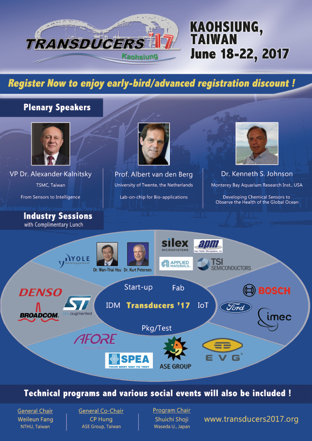
Transducers ’17

Digg This
Reddit This
Stumble Now!
Share on Facebook
Bookmark this on Delicious
Share on LinkedIn
Bookmark this on Technorati
Post on Twitter
Google Buzz (aka. Google Reader)

New YouTube Videos

Liquid-capped encoded microcapsules for multiplex assays


Pinch-off Dynamics and Dripping-onto-Substrate (DoS) Rheometry of Complex Fluids


3D microvascular model recapitulates the diffuse large B-cell lymphoma tumor microenvironment in vitro

Digg This
Reddit This
Stumble Now!
Share on Facebook
Bookmark this on Delicious
Share on LinkedIn
Bookmark this on Technorati
Post on Twitter
Google Buzz (aka. Google Reader)

Amy E. Herr – Our New Advisory Board Member!


We are delighted to announce our new Advisory Board member – Amy E. Herr!

Amy E. Herr is the Lester John & Lynne Dewar Lloyd Distinguished Professor of Bioengineering at the University of California, Berkeley and a Chan Zuckerberg (CZ) Biohub Investigator. Prior to joining UC Berkeley, she was a staff member at Sandia National Laboratories (Livermore, CA), earned Ph.D. and M.S. degrees in Mechanical Engineering from Stanford University, and completed her B.S. in Engineering and Applied Science with honors from the California Institute of Technology. Her research has been recognized by the NIH New Innovator Award, NSF CAREER Award, Alfred P. Sloan Fellowship (Chemistry), and DARPA Young Faculty Award. Professor Herr has chaired the Gordon Research Conference (GRC) on the Physics & Chemistry of Microfluidics. She is an elected Fellow of the American Institute of Medical and Biological Engineering (AIMBE), an entrepreneur, and was recently elected to the US National Academy of Inventors.  Her research program lies at the intersection of engineering design, analytical chemistry, and targeted proteomics – with a recent focus on cytometry spanning fundamental biological to clinical questions.


Read Amy’s recent papers in Lab an a Chip:


High-selectivity cytology via lab-on-a-disc western blotting of individual cells

John J. Kim, Elly Sinkala and Amy E. Herr

A lateral electrophoretic flow diagnostic assay

Robert Lin, Arunan Skandarajah, Rachel E. Gerver, Hector D. Neira, Daniel A. Fletcher and Amy E. Herr


Digg This
Reddit This
Stumble Now!
Share on Facebook
Bookmark this on Delicious
Share on LinkedIn
Bookmark this on Technorati
Post on Twitter
Google Buzz (aka. Google Reader)

2017 MicroTAS Video Competition

Lab on a Chip is pleased to announce the 2017 MicroTAS Video Competition in partnership with Dolomite Microfluidics and supported by μTAS and Chemical and Biological Microsystems Society (CBMS).

We invite registered μTAS participants to submit short videos (see terms and conditions below) that are either scientifically or educationally focused. Videos may be fun, artistic or just surprising and unusual in order to meet these criteria.

Dolomite Microfluidics, innovators in microfluidic solutions, are sponsoring this competition with the prize of $1500 worth of Dolomite equipment.

If you have an idea for a video that you would like to share with the μTAS community read the entry conditions below!


Deadline: 23rd October 2017


Terms and Conditions

  • Only participants registered for the MicroTAS conference can take part and submit videos.
  • Videos must be either scientific (demonstrating interesting aspects) or educational (enhancing understanding) with respect to micro- or nanofluidics.
  • Videos can be enhanced by audio, animations, or annotations.
  • Videos should be no longer than 2 minutes with a file size less than 25 Mbytes (please use appropriate video compression).
  • Videos must be viewable on a PC without special software (.mpg, .mp4, .mov, .avi or .wmv).
  • All videos are submitted on the basis that they may be used by LOC and/or CBMS for promotional purposes in any form.
  • Assessment by an international panel of judges will take place at MicroTAS 2017 and the judges’ decision will be final.
  • The prize will be awarded at MicroTAS 2017, and a voucher for the equipment will be presented to the person submitting the winning entry.
  • The video submission deadline is the end of Monday, 23rd October, 2017 (Honolulu, Hawaii, USA time).


Video Award Submission Process – Easy 3 Step Process

Step 1. Sign-In to the Electronic Form Using Your Registration Number

Please have your Registration Number accessible. If you are unable to locate your Registration Number, please contact info@microtas2017.org.

Step 2. Fill in information on Electronic Submission Form

Please fill in information on the electronic submission form including title of image and your caption.

Step 3. Upload Your Video

All entries are to be submitted online via this website  as .mpg, .mp4, .mov, .avi or .wmv. Once your entry has been successfully uploaded and submitted, you will be given an entry number and you will be sent a confirmation email with the information you provided, minus the video. The ability to submit a video will close at the end of Monday, 23rd October 2017 (Honolulu, Hawaii, USA time).

Good Luck!


Previous winners:

MicroTAS 2016 Conference, Dublin, Ireland
Micropillars Chocolate Cake
Enrica Rollo
EPFL, Switzerland

MicroTAS 2015 Conference, Gyeongju, Korea
Spin Me Right Round

David Kinahan, Ducrée Labs, Dublin City University, Ireland

MicroTAS 2014 Conference, San Antonio, Texas, USA
Magnetotactic Bacteria
Tijmen Hageman, KIST Europe GmbH, Germany

Digg This
Reddit This
Stumble Now!
Share on Facebook
Bookmark this on Delicious
Share on LinkedIn
Bookmark this on Technorati
Post on Twitter
Google Buzz (aka. Google Reader)

Pioneers of Miniaturization Lectureship 2017

Lab on a Chip and Dolomite are proud to sponsor the twelfth Pioneers of Miniaturization Lectureship, to honour and support the up and coming, next generation of scientists who have significantly contributed to the understanding or development of miniaturised systems. This year’s Lectureship will be presented at the µTAS 2017 Conference in Savannah, Georgia, USA with the recipient receiving a prize of US$2,000.

Who should you nominate?

Early to mid-career scientists (maximum 15 years post completion of PhD).

Scientists who have demonstrated extraordinary contributions to the understanding or development of miniaturised systems.

How do you nominate?

Submit your nominations to Lab on a Chip Editor Sam Keltie at LOC-RSC@rsc.org

Nominations should include:

  • Full contact and affiliation details of the person making the nomination.
  • A letter of nomination with the candidate’s accomplishments and why the lectureship is deserved. (The nominee must be aware that he/she has been nominated for this lectureship.)
  • A list of the candidate’s relevant publications or recent work (all work should be original).
  • Candidate’s scientific CV stating PhD completion date; address; and full contact details.

Nomination Deadline: 15 May 2017

Who has won the Pioneers of Miniaturization Lectureship in the past?

  • 2016: Professor Daniel Irimia, Massachusetts General Hospital, USA
  • 2015: Professor Dino Di Carlo, University of California, Los Angeles, USA
  • 2014: Professor Sangeeta N. Bhatia, Massachusetts Institute of Technology, USA
  • 2013: Professor Shuichi Takayama, University of Michigan, USA
  • 2012: Professor Andrew deMello, ETH Zürich, Switzerland
  • 2011: Professor Ali Khademhosseini, Massachusetts Institute of Technology, USA
  • 2010: Professor Stephen Quake, Stanford University, USA
  • 2009: Professor Abe Lee, University of California, Irvine, USA
  • 2008: Dr Patrick Doyle, Massachusetts Institute of Technology, USA
  • 2007: Dr Manabu Tokeshi, Nagoya University, Japan
  • 2006: Dr David Beebe, University of Wisconsin, USA

Terms and Conditions

The Lectureship consists of the following elements:

  • A prize of US$2,000. No other financial contribution will be offered
  • A certificate recognising the winner of the lectureship
  • The awardee is required to give a short lecture at the 2017 µTAS Conference

The award is for early to mid-career scientists (maximum 15 years post completion of PhD).

The award is for extraordinary or outstanding contributions to the understanding or development of miniaturised systems. This will be judged mainly through their top 1-3 papers and/or an invention documented by patents/or a commercial product. Awards and honorary memberships may also be considered.

The winner will be expected to submit at least two significant publications to Lab on a Chip in the 12 months after the lectureship is awarded.

Nominations from students and self-nominations are not permissible.

The decision on the winner of the lectureship will be made by a panel of judges coordinated by the Editor, and this decision will be final.

Sponsors

Dolomite

Dolomite Microfluidics has grown to be the world leader in the design and manufacture of innovative microfluidic products. Modularity, ease of use and scalability are common to all Dolomite products, which are used across a broad range of applications in biology, drug discovery, chemistry, food, cosmetics and academia. Dolomite is a world leader in Productizing Science™ which means creating marketable and commercially successful products from scientific discovery. Dolomite is a part of the Blacktrace group of companies.

Lab on a Chip

Lab on a Chip provides a unique forum for the publication of significant and original work related to miniaturisation, at the micro- and nano-scale, of interest to a multidisciplinary readership. The journal seeks to publish work at the interface between physical technological advancements and high impact applications that are of direct interest to a broad audience.


Digg This
Reddit This
Stumble Now!
Share on Facebook
Bookmark this on Delicious
Share on LinkedIn
Bookmark this on Technorati
Post on Twitter
Google Buzz (aka. Google Reader)

Register now for the Flow17 conference on microfluidics

Flow17 will be hosted by Pierre Gilles de Gennes Institute at Pierre-and-Marie-Curie University, Paris on 3rd – 5th July 2017.


An excellent scientific program will include three parallel sessions, plenary talks, pitches and a very strong poster exhibition. Among the speakers are Lab on a Chip Editorial Board Member Shoji Takeuchi and Advisory Board Members George Whitesides and Albert Folch. The social program surrounding the sessions should allow for stimulating scientific exchange with your colleagues.

The aim of this three-day international conference is to develop the fundamentals of micro- and nanofluidics by stimulating exchanges within the community, providing them a unique opportunity to meet and be inspired by the exciting applications that are currently driving the field.


Main topics will include:

  • Droplets & Emulsion
  • Interface & Wetting
  • Microreactors
  • Nanofluidics
  • Modeling / Numerical Simulation / Theory
  • Cells & Tissues
  • Physicals concepts in technology / Paper microfluidics
  • Fluids transports (actuation & porous)
  • Biological flows


IMPORTANT DATES

17th March 2017: Abstract submission closes

24th April 2017: Notification of authors

15th May 2017: Early bird registration


Register now on the Flow17 website.


Digg This
Reddit This
Stumble Now!
Share on Facebook
Bookmark this on Delicious
Share on LinkedIn
Bookmark this on Technorati
Post on Twitter
Google Buzz (aka. Google Reader)