Art in Science Competition Winner Announced at MicroTAS 2017

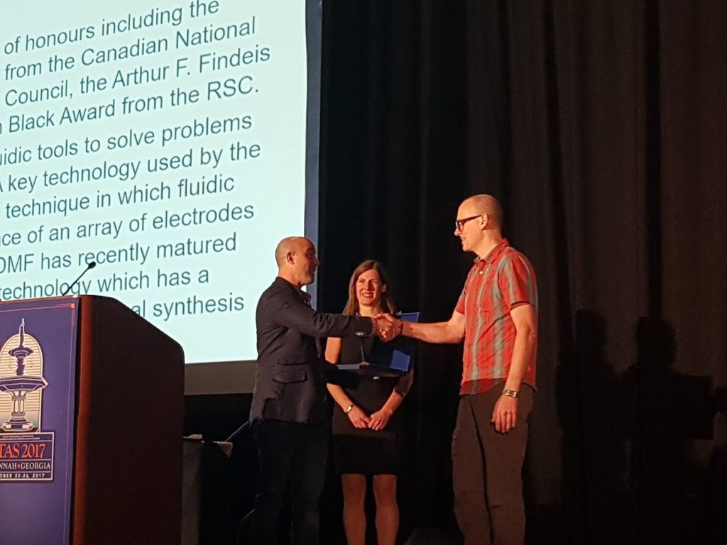
Lab on a Chip and the National Institute of Standards Technology (NIST) were pleased to present the Art in Science award at the µTAS 2017 Conference on 26 October 2017. The award highlights the aesthetic value in scientific illustrations while still conveying scientific merit. Many fantastic submissions were received this year with the winner selected by Maria SouthallLab on a Chip Deputy EditorDarwin Reyes, NIST and Petra DittrichLab on a Chip Associate Editor member.

And the winner is…

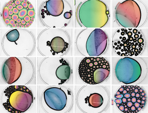
Give Bubbles a Chance
Maria Cristina Letizia, EPFL, SWITZERLAND


Runners up are…

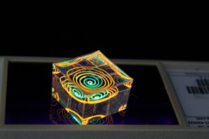
Reflections
Dorothea Helmer, Karlsruhe Institute of Technology, GERMANY

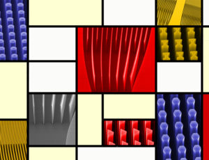
Mondrian’s Micropillars
Eloise Pariset, CEA – LETI, FRANCE

A big thank you to all the contributors this year!

Digg This
Reddit This
Stumble Now!
Share on Facebook
Bookmark this on Delicious
Share on LinkedIn
Bookmark this on Technorati
Post on Twitter
Google Buzz (aka. Google Reader)

MicroTAS “Late News” Posters

This year’s 21st International Conference on Miniaturized Systems for Chemistry and Life Sciences (more commonly known as MicroTAS) was held in Savannah, Georgia. As with previous MicroTAS conferences, the event brought together the international microfluidics and lab-on-a-chip community for an outstanding week of talks and posters. Rather than starting with a talk, MicroTAS 2017 opened with a conversation with George Whitesides moderated by Thomas Laurell, who asked questions pre-selected from the conference attendees. Whitesides, one of the pioneers of microfluidics, provided salient and humorous takes on the past, present, and future of our field. Two of Whitesides’ memorable takeaways were: (1) keep things simple, and (2) make sure you can answer the question “who cares?” The spirit of open discussion that started at the beginning of the conference continued through the many oral presentations and poster sessions at the conference. While the talks and posters represented the usual gamut of microfluidic technologies, 3D printing made a big splash this year. There was a 3D printing session, with a great keynote from Greg Nordin, as well as posters and companies featuring 3D-printed microfluidics. The sense of community was also palpable during many ferry rides to the Savannah International Trade & Convention Centre, the student trivia night, female faculty mixer event, and the conference ending banquet (and unofficial after party!).

In addition to all the great talks, we’ve highlighted some of our favourite “Late News” posters for our readers:


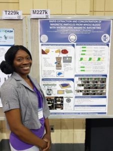
Rapid Extraction and Concentration of Magnetic Particles from Whole Blood with Microfluidic Magnetic Ratcheting, 

Oladunni Adeyiga, Coleman Murray, Dino Di Carlo

Magnetic particles are a useful tool for extracting and concentrating target analytes. Conventionally, pipetting, centrifuge tubes, and magnetic racks are used, but this approach is prone to loss of particles and errors from pipetting. Dunni presented a new microfluidic device that concentrates magnetic particles using magnetic ratcheting. A rotating permanent magnet was used to induce magnetic fields in permalloy micropillars embedded within the devices, which could then move magnetic particles along like on a conveyor belt. At the end of the process, the particles are concentrated into a drop within an immiscible fluid. While demonstrated as a standalone device, this tool could also function as a pre-concentrator in an integrated microfluidic device.


High Density, Reversible 3D Printed Microfluidic Interconnects, 

Hua Gong, Adam T. Woolley, Greg P. Nordin

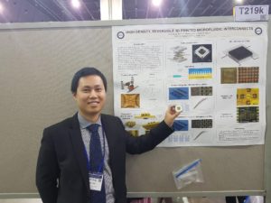
Earlier this year, Lab on a Chip published a paper describing the first 3D printed microchannels that truly had microscale dimensions. You can read the blog post summarizing the work here. It was nice to see this poster from Hua demonstrating the capabilities of their custom 3D printer, including printing complex valves. Hua explained how their 3D printer has a very small print area, and so one of the challenges to overcome was how to fit all the interconnects needed to control the valves into such a small space. This was achieved by printing a world-to-chip interface that connects tubing to the smaller, more densely packed inlets and outlets on the microfluidic chip.


3D-printing, Ink Casting and Lamination (3-D PICL): A Rapid, Robust, and Cost Effective Process Technology Toward The Fabrication of Microfluidic and Biological Devices, 

Tariq Ausaf, Avra Kundu, and Swaminathan Rajaraman

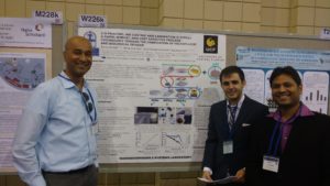
Tariq, Avra, and Swami presented an application of 3D-printing aimed at developing microelectrode arrays, microneedles, and microfluidic chips without cleanroom facilities. This work was motivated by a desire for disposable microelectrode arrays that can be developed from concept to functional prototype in < 24 hours. The authors integrated stereolithographic 3D-printing to form the device body, selective ink casting to define conductive traces, lamination of an insulating layer, and micromachining of electrodes and connecting layers. Combining multiple benchtop fabrication techniques could increase the functionality of the developed microdevices and could speed up the transition from prototype to final product in a cost effective manner.


An Automated Modular Microsystem For Enzymatic Digestion With Gut-On-A-Chip Applications
Pim de Haan, Margaryta A. Ianovska, Klaus Mathwig, Hans Bouwmeester and Elisabeth Verpoorte.

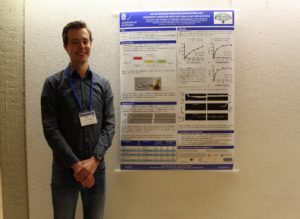
What is the primary function of the gastrointestinal tract? Digestion. However, most human gut-on-a-chip models tend to focus on cultured cells in one region of the gut (typically the small intestine) and do not model the initial digestive processes that take place in the mouth, stomach, and small intestine. Pim’s work is focused on developing bioreactors to study the conversion of food into chyme, as it moves from the mouth, through the stomach, and into the small intestine. This requires accurate modelling and realization of the vast pH differences that take place from one region of the gut to the next and verifying enzyme activity within his gut-on-a-chip model. Future work will focus on integration of this upstream digestion model with the downstream study of absorption of nutrients across an intestinal cell layer.


__________

About the Webwriters

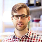
Darius Rackus (Right) is a postdoctoral researcher at the University of Toronto working in the Wheeler Lab. His research interests are in combining sensors with digital microfluidics for healthcare applications.


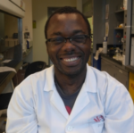
Ayokunle Olanrewaju (Left) is an industrial postdoctoral fellow at McGill University working in the Juncker lab (dj.lab.mcgill.ca). He is excited about projects that use engineering design to effect real world change, especially in healthcare. Currently, he builds portable and self-powered microchips that rapidly detect bacteria in urine.


Digg This
Reddit This
Stumble Now!
Share on Facebook
Bookmark this on Delicious
Share on LinkedIn
Bookmark this on Technorati
Post on Twitter
Google Buzz (aka. Google Reader)

Magnetic separation of circulating tumor cells…but it’s not what you think

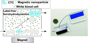
Cancers typically originate in one organ yet can spread to distant regions of the body forming secondary tumours called metastases. This happens as cells from the primary tumour migrate into the circulatory system and then travel to other organs. These cells, which are a very rare population within the circulatory system, are termed circulating tumor cells (CTCs). Because of their role in cancer biology, they have garnered a lot of interest lately. Their detection and isolation present several analytical challenges. For one, they are the proverbial “needle in a haystack”, with counts on the order of one CTC for every billion blood cells. This has traditionally led to a paradox: these rare cells are best handled in microscale systems but the world-to-chip mismatch limits microfluidic devices from rapidly processing the large (> 5 mL) samples necessary. Second, recent studies have revealed CTCs to be very heterogeneous populations, limiting the use of surface markers for labelling and capturing a broad range of CTCs. Because there is much still to learn about CTCs, there’s also an interest in recovering viable CTCs for further analysis. In their recent report, Zhao et al. demonstrate a microfluidic device capable of enriching CTCs using magnetic separation. But it’s not that typical magnetophoretic separations you may be familiar with!

Magnetic separation of circulating tumor cells - nanoparticles

Rather than using magnetic particles to bind to surface antigens and eventually separate out CTCs, they capitalize on a phenomenon known as “negative magnetophoresis”. Cells are suspended within a uniformly magnetic medium and application of a non-uniform magnetic field results in a magnetic buoyant force. (This is akin to how negative dielectrophoresis exerts a force on particles in a non-uniform electric field.) The advantage of this method is that the “working principle applies to every non-magnetic material,” according to Prof. Leidong Mao. “Naturally,” he thought “it could apply to CTC enrichment.” However, despite previous work separating different cell populations with negative magnetophoresis, moving to CTC enrichment is not so straightforward. CTC enrichment is the most challenging separation. “All previous applications in our group are with cells at high concentrations,” mentioned Prof. Mao. The main challenge in developing the chip was trying to preserve the characteristics of an “ideal” CTC enrichment device; one that could process a significant amount of blood quickly, have a high recovery rate of CTCs, give reasonable purity of isolated CTCs, and retain cell integrity and viability for further analysis.

With this method, heterogeneous populations of CTCs can be enriched as selection is size dependent rather than based on expression of certain surface markers. This also avoids the costs associated with traditional magnetic labelling – typically used to label and deplete the millions of white blood cells. The device is capable of working at flow rates of 5-7 mL/hr, which is what is necessary to process an entire blood sample and can achieve high recovery rates (>90%). While the authors report purities that appear low (10-12%), they are working on improving purity. One strategy they suggest in their report is to follow the route of the iChip and combine size based separations with magnetic WBC depletion.

To read the full paper for free*, click the link below:

Label-free ferrohydrodynamic cell separation of circulating tumor cells
DOI: 10.1039/C7LC00680B (Paper) Lab Chip, 2017, 17, 3097-3111

—————-

About the Webwriter

Darius Rackus is a postdoctoral researcher at the University of Toronto working in the Wheeler Lab. His research interests are in combining sensors with digital microfluidics for healthcare applications.

*free to access until 14th December 2017


Digg This
Reddit This
Stumble Now!
Share on Facebook
Bookmark this on Delicious
Share on LinkedIn
Bookmark this on Technorati
Post on Twitter
Google Buzz (aka. Google Reader)

Emerging Investigator Series – Wilbur Lam

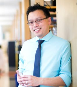
We are delighted to introduce our latest Lab on a Chip Emerging Investigator, Wilbur Lam!

Wilbur A. Lam, MD, PhD is an Associate Professor of Biomedical Engineering and Pediatrics at the Georgia Institute of Technology and Emory University and has a unique background as a physician-scientist-engineer trained in pediatric hematology/oncology and bioengineering. Dr. Lam’s interdisciplinary laboratory, located at both Emory and Georgia Tech, includes engineers, biologists, biophysicists, chemists and physicians. Our laboratory serves as a unique “one-stop shop” in which we develop in vitro microsystems to study hematologic processes in both health and disease and then immediately bring those technologies to the patient bedside. More specifically, the Lam laboratory’s research interests involve the development and application of microsystems to enable research in pathologic biophysical blood cell interactions that occur in diseases such as sickle cell disease and thrombosis, as well as further translating those systems into novel therapeutics and diagnostic devices.

Read Wilbur’s Emerging Investigator series paper “Probing blood cell mechanics of hematologic processes at the single micron level” and find out more about him in the interview below:


Your recent Emerging Investigator Series paper focuses on probing blood cell mechanics of hematologic processes. How has your research evolved from your first article to this most recent article?

As a bioengineer and a physician specializing in paediatric haematology, my initial goals for our lab were really twofold: 1) to leverage microscale technologies to apply cell mechanics principles towards investigating clinically relevant biologic processes and 2) to convince the medical and clinical haematology fields that physical phenomena such as shear stress and the mechanical properties of the microenvironment can directly mediate the biologic processes of blood cells and pathophysiology of blood diseases such as sickle cell disease and thrombosis. Over the last few years, I think our lab and other groups have accomplished that and we’re now concentrating on not only the basic science questions of blood cell mechanics but how to apply the microtechnologies we develop as potential diagnostics and even therapeutic systems for patients with blood diseases.

What aspect of your work are you most excited about at the moment?

Along those same lines, we’ve been at this game long enough to see some of our microtechnologies translate to my patients with blood diseases, which we find pretty exciting. For example, we just received FDA clearance for a point-of-care anaemia diagnostic our lab developed. We also have several other blood cell mechanics-based microtechnologies that are in our lab’s translational pipeline we’re pretty excited about. For instance, we’ve developed a microfluidic system that can assess platelet contraction forces at the single cell level and we’re now trying to determine whether that system can be used to diagnose patients with bleeding disorders, a clinical interest of mine. At the same time, we’re also trying to leverage that mechanical phenomenon of platelet contraction as a “Trojan Horse” strategy of targeted drug delivery for haemophilia, which we find to be pretty sci-fi and exciting. Moreover, using fairly simple microfluidic devices, we determined that intravenous hydration, a standard first-line therapy for sickle cell disease, may have beneficial as well as deleterious effects on red cell mechanics, which could potentially alter how we treat this disease. We’ve also developed technologies that can enable the translation of other technologies including a microfluidic device that can improve the transduction efficiency of lentiviruses for gene therapy and CAR T-cell therapy, which are both making significant clinical impacts right now. That said, we still remain very interested in the basic science of blood cell mechanics, which is evidenced by this current paper of ours, and we have active projects including but not limited to investigating leukocyte mechanics and are developing new microfluidic strategies to study and model bleeding.

In your opinion, what is the biggest potential impact the results of this research will have on blood disorder diagnostics?

I think impact of our research regarding blood disorder diagnostics can be measured in several ways. First, we obviously want to positively affect and help as many patients and people as possible, which is why we’re clearly excited about the recent FDA 510K clearance of our point-of-care anaemia diagnostic. For that project, we’re currently planning our manufacturing and distribution strategy with different partners to commercialize and disseminate this technology in the US and abroad to impact as many people globally as we can. In addition, we can also measure impact on a more personal level. One of the reasons why our lab has been fortunate enough to be able to recruit the best graduate students and postdoctoral fellows is because of our unique setup in which our lab is located both on the engineering side at Georgia Tech as well as the clinical side at Emory University and Children’s Healthcare of Atlanta, where I am a practicing physician. I’ve truly been blessed to work with the best and most talented students and postdocs in the field of bioengineering and I think they enjoy the fact that they can design a device and literally see it be used on a real live patient within a relatively short time frame. This is great for my patients as well, who are able to witness firsthand how medical technology develops and the potential of how it can improve their own lives in the near future. So, in essence, our lab has developed a “basement-to-bench-to-bedside” approach in which we design and develop microtechnologies to not only study disease but also directly translate these to the patients I care for and even conduct clinical assessments and trials for those devices – all under one roof. By the way, if your readers can help me come up with a better word than “basement” while still maintaining the alliteration as well as the overall message of the phrase, my lab and I will be extremely grateful.

What do you find most challenging about your research?

Like many other bioengineering laboratories among your readership, successful design and development of a novel micro/nanoscale technology is only half the battle. Then you have to do the actual experiment to demonstrate its utility and hope it delivers some type of value. This can be frustrating at times, as our projects can take twice as long as average, but the hope is that our impact will be doubled as well.

In which upcoming conferences or events may our readers meet you?

I attend the annual American Society of Hematology and International Society on Thrombosis and Haemostasis meetings as I serve on the scientific committees for both of them. I also frequently attend the annual Biomedical Engineering Society and MicroTAS meetings as well.

How do you spend your spare time?

I answer interview questions for scientific blogs! Seriously, I’m a huge fan of pop culture and pop music. I even have a lifetime subscription to Rolling Stone, which I’ve been starting to regret in recent years as I’ve grown older as the covers of that magazine have progressively gotten more risqué (or maybe that’s just my perception as whatever semblance of hipness and coolness I ever had is exponentially decreasing with time). I also still pick up my guitar every now and then – especially when I’m procrastinating writing a grant or submitting grades.

Which profession would you choose if you were not a scientist?

I find that when most people are asked this question, they give an idealized answer, so I’ll do the same. I’d like to say that I’d be a successful songwriter/musician as I’ve played guitar in bands from my teenage years until my first faculty position (during my clinical training, I played in an all-paediatrician band named “Booster Shot”). However, to be frank, I really wasn’t that good and seriously doubt I could have made it as a professional musician. So, I think the realistic answer is that my most likely alternative profession would be that of a disgruntled sales associate at a Guitar Center somewhere or working as a roadie for artists a fraction of my age and whom I most likely would have despised. So, it’s good that this science and medicine thing worked out…

Can you share one piece of career-related advice or wisdom with other early career scientists?

Hone your writing skills, start early and keep practicing. I am constantly amazed at how much of my time is devoted to writing and how important that skill is to be a successful scientist. Whether it’s writing or editing papers, lectures, or grants, I find I actually spend most of my time writing. In retrospect, this makes sense as communication really is the currency of science, but there’s no way my younger self would believe me if I were to travel back in time to let him what was ahead of him. I’ve had many young people tell me they want to go into science specifically because they dislike writing, which is obviously a misperception I’m quick to point out. In fact, one joke (and not a very good one, admittedly) I often share with my students is that as a scientist, I actually write really bland non-fiction for a living and for a very small audience, and when I’m grantwriting, I’m only writing to an audience of three people.

Digg This
Reddit This
Stumble Now!
Share on Facebook
Bookmark this on Delicious
Share on LinkedIn
Bookmark this on Technorati
Post on Twitter
Google Buzz (aka. Google Reader)

Lab on a Chip awards prestigious prizes at MicroTAS 2017

The µTAS 2017 Conference was held during 22 – 26th October in Savannah, Georgia, USA.  Maria Southall,  Deputy Editor of Lab on a Chip, attended this conference and announced the prestigious Lab on a Chip awards which include the Pioneers of Miniaturization Lectureship (in partnership with Dolomite Microfluidics), the Widmer Young Researcher Poster Prize, the Art in Science competition (in partnership with NIST) and the µTAS video competition (in partnership with Dolomite Microfluidics). The competition was tough, but we are pleased to announce this year’s Prize Winners below.

“Pioneers of Miniaturization” Lectureship

Professor Aaron Wheeler (University of Toronto) was announced as the winner of the 12th “Pioneers of Miniaturization” Lectureship, sponsored by Dolomite and Lab on a Chip. The “Pioneers of Miniaturization” Lectureship rewards early to mid-career scientists who have made extraordinary or outstanding contributions to the understanding or development of miniaturised systems. Professor Aaron Wheeler received a certificate and a monetary award, and delivered a short lecture titled “A Pioneer’s Trail: from Savannah to Toronto to Kakuma and Beyond” at the conference.

Left to Right – Aaron Wheeler (Winner), Maria Southall (Lab on a Chip), Mark Gilligan (Dolomite). Photo taken by Darius Rackus.

Art in Science Competition

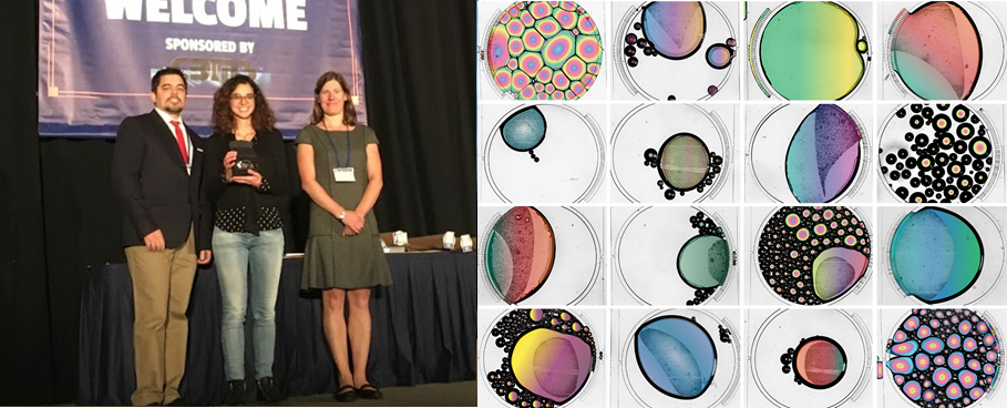
Darwin Reyes from the National Institute of Standards Technology (NIST) and Lab on a Chip presented the Art in Science award to Maria Cristina Letizia (EPFL, Switzerland). The award aims to highlight the aesthetic value in scientific illustrations while still conveying scientific merit. Check our her winning photograph “Give Bubbles a Chance” below.

Left to right: Darwin Reyes (NIST), Maria Cristina Letizia (Winner), Maria Southall (Lab on a Chip), Winning photo “Give Bubbles a Chance”

µTAS Video Competition

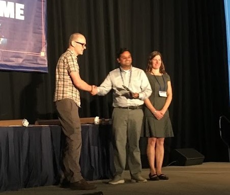
Dolomite and Lab on a Chip announced Aniruddha Kaushik (Johns Hopkins University) as the winner of the 2017 µTAS video competition. µTAS participants were invited to submit short videos with a scientific or educational focus. The winning video “Droplet Microfluidics Rap” can be viewed on our YouTube channel, along with the runner up video “Bubbles in Complex Microgeometries at Large Capillary Numbers” by Martin Sauzade (Stony Brook University). Mark Gilligan of Dolomite presented the winner with a voucher for Dolomite equipment.

Left to right: Mark Gilligan (Dolomite), Aniruddha Kaushik (Winner), Maria Southall (Lab on a Chip)

Widmer Young Researcher Poster Prize

The Widmer Young Researcher Poster Prize was awarded to Jin Ko, PhD student at the University of Pennsylvania for their poster on the prognosis of traumatic brain injury using machine learning based miRNA signatures in nanomagnetically isolated brain-derived exosomes.

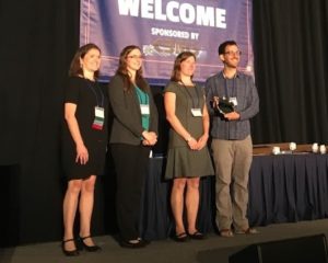
Left to right: Séverine Le Gac and Ashleigh Theberge (Poster Award Chairs), Maria Southall (Lab on a Chip), David Issadore (PhD supervisor of award winner)

Congratulations to all the winners at the conference! We look forward to seeing you at µTAS 2018 in Kaohsiung, Taiwan!

Digg This
Reddit This
Stumble Now!
Share on Facebook
Bookmark this on Delicious
Share on LinkedIn
Bookmark this on Technorati
Post on Twitter
Google Buzz (aka. Google Reader)

Emerging Investigator series – Pouya Rezai

We are delighted to introduce our latest Lab on a Chip Emerging Investigator, Pouya Rezai!

Dr. Pouya Rezai is an emerging investigator in the fields of microfluidics and Lab-on-Chips (LoC). He received his PhD in Mechanical Engineering from McMaster University in 2012. Dr. Rezai was an NSERC Visiting Fellow at Public Health Agency of Canada before joining York University in July 2013 as an Assistant Professor. He is the Graduate Program Director of the Department of Mechanical Engineering at York University and the Editor of the Canadian Society for Mechanical Engineering (CSME) bulletin. The overarching goal of his research program is to expand fundamental understanding of the interactions between fluids and nano- to micro-scale biological substances in their micro-environments and to employ this knowledge to devise efficient microsystems for facilitating research and development in human health-related applications

Read Pouya’s Emerging Investigator series paper “A microfluidic device for partial immobilization, chemical exposure and behavioural screening of zebrafish larvae” and find out more about him in the interview below:


Your recent Emerging Investigator Series paper focuses on a microfluidic device to partially immobilise Zebrafish Larvae for behavioural screening. How has your research evolved from your first article to this most recent article?

I started my work on organisms-on-a-chip with the C. elegans worm model. Our work was initially focused on C. elegans response to electric field in microchannels, a phenomenon called electrotaxis. We then became interested in using electrotaxis as a tool to screen movement of worms under exposure to chemicals. Recently, we have become interested in not only electrotaxis, but also chemotaxis of C. elegans and other model organisms such as Drosophila melanogaster and Danio rerio in lab-on-chips. We have developed multiple microfluidic devices for neurobehavioral screening of these organisms. Our goal is to continue working on the same models at the behavioural level but also focus on their cellular responses to stimuli. Our long term objective is to use our organism-on-a-chip devices for drug screening and toxicology studies in collaboration with academia and industry.

What aspect of your work are you most excited about at the moment?

Two major aspects of my work as a professor excites me the most. First and foremost is being able to teach and train young students in the fields of Mechanical and Biomedical Engineering that I am passionate about; and second is having the opportunity to apply my knowledge as an engineer directly to human health related issues. Due to the interdisciplinary nature of our research, I find the collaborative aspects of our work very rewarding for me and my trainees.

In your opinion, what is the biggest benefit of immobilising Zebrafish Larvae for analysis over the conventional droplet-based technique?

The most significant benefit is achieving higher sensitivity in quantifying subtle movement behavioural phenotypes of zebrafish in an easier and faster way. In droplets, you either need an expert to monitor movement manually or complex setups for tracking larvae’s movement in the droplet. With our technique, minimally trained personnel can quickly gain the ability to assay zebrafish movement under exposure to various chemicals.

What do you find most challenging about your research?

We develop devices for live organisms with the capability of making voluntary decisions while in the chip. One does not encounter this challenge with cultured cells or molecules. Decision-making generates opportunities to study sensory-motor responses at the whole-organism level, but also produces a wide variety of challenges in designing microfluidic devices and quantification of the desired biological processes. Another challenge is the general trend in the field to move towards biological models that better mimic human diseases and disorders. This has generated significant momentum towards the use of human-derived cells in biomimetic microfluidic devices. However, I still think there are many unanswered questions that can be addressed by small scale organisms.

In which upcoming conferences or events may our readers meet you?

The International Conference on Miniaturized Systems for Chemistry and Life Sciences (MicroTAS) is my favorite conference to go to every year. Some other events that we attend are the MEMS and Transducers conferences.

How do you spend your spare time?

Time is the most precious thing in the world. The nature of an academic job requires a significant dedication of time to professional development, especially at the initial stages of your career. Is this a correct path that we have taken? Let’s not get there for now!
Like many of my colleagues who are my role models and mentors, I rarely have any spare time to be spent on my hobbies. But the most important activity that I enjoy doing, and I wish I could do much more, is to spend quality time with my lovely family. After this, I enjoy nature and listening to good music.

Which profession would you choose if you were not a scientist?

I would pursue the entrepreneurship path and start my own business to help resolve a pressing health related issue.

Can you share one piece of career-related advice or wisdom with other early career scientists?

One of the most influential factors during my professional career has been the opportunity to have excellent mentors and role models who have guided me throughout my postgraduate studies and academic career. The leaders in the field and their path to success have been very inspiring to me in the recent past. I recommend early career scientists to reach out to their communities and seek professional advise on their research and teaching activities.

Digg This
Reddit This
Stumble Now!
Share on Facebook
Bookmark this on Delicious
Share on LinkedIn
Bookmark this on Technorati
Post on Twitter
Google Buzz (aka. Google Reader)

Emerging Investigators series – Milad Abolhasani

We are delighted to introduce our latest Lab on a Chip Emerging Investigator – Milad Abolhasani!

Milad Abolhasani is currently an Assistant Professor in the Department of Chemical and Biomolecular Engineering at North Carolina State University. He received his B.Sc. (2008) and M.A.Sc. (2010) degrees in Mechanical Engineering from Sharif University of Technology and the University of British Columbia, respectively. He then obtained his Ph.D. degree (2014) from the Department of Mechanical and Industrial Engineering in collaboration with the Departments of Chemistry and Chemical Engineering at the University of Toronto. Prior to joining NC State University, he was a postdoctoral fellow in the Department of Chemical Engineering at MIT (Jensen group, 2014-2016), where he developed a modular flow chemistry strategy for in-situ mass transfer and kinetic studies of single/multi-phase chemical processes including bi-phasic cross-coupling reactions and colloidal synthesis and ligand exchange of semiconductor nanocrystals. Dr. Abolhasani‘s research interests include the development of microfluidic technologies tailored for solution-phase processing of energy harvesting nanomaterials and for fundamental studies of transport mechanisms involved in CO2 capture, recovery, and utilization in green chemistry (enabled by switchable solvents). Over the course of his doctoral and postdoctoral research, Dr. Abolhasani received numerous fellowships and awards including NSERC Postdoctoral Fellowship, CSME 2014 Best Graduate Student Paper Award, Bert Wasmund Graduate Fellowship in Sustainable Energy Research, and Russell A. Reynolds Graduate Fellowship in Thermodynamics.

Read his Emerging Investigators paper “Automated microfluidic platform for systematic studies of colloidal perovskite nanocrystals: towards continuous nano-manufacturing” and find out more about his research in the interview below:

Your recent Emerging Investigator Series paper focuses on studying colloidal perovskite nanocrystals. How has your research evolved from your first article to this most recent article?

Well, my first research article as a graduate student (published in Lab on a Chip) was focused on the development of an inexpensive approach for rapid determination of thermodynamic characteristics of gas-liquid reactions using an image processing technique. Since then, I’ve expanded my expertise in multi-phase microfluidic systems with a focus on integrated systems with in-situ spectroscopy and/or in-line analytical characterization capabilities for material- and time-efficient studies of various physical/chemical processes. Few examples of such processes include homogenous catalytic reactions, partition coefficient of pharmaceutical compounds, colloidal synthesis of semiconductor nanocrystals, and hydrophilicity switching of switchable solvents. Despite different applications, the common theme among all multi-phase microfluidic technologies that I’ve developed so far has been the focus on realizing the early promise of microfluidics on minimizing the reagents volume used for each experimental condition while maximizing the amount of data obtained. My latest article builds on my experience in integrated microfluidic systems and in-situ spectroscopy techniques to study the effect of early stage mixing times on the optical properties of in-flow synthesized colloidal perovskite nanocrystals.

What aspect of your work are you most excited about at the moment?

Development of microfluidic technologies to contribute towards the next generation of energy-efficient and solution phase-processed photovoltaics.

In your opinion, what is the biggest advantage to using colloidal organic/inorganic metal-halide perovskite nanocrystals for photovoltaics over the current materials?

From materials perspective, hybrid organic/inorganic perovskite nanocrystals are inexpensive and can be manufactured using solution-phase processing techniques. In addition, these shiny nanocrystals possess high surface defect tolerance, high and broad absorption coefficient, high quantum-yield, and long charge carrier lifetime and diffusion length. Combining the superior physicochemical properties of colloidal perovskite nanocrystals with precise band-gap engineering and unparalleled experimental parameter control offered by multi-phase microfluidic platforms make them a promising candidate for the next generation photovoltaics and LED displays.

What do you find most challenging about your research?

Learning about details of different steps involved in manufacturing thin-film solar cells. It is challenging but fascinating to learn.

In which upcoming conferences or events may our readers meet you?

I am currently conducting this interview from MicroTAS 2017 conference in Savannah, GA. I will be attending the Annual AIChE meeting in Minneapolis, MN, between Oct 29 – Nov 3.

How do you spend your spare time?

Catching up with our favorite TV shows (TWD and GOT) with my wife

Which profession would you choose if you were not a scientist?

I would most probably choose to become an architect. I was (and am) always fascinated by “futuristic looking buildings” around the world such as Galaxy Soho in Beijing and Notre Dame du Haut in Ronchamp. The level of attention to details and precision in engineering are just mind-blowing.

Can you share one piece of career-related advice or wisdom with other early career scientists?

In my opinion, focusing on one long-term visionary project that fits your research interests and expertise should be the main goal of a junior faculty. There are so many interesting problems around you, but there is probably only ONE big-impact problem that would truly fit your background which you (hopefully) can solve within your precious pre-tenure adventures.

Digg This
Reddit This
Stumble Now!
Share on Facebook
Bookmark this on Delicious
Share on LinkedIn
Bookmark this on Technorati
Post on Twitter
Google Buzz (aka. Google Reader)

Early Career Researcher workshop on Diagnostics for Antimicrobial Resistance

Early Career Researcher workshop on Diagnostics for Antimicrobial Resistance

20 November 2017, London, UK           

Join a diverse delegate list of early career researchers and invited experts to discuss the barriers and opportunities facing the development of rapid diagnostics for infectious disease.

Our speakers include:

  • Jim Huggett LGC & University of Surrey, United Kingdom
  • David H Persing Executive VP, Chief Medical & Technology Officer, Cepheid, United States
  • Bhargavi Rao Médecins Sans Frontières, Switzerland
  • Tim Rawson Imperial College London, United Kingdom
  • Annegret Schneider University College London, United Kingdom
  • Chris Walton Cranfield University, United Kingdom

The main themes identified at this workshop will be shared with various research funders and stakeholders. Don’t miss this chance to discuss some of the exciting developments in diagnostics for AMR and to share your thoughts about how to support early career researchers working in this field.

Register by 6th November to attend!

To find out more and register, please visit: http://rsc.li/diagnostics4AMR

Digg This
Reddit This
Stumble Now!
Share on Facebook
Bookmark this on Delicious
Share on LinkedIn
Bookmark this on Technorati
Post on Twitter
Google Buzz (aka. Google Reader)

Zenith in “artery”

When cutting a finger, thrombocytes and fibrin in the blood make up the blood-clotting mechanism, aka. haemostasis, to stop the blood loss. Another way to trigger this mechanism is having an artery damaged by atherosclerosis, which is often caused by several genetic or acquired factors. In the latter case, thrombosis develops within a vein or artery, obstructing or stopping the blood flow to major organs like the heart and eventually causing heart attack. Considering every year over 14 million lives worldwide are lost to heart attacks, more investigation on this topic is needed without any doubt.

Recently, a research team led by Andries van der Meer published a research article in Lab on a Chip on mimicking arterial thrombosis in 3D vascular structures, representing a major step forward in the development of accurate and faster methods of studying arterial thrombosis without using animals. The authors highlighted the inconvenience of using animal models to predict arterial thrombosis in humans. This is mainly due to fundamental differences between human and animal physiology, the researchers explain. For instance, rodent platelet biology, coagulation dynamics, and shear stress in mice arteries significantly vary between humans and mice.

thrombosis on chip

Figure 1. Three-dimensional models of a healthy and stenotic vessels and thrombosis formation upon blood perfusion through the channels.

The paper uses miniaturized vascular structures mimicking 3D architectures found in both healthy and stenotic blood vessels in-vitro (Figure 1). They combined stereolithography and 3D printing of computed tomography angiography data to construct 3D-printed templates of vessels in PDMS microchips. The 3D printed vessels are then coated with human umbilical vein endothelial cells, forming a monolayer fully covering the surface. In the next step, the artificial vessels are perfused with blood at normal arterial shear rates, allowing a blood clot to form as it would happen in the human body. The 3D printed vessel is clinically more relevant when compared to 2D vessel models, since the realistic flow profiles of blood and even distribution of shear stress across the vessel are of great importance when researching arterial thrombosis. Hugo Albers, the co-first author of the paper explains what led the team to try 3D models: “Other groups have worked on thrombosis-on-a-chip before, but we wanted to incorporate flow profiles that are similar to what one would find in-vivo. So we opt for a round and thus 3D shape. Since the stenotic geometry is an important part of this work, we wanted to find a technique that allowed us to make almost any shape we could come up with. Thus 3D-printing seemed to be the way to go.”

When it comes to defining the challenges in 3D organ-on-chip modeling and fabrication, “we needed to replicate the cellular environment using human endothelial cells and human whole blood to fully mimic the nature of vasculature” says Albers. “Incorporating the shape of vasculature to recreate the flow profiles found in-vivo and recreating the shape of vasculature on a small scale was quite challenging, since the resolution of 3D-printing quickly started to be the limiting factor. Furthermore, we ran into problems related to working with whole blood. We had to figure out how to perfuse small channels with blood without instigating thrombosis outside of the microfluidic channel.” The researchers successfully overcame the challenges mentioned by Albers and mimicked the formation of thrombosis in a stenotic vessel model as seen in Figure 1 (bottom).

The researchers note that the next step involves co-culturing arterial endothelial cells and smooth muscle cells with human umbilical vein endothelial cells or moving to different cell lines such as differentiated human induced pluripotent stem cells. “I think we can also apply the 3D-printing technique to create thrombosis-on-a-chip devices with different geometries, e.g. aneurysms or bifurcated geometries”, says Albers.

To download the full article for free* click the link below:

Mimicking arterial thrombosis in a 3D-printed microfluidic in vitro vascular model based on computed tomography angiography data

Pedro F. Costa, Hugo J. Albers, John E. A. Linssen, Heleen H. T. Middelkamp, Linda van der Hout, Robert Passier, Albert van den Berg, Jos Malda and Andries D. van der Meer

Lab Chip, 2017, Paper

DOI: 10.1039/C7LC00202E

This paper is included in our Organ-, Body- and Disease-on-a-Chip Thematic Collection. To read other articles in the collection, visit – rsc.li/organonachip

About the Webwriter

Burcu Gumuscu is a postdoctoral fellow in Herr Lab at UC Berkeley in the United States. Her research interests include development of microfluidic devices for quantitative analysis of proteins from single-cells, next generation sequencing, compartmentalized organ-on-chip studies, and desalination of water on the microscale.

Digg This
Reddit This
Stumble Now!
Share on Facebook
Bookmark this on Delicious
Share on LinkedIn
Bookmark this on Technorati
Post on Twitter
Google Buzz (aka. Google Reader)

Emerging Investigators series – Ian Wong

We are delighted to introduce our latest Lab on a Chip Emerging Investigator – Ian Wong!

Ian Y. Wong is currently an Assistant Professor of Engineering and of Medical Science at Brown University. He completed an A.B. in Applied Mathematics from Harvard University in 2003, Ph.D. in Materials Science and Engineering with Nick Melosh at Stanford University in 2010, and postdoctoral training at Massachusetts General Hospital with Mehmet Toner and Daniel Irimia in 2013. He has been recognized with an NSF Graduate Research Fellowship, a Damon Runyon Cancer Research Fellowship, the Brown University Pierrepont Prize for Outstanding Advising, as well as a Biomaterials Science Emerging Investigator. His research interests include the development of miniaturized technologies to investigate cancer cell invasion, phenotypic plasticity and drug resistance. Moreover, his group engineers unconventional fabrication techniques for printing and patterning nano/bio materials.

Read his Emerging Investigators paper “Stereolithographic printing of ionically-crosslinked alginate hydrogels for degradable biomaterials and microfluidics“, watch the associated video and find out more about his research in the interview below:

Your recent Emerging Investigator Series paper focuses on stereolithographic printing of ionically-crosslinked alginate hydrogels. How has your research evolved from your first article to this most recent article?

I give complete credit to my graduate student, Tom Valentin, who came up with this approach to light-based 3D printing via ionic crosslinking – and then actually got it to work. In retrospect, my Ph.D. thesis focused on biomolecular self-assembly based on ionic interactions, so it’s serendipitous that my current research has circled back to some of these concepts.

 What aspect of your work are you most excited about at the moment?

My lab integrates biomaterials, microfluidics, and computer vision to investigate cancer cell migration and drug resistance. Our first few papers set down the foundations for these different technologies, but now we’re starting to put these pieces together to gain some fascinating insights into cancer biology.

 In your opinion, what is the biggest advantage of stereolithographic printing of hydrogels over other printing techniques?

 Conventional light-based 3D printing of soft materials is based on covalent crosslinking, which results in strong but irreversible bonds. Our demonstration of light-based patterning using reversible ionic crosslinks should enable smart and “biomimetic” properties such as self-healing and stimuli-responsiveness. These properties have been previously demonstrated in bulk hydrogels, but remain relatively nascent for 3D printed structures.

What do you find most challenging about your research?

I work at the interface of engineering and cancer biology, and I find that it takes a lot of effort to bridge between these two communities and become fluent in both disciplines. Moreover, there are twice as many things that can go wrong with the experiments! Nevertheless, it has been extremely worthwhile to see how our technologies could potentially make an immediate and highly meaningful impact.

In which upcoming conferences or events may our readers meet you?

I will be attending BMES this October in Phoenix, AZ.

How do you spend your spare time?

Whenever possible, I enjoy dining out with my wife. I also enjoy cycling, which helps to burn off all those calories

Which profession would you choose if you were not a scientist?

I’ve always been interested in entrepreneurship, and this is something I will likely revisit once my lab and technologies become more established.

Can you share one piece of career-related advice or wisdom with other early career scientists?

Early career scientists are constantly pulled in many directions and have limited time to commit to anything. Nevertheless, I try my best to spend a lot of time with my students and postdocs early on. Such mentoring helps trainees transition towards independence and can also catch problems before they become serious, so it is incredibly worthwhile in the long run.

Digg This
Reddit This
Stumble Now!
Share on Facebook
Bookmark this on Delicious
Share on LinkedIn
Bookmark this on Technorati
Post on Twitter
Google Buzz (aka. Google Reader)