Archive for the ‘Hot Articles’ Category

Chlorophyll lasers

From space shuttles to military equipment, or even the kitchenware that we use on daily basis, lasers have found use in more places than we often realise. Interestingly, the solid-state lasers were believed to be a solution to an unknown problem after their invention in 1950s. In that time nobody—including their developer Charles Townes—noticed that the lasers were one of the game changer inventions in the world’s history. After tens of years, in 1964, the laser technology was awarded with a Nobel prize when its potential for diverse applications were realized.

So far we have only discussed solid-state lasers, but there is definitely capacity in the laser world to improve the performance and versatility with the help of little twists, such as optofluidic lasers. “Optofluidics” is a synergic combination of optical systems and microfluidics. In other words, optical systems are built or synthesized from liquids, aiming to serve as good as their solid-state equivalents. For example, two immiscible liquids form a smooth surface in their interface, leading to a laser cavity or an optical resonator with a very high Q-factor that allows for operating at low energy levels. This emerging field gains importance when considering most of the biochemical reactions that occur in aqueous environments. Optofluidic laser systems are flexible to change their optical properties by just replacing the liquid media; and with this twist, lasers have new application areas including diagnosis of genetic disorders at the cellular level and in vivo biosensing.

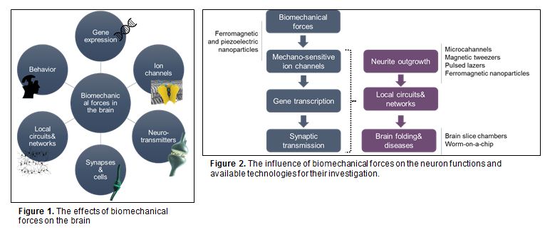
What is more exciting about the optofluidic lasers is that they can be biodegradable and easily tunable in microenvironments. Researchers in University of Michigan recently showed that one of the most abundant pigments on earth, chlorophylls, can maintain both biodegradability and tunability in optical systems owing to their fluorescence capabilities. Chlorophylls have a high Q-factor, dual-absorption bands in the visible spectrum, and a large shift between absorption and emission bands, suggesting that chlorophylls can be used as donors in fluorescence resonance energy transfer (FRET) laser (Figure 1). In this study, chlorophyll a was isolated from spinach leaf and used as the gain medium and the donor to develop a novel optofluidic laser. Two lasing bands of chlorophyll a was investigated by both theoretical and experimental means. Concentration-dependent studies enabled more insight for the mechanism determining when, where, and why the laser emission band appears. This new technique seems to gain increasing attention for applications in in vivo and in vitro biosensing, solar lighting and energy harvesting.

This article, published on 12th May 2016, is included in the Lab on a Chip Recent HOT Articles themed collection.

To download the full article for free* click the link below:

Optofluidic chlorophyll lasers
Yu-Cheng Chen, Qiushu Chena, Xudong Fan
Lab Chip, 2016, 16, 2228-2235
DOI: 10.1039/C6LC00512H

About the Webwriter

Burcu Gumuscu is a PhD researcher in BIOS Lab on a Chip Group at University of Twente in The Netherlands. Her research interests include development of microfluidic devices for second generation sequencing, organ-on-chip development, and desalination of water on the micron-scale.

*Access is free until 23/09/2016 through a registered RSC account.

Digg This
Reddit This
Stumble Now!
Share on Facebook
Bookmark this on Delicious
Share on LinkedIn
Bookmark this on Technorati
Post on Twitter
Google Buzz (aka. Google Reader)

What is the best way to study brain?

Before the 1700’s, when dissection techniques were not yet available, the cause of mood changes were thought to be the replacement of liquids and vapors in the body. The biology of the brain has been better understood since the discovery of research and test tools. However, occupying only about 1/50 of the body mass, the brain is perhaps the most complicated organ to study. National Institute of Neurological Disorders and Stroke’s list of over 400 neurological disorders can be seen as a sound proof of this exciting- and frustrating- fact.

Figure 1. The effects of biomechanical forces on the brain

Biomechanical forces on neurons play a fundamental role in neuronal physiology, which, in turn, affect brain development and disorders. During the growth of neurons, the tension created by the biomechanical forces are suggested to influence the cells’ motor activities, gene expression, neurotransmitter release, together with neurite growth and network connections (Figure 1). Research on the biomechanical forces can definitely help us to understand how the brain works, but many questions related to these forces remain unanswered. Quantitative measurements of the cell activity seem to be the only possible path to find satisfying answers to those questions.

A comprehensive list of experimental techniques involving both conventional and alternative micro&nanotechnology approaches have been recently brought to the attention of scientific community by Di Carlo and his coauthors. In their recent critical review, both advantages and disadvantages of conventional toolsnamely motor-driven pressure, patch membrane pressure, osmotic pressure, fluid shear stresses, and deformation of flexible elastomers—, microtechnology toolsincluding atomic force microscopy, micropatterning, and some other potential techniques—, and nanotechnology toolssuch as ferromagnetic and piezoelectric nanoparticles— are discussed.

The literature reports provided in the paper suggest that micro and nanotechnology tools offer better spatiotemporal resolution and throughput when compared to conventional techniques. The cellular functions and the possible technologies for the characterization of those functions are further described (Figure 2). For instance, behind-the-scene biological mechanisms for recovery in traumatic brain injuries can be determined by applying the biomechanical forces at the right place and right time to ultimately mitigate the injuries.

Figure 2. The influence of biomechanical forces on the neuron functions and available technologies for their investigation.

This article, published on 26 April 2016, is included in the Lab on a Chip Recent HOT Articles themed collection.

To download the full article for free* click the link below:

Micro- and nano-technologies to probe the mechano-biology of the brain
Andy Tay, Felix E. Schweizer, and Dino Di Carlo
Lab Chip, 2016,16, 1962-1977
DOI: 10.1039/C6LC00349D, Critical Review

—————-

About the webwriter

Burcu Gumuscu is a PhD researcher in BIOS Lab on a Chip Group at University of Twente in The Netherlands. Her research interests include development of microfluidic devices for second generation sequencing, organ-on-chip development, and desalination of water on the micron-scale.

—————-

*Access is free until 15/08/2016 through a registered RSC account.

Digg This
Reddit This
Stumble Now!
Share on Facebook
Bookmark this on Delicious
Share on LinkedIn
Bookmark this on Technorati
Post on Twitter
Google Buzz (aka. Google Reader)

Implanted tumour within a transparent chamber allows analysis of tumours in vivo

Lance Munn and co-workers at Massachusetts General Hospital have developed tissue isolation chambers that can be implanted into the brain or skin of mice beneath a transparent window, to allow host-tumour interactions to be observed over a timescale of weeks to months. A small tumour fragment from a donor mouse was placed within the shallow ‘tumour isolation chamber’ and implanted into another mouse, forcing any vasculature and connective tissue (stroma) to occur in an essentially 2D space. Fluorescent reporters were then used to visualise specific components of the tissue.

Different implantable tissue isolation chambers that were developed. a) 'raft' model; b) 'hole' model; c) 'pillar' model; d) transparent window models in the dorsal skin or brain

By using a shallow chamber, this new method overcomes some of the limitations of other systems used for studying tumour microenvironments and stromal remodelling processes. Other in vivo mouse models use fluorescent reporter and even transparent windows, however the penetration depth of optical microscopy is only a few hundred micrometers, preventing observations below this depth in the tissue. By using tissue chambers of around 150 µm, this issue is circumvented. Another problem can be in visualising structures that extend in the direction, as they may overlap and be hidden; this is overcome in this work by allowing freedom of movement in the x-y plane, while restricting movement or growth in the z direction.

In the studies carried out by the authors, tumour angiogenesis was clearly observed and was found to show the same properties usually observed in tumours. It was also found that migrating blood vessel sprouts were closely associated with bundles of collagen fibres, providing the first evidence for matrix-guided sprouting in tumour angiogenesis. The tissue isolation chambers also allowed analysis of processes that are difficult to study through other methods, due to either the short distances involved, low frequency of occurrence, or rapid dynamics.

Image sequence showing the expansion of vessel sprouts and vascular loops in the tumour isolation chamber. D1=day 1, etc. Pillar structure is indicated by an asterix.

One potential application of this technology highlighted by the authors, is to provide vascularised tissues for transplantation, allowing good blood supply to the transplanted tissue immediately after implantation. In the experiments reported in this paper, stable and mature vasculature was formed that remained functional for more than 2 months after the tissue chambers were implanted. Although these initial findings are very positive, further studies would need to be carried out on a wide range of tissue types.

To download the full article for free* click the link below:

Implantable tissue isolation chambers for analyzing tumor dynamics in vivo
Gabriel Gruionu, Despina Bazou, Nir Maimon, Mara Onita-Lenco, Lucian G. Gruionu, Peigen Huang and Lance L. Munn
Lab Chip
, 2016,16, 1840-1851
DOI:
10.1039/C6LC00237D

—————-

About the webwriter

Claire Weston is a PhD student in the Fuchter Group, at Imperial College London. Her work is focused on developing novel photoswitches and photoswitchable inhibitors.

—————-

*Access is free until 30/06/2016 through a registered RSC account – click here to register

Digg This
Reddit This
Stumble Now!
Share on Facebook
Bookmark this on Delicious
Share on LinkedIn
Bookmark this on Technorati
Post on Twitter
Google Buzz (aka. Google Reader)

One big step towards building “body-on-a-chip”

It takes about 14 years and 2 billion dollars to bring a successful drug from laboratory to clinic. A large portion of this time period includes in vitro culture tests, animal tests, and clinical trials. The overall success rate of a new drug molecule making it through this entire process is only around 10%. To improve this situation, there has been a tremendous amount of work in recent years on developing in vitro organ-on-chip models. Many organ-on-chip platforms (including heart, lung, kidney, liver, and intestine) have shown to mimic organ functions on the microscale, offering the possibility to eliminate animal testing, shorten long development times, and reduce costs. More importantly, such platforms can offer personalized medicine, enabling drug molecules to be tested directly on individual patient cells without adverse side-effects or harm.

Figure 1. The concept of elastomeric endothelialized blood vessels for interconnecting multiple organs on chip systems (liver, heart, and lung modules as illustrated).

Although existing organ-on-chip models have been shown to function well individually, integrating all models into a single fluidic circuitry (or “body-on-a-chip”) remains a necessary goal to recapitulate multi-physiological functions (Figure 1). Passive tube connections and chip-based vessels have thus far been utilized for this purpose. However, bulky dead volumes created in connections, and unbalanced scaling of the volumes between organ models and chip-based vessels, seem counterintuitive to the miniaturized nature of microscale platforms. These methods may also result in miscommunication between the organ models due to the dilution of the signal molecules secreted by the cells.

This fundamental problem has recently been addressed in a practical way by Khademhosseini and co-workers, who are the first to develop polydimethylsiloxane (PDMS) hollow tubes in a range of different sizes and wall thicknesses which mimic the physio-anatomical properties of blood vessels. The fabrication of the PDMS tubes was enabled by two different strategies, including both hard and soft templating (Figure 2a). After fabrication, the tube’s interior surface was coated with human umbilical vein endothelial cells (HUVEC) to introduce biological functions (Figure 2b). The biofunctionality of the elastomeric blood vessels was demonstrated by the expression of an endothelial biomarker and dose-dependent responses in the secretion of von Willebrand factor. The endothelialized PDMS tubes were also utilized for assessing a panel of drugs, including the anti-cancer drug doxorubicin, immunosuppressive drug rapamycin, and vasodilator medication minoxidil, as well as amiodarone, acetaminophen, and histamine (Figure 2c). Functional elastomeric blood vessels can be fabricated up to 20 cm in length, which is sufficient for interconnecting the organ-on-chip models. Moreover, tailorable wall thicknesses enable the opportunity to study various disease models, such as the effect of diabetes or hyperlipidemia on blood vessels. The elastomeric blood vessels are expected to replace the current technologies in assembling human organ-on-chip models.

Figure 2. (a) The elastomeric PDMS blood vessels fabricated using hard and soft templating. (b) A blood vessel template with 0.28 mm diameter is used to culture HUVEC. F-actin (green) and DAPI (blue) staining are performed to visualize the cytoskeletons and the nuclei of the cells. Scale bars are 200 μm. (c) The cell growth in the templates are further shown by live (green) and dead (red) staining under application of several drugs, including Doxorubicin (anti-cancer drug) and Minoxidil (a vasodilator usually used for treatment of severe hypertension). Scale bars are 50 μm.

To download the full article for free* click the link below:

Elastomeric free-form blood vessels for interconnecting organs on chip systems
Weijia Zhang, Yu Shrike Zhang, Syeda Mahwish Bakht, Julio Aleman, Kan Yue, Su-Ryon Shin, Marco Sica, João Ribas, Margaux Duchamp, Jie Ju, Ramin Banan Sadeghian, Duckjin Kim, Mehmet Remzi Dokmeci, Anthony Atala, and Ali Khademhosseini
Lab Chip
, 2016, 16, 1579-1586
DOI: 10.1039/C6LC00001K, Advance Article

—————-

About the webwriter

Burcu Gumuscu is a PhD researcher in BIOS Lab on a Chip Group at University of Twente in The Netherlands. Her research interests include development of microfluidic devices for second generation sequencing, organ-on-chip development, and desalination of water on the micron-scale.

—————-

*Access is free until 17/06/2016 through a registered RSC account.

Digg This
Reddit This
Stumble Now!
Share on Facebook
Bookmark this on Delicious
Share on LinkedIn
Bookmark this on Technorati
Post on Twitter
Google Buzz (aka. Google Reader)

Comparison of colorimetric methods for paper-based immunoassays

Shefali Lathwal and Hadley Sikes at Massachusetts Institute of Technology have carried out an in-depth study published in Lab on a Chip comparing different colorimetric paper-based immunoassays (where a positive or negative result is shown by the appearance or absence of a certain colour). The assays used were all for the detection of an enzyme found in P. falciparum in order to diagnose malaria. The authors sought to identify the optimal readout times for the different methods in order to prevent false positives.

HRP = P. falciparum histone rich protein 2; DAB = 3,3'-diaminobenzidine; TMB = 3,3′,5,5′-tetramethylbenzidine; ALP = alkaline phosphatase;NBT = nitro-blue tetrazolium;BCIP = 5-bromo-4-chloro-3-indolyl phosphate

Time course for colour generation on negative and positive surfaces using different colorimetric methods

One of the main purposes of this study was to compare a new paper-based assay that had recently been developed by Sikes in collaboration with George Whitesides at Harvard (Lab on a Chip, 2015) with other state-of-the-art methods.

The new method in question is a colorimetric assay that utilises a photo-initiated polymerisation reaction to amplify the signal when the P. falciparum enzyme is present. The reaction only occurs when the sample is being irradiated, so by using an automated timing switch the reaction time can be accurately controlled and no further signal amplification will take place once the light has been turned off. In contrast, other methods require accurate manual time keeping as they are thermally rather than photochemically controlled. This means that if the sample is left too long, it may lead to false positives due to colour forming in a negative sample.

This can be seen in the figure on the right, where positive and negative controls detected using the different colorimetric methods were photographed at various time points. All the methods tested had the same binding events in order to allow a fair comparison (i.e., all assay steps were the same apart from the detection method). A diagram included in the manuscript (Scheme 2) shows the key steps to all the assays, and how the colour is formed.

Three of the methods were enzymatic amplifications; for these reactions t=0 was taken as when the substrate solution was added to the surface of the paper. Another method was silver deposition, and t=0 was taken as when the silver enhancement solution was added to the surface. For the polymerisation-based amplification (PBA) method, the aqueous monomer was added to the surface and after illumination a basic solution was added, which led to formation of colour in the positive samples; t=0 was taken as when the basic solution was added.

In all cases other than the photo-controlled reaction, colour developed in the negative controls over time, leading to very similar results as in the positive controls. These assays are usually used at the point of care in resource limited settings, therefore the readouts are carried out by eye and there is often not a negative control to compare to. Instead, the result is compared to a colour chart, making it even easier to obtain a false positive if the readout time is not correct.

For the enzymatic amplification and silver deposition the optimal readout times varied considerably and in some cases the time window was very narrow, in order to prevent false positives. In the PBA reaction however, no colour developed in the negative control over 40 minutes, and at all time intervals there was a clear difference between the positive and negative controls. In addition to this, the visual limit of detection for the PBA reaction was much higher than that of the enzymatic amplifications and silver deposition.

This study is the first to compare multiple colorimetric methods for paper-based immunoassays with carefully controlled variables. Previously, different binding reagents, imaging techniques and methods of quantification have meant that meaningful comparisons could not be obtained. The results clearly highlight the benefits of using a photo-controlled reaction, where the reaction time can be carefully controlled with an automated timer without the requirement of accurate manual time keeping.

To download the full article for free* click the link below:

Assessment of colorimetric amplification methods in a paper-based immunoassay for diagnosis of malaria
Shefali Lathwal and Hadley D. Sikes
Lab Chip, 2016, Advance Article

DOI: 10.1039/C6LC00058D
—————-

About the webwriter

Claire Weston is a PhD student in the Fuchter Group, at Imperial College London. Her work is focused on developing novel photoswitches and photoswitchable inhibitors.

—————-

*Access is free until 27/04/2016 through a registered RSC account – click here to register

Digg This
Reddit This
Stumble Now!
Share on Facebook
Bookmark this on Delicious
Share on LinkedIn
Bookmark this on Technorati
Post on Twitter
Google Buzz (aka. Google Reader)

When a droplet of water meets with sound

Water is a simple element found in abundance throughout nature, and for many, its commonplace nature may mask its importance. Even a single droplet of water is of high value in the science world: when manipulated with sound waves, a water droplet can be used in a tremendous number of applications. For example, this simple technique can contribute to reducing the high costs of diagnosis tools, ensure the correct dosage of many drugs for effective treatment, and detect contaminants or pathogenic threats in food industry. This actuation of droplets using sound waves on the micron-scale is known as acoustofluidics.

Fig. 1 Screenshots of the particle-laden sessile drops were captured before (a-d) and after (e-h) the SAW exposures

The science behind the above-mentioned advances lies in the handling of droplets with surface acoustic waves (SAW), produced by an interdigitated transducer interacting with a sessile water droplet. This interaction leads the droplets to dissipate the energy absorbed from SAW, giving rise to acoustic streaming flow (ASF) and acoustic radiation force (ARF). As a result, sound waves with different amplitudes travel across the droplet and create micro-streams. Mixing, merging, and even sorting of the suspended particles are therefore enabled due to these micro-streams. To achieve this, an in-depth understanding of the generation of these micro-streams and their effect on the suspended particles are crucial for better controlled manipulation of these on-demand applications.

Luckily, scientists in the Flow Control Laboratory at KAIST have recently published a comprehensive study in Lab on a Chip on the fate of different sized microparticles inside a droplet of water actuated by SAW. In this work, for the first time, polystyrene microparticles were reported to go under four different unexplored modes at high frequencies. The elastic character of the polystyrene particles exhibits significantly different behaviors under SAW applied with different frequencies. For example, large particles were found to concentrate at the center of the droplet while the smaller ones form a ring structure around the periphery.

Fig. 2 Separation of red 3 and green 5 µm polystyrene particles inside a (a) 5 and (b) 10 µm droplets

Other intermediate modes include particle concentration at the side of the droplet and ring formation close to the droplet center: all dependent on the particle size, applied frequency, ASF, and ARF. Flow Control Laboratory further explored this interesting behavior using microparticles in the range of 1-30 µm at nominal frequencies of 10, 20, 80, and 133 MHz. Figure 1 shows before and after manipulation of the water droplets. Better yet, SAW applied at high frequencies allowed the separation of different-sized microparticles in a water droplet as seen in Figure 2.

For a real demonstration of what happens when a droplet of water meets with sound, the movie appended to the article and included below is well worth the watch. Thanks to the improved understanding of the physics behind the technique, this self-contained microcentrifuge technology seems promising to further widen our horizons in both clinical and biological applications.


To download the full article for free* click the link below:

Acoustofluidic particle manipulation inside a sessile droplet: four distinct regimes of particle concentration
Ghulam Destgeer, Hyunjun Cho, Byung Hang Ha, Jin Ho Jung, Jinsoo Park and Hyung Jin Sung
Lab Chip
, 2016, 16, 660-667
DOI: 10.1039/C5LC01104C, Paper

—————-

About the webwriter

Burcu Gumuscu is a PhD researcher in BIOS Lab on a Chip Group at University of Twente in The Netherlands. Her research interests include development of microfluidic devices for second generation sequencing, organ-on-chip development, and desalination of water on the micron-scale.

—————-

*Access is free until 02/05/2016 through a registered RSC account.

Digg This
Reddit This
Stumble Now!
Share on Facebook
Bookmark this on Delicious
Share on LinkedIn
Bookmark this on Technorati
Post on Twitter
Google Buzz (aka. Google Reader)

New microfluidic system for intracochlear drug delivery

William Sewell at Massachusetts Eye and Ear Infirmary and Harvard Medical School and Jeffrey Borenstein at the Charles Stark Draper Laboratory in Massachusetts have developed an automated micropump device for direct delivery of drugs into the perilymph fluid within the cochlea. This has potential for use in the treatment of sensorineural hearing loss and would remove the toxicity issues that are common when drugs are administered systemically. This is one of the most common forms of hearing loss and is caused by damage to the sensory hair cells or to the auditory nerve.

Components of the device and process flow for one drug delivery cycle

Due to the small volumes of perilymph fluid within the cochlea (~ 0.2 mL) and the sensitivity of the ear, the authors have developed a reciprocating delivery system, where an accurate volume of the concentrated drug can be infused and, once given time to distribute, the same volume of fluid can be withdrawn, resulting in zero overall net increase in cochlear fluid. The specific design also minimised the dead volume present in the device in order to reduce the amount of pumping needed, and by incorporating capacitors, prevented high flow rates during pumping, which can lead to cochlear damage.

The authors emphasise the need for a device that is small and lightweight enough to be implanted near to the cochlea and that is also able to administer precise sub-microliter volumes of fluid over several days or months. The microfluidic device presented in Lab on a Chip has been fabricated onto a ~4 x 3 cm chip and is capable of delivering accurate and repeatable volumes of fluid over more than 1000 pump strokes. The authors highlight that by incorporating the device onto a head mount, this particular design could be used in animal models for preclinical drug characterisation, where extensive studies are required.

All the fluidic components of this system have been incorporated into the chip, so that, if battery operated, it could be used as a stand-alone device. In this design, a separate controller was used; however, it is stated that the control circuitry could also be miniaturised and incorporated into the chip, for use with a battery. Efforts were also made to minimise the power consumption of the pump for this purpose. The main components of the device are a drug reservoir, a fluid storage capacitor which contains artificial perilymph for flushing the system, an infuse-withdraw line, and multiple valves to control the different steps of the drug delivery process, as shown in the diagram.

Dose control was successfully demonstrated by loading the pump with fluorescein as the test drug and monitoring the fluorescence of the aliquots collected following different dosage schemes. Several studies were also carried out on guinea pigs using a glutamate receptor antagonist as the test drug. This compound reversibly suppresses compound action potentials (CAPs) in the cochlea – monitoring changes in CAP amplitude and threshold can be used to test for hearing loss.

The results showed that fully reversible hearing loss was induced and this was used to estimate the optimum wait time between infusion and withdrawal for the reciprocating delivery. The distribution of the drug in the ear was also monitored by measuring changes to CAPs at different frequencies and comparing these to the known tonotopic organisation of the cochlea. To test for cochlear damage, the authors monitored another hearing response (distortion product otoacoustic emission) that was not expected to change, and determined that there was no acute mechanical damage.

This drug delivery system has excellent potential for use in clinical and preclinical trials and also for long term treatment of hearing loss using existing drugs. The potential for battery operation is particularly important, and is an aspect that the authors are now focusing on for future work.


To download the full article for free* click the link below:

Microfabricated reciprocating micropump for intracochlear drug delivery with integrated drug/fluid storage and electronically controlled dosing
Vishal Tandon, Woo Seok Kang, Tremaan A. Robbins, Abigail J. Spencer, Ernest S. Kim, Michael J. McKenna, Sharon G. Kujawa, Jason Fiering,  Erin E. L. Pararas, Mark J. Mescher, William F. Sewell, Jeffrey T. Borenstein
Lab Chip, 2016, 16, 829-846
DOI: 10.1039/C5LC01396H

—————-

About the webwriter

Claire Weston is a PhD student in the Fuchter Group, at Imperial College London. Her work is focused on developing novel photoswitches and photoswitchable inhibitors.

—————-

*Access is free until 05/04/2016 through a registered RSC account.

Digg This
Reddit This
Stumble Now!
Share on Facebook
Bookmark this on Delicious
Share on LinkedIn
Bookmark this on Technorati
Post on Twitter
Google Buzz (aka. Google Reader)

Optimising the conditions for biocrude formation using microfluidics

If the Lab on a Chip HOT articles are anything to go by, using microalgae as a feedstock for biofuel is definitely a flourishing research area.  Microalgae is a particularly attractive feedstock as it grows rapidly, has a large oil content, and can be grown pretty much anywhere.

David Sinton and co-workers at the University of Toronto have previously published in Lab on a Chip on this topic and have now reported their work on optimising the conditions for converting microalgae ‘biomass’ into crude biofuel (‘biocrude’). The process by which this is achieved is known as hydrothermal liquefaction. High temperatures and pressures are employed to break down the organic compounds from the biomass into the oils that make up biocrude.

a) Fluorescence images at increasing reaction time; b) Fluorescence and dark-field imaging of fluids at inlet and outlet.

The Sinton lab have developed a microfluidic chip in order to accurately control the reaction conditions of this process and also to study the effect of changing conditions on the biofuel that is formed. The continuous flow and small volume of the chip allow very fast heating of the algal slurry so reaction times can be accurately studied – in fact the heating rate achieved is the fastest reported to date. The slurry was analysed in situ by fluorescence imaging and changes to the fluorescence signature were monitored. Over the course of the reaction, the fluorescence signal due to chlorophyll disappeared and a new peak developed, indicating the formation of the aromatic compounds that are a characteristic component of crude oil and plant based oils.

Further analysis of the samples collected from the chip outlet found that the energy content (measured by the elemental composition) of the biocrude reached saturation after short reaction times – much before the fluorescence signal stopped changing. In addition to this, non-fluorescent droplets could be seen inside the reaction chamber, as shown in the diagram on the left, which were presumed to comprise of aliphatic oils. These findings indicate that analysis of the elemental composition alone is insufficient to measure chemical conversion to biocrude and methods such as fluorescence imaging should also be employed.

This work is the first example of using a microfluidic platform in hydrothermal liquefaction research and just goes to highlight the versatility of lab-on-a-chip systems.

To download the full article for free* click the link below:

Biomass-to-biocrude on a chip via hydrothermal liquefaction of algae
Xiang Cheng, Matthew D. Ooms and David Sinton
Lab Chip, 2016, 16, 256-260
DOI: 10.1039/C5LC01369K

—————-

About the webwriter

Claire Weston is a PhD student in the Fuchter Group, at Imperial College London. Her work is focused on developing novel photoswitches and photoswitchable inhibitors.

—————-

*Access is free through a registered RSC account until 29/02/2016.

Digg This
Reddit This
Stumble Now!
Share on Facebook
Bookmark this on Delicious
Share on LinkedIn
Bookmark this on Technorati
Post on Twitter
Google Buzz (aka. Google Reader)

Differentiation of stem cells into beating cardiac tissues on paper!

The Biomicrofluidics System Group at the Dalian Institute of Chemical Physics in China have published an exciting paper in Lab on a Chip where they have used paper as a material to grow and differentiate human pluripotent stem cells.

Recently, there has been much research into generating biocompatible materials for creating microenvironments for the growth of stem cells, with the aim of improving their regenerative potential. Using paper as the material has several advantages over the conventional polymers – it is cheap and readily available, it is biocompatible, and the bundles of cellulose microfibers that make up paper provide a porous 3D structure.

Identification of cardiomyocytes derived from pluripotent stem cells on paper

The authors used three different types of paper to identify which were best for stem cell growth – printing paper, filter paper, and nitrocellulose membrane. The paper was pre-coated with the required gels and the stem cells were seeded onto the surface. Initially, the stem cells were differentiated into cardiomyocytes prior to being added to the paper to test if the differentiated cells were able to grow on the different types of paper. The cells aggregated on both printing and filter paper and demonstrated spontaneous beating function, but not on the nitrocellulose membrane. These tissues also maintained their beating function for up to three months. The stem cells were then added to the paper prior to differentiation and the required cardiac differentiation procedures were carried out. The cells differentiated to the cardiomyocytes on all three paper types, however the cardiac-specific marker was only expressed weakly on the nitrocellulose membrane. Within two weeks a strong beating function was observed for the printing paper, but not the other paper types. The authors suggest the printing paper had a better pore size to support the cells than the filter paper, while the nitrocellulose membrane didn’t have a favourable microstructure to support growth of cardiac tissue.

Along with this article, there are some impressive videos showing the cardiac tissue beating that are well worth a watch!

To download the full article for free* click the link below:

Human induced pluripotent stem cell-derived beating cardiac tissues on paper
Li Wang, Cong Xu, Yujuan Zhu, Yue Yu, Ning Sun, Xiaoqing Zhang, Ke Feng and Jianhua Qin
Lab Chip,
2015, 15 , 4283-4290
DOI:
10.1039/C5LC00919G

—————-

About the webwriter

Claire Weston is a PhD student in the Fuchter Group, at Imperial College London. Her work is focused on developing novel photoswitches and photoswitchable inhibitors.

—————-

*Access is free through a registered RSC account until 18/12/2015  – click here to register

Digg This
Reddit This
Stumble Now!
Share on Facebook
Bookmark this on Delicious
Share on LinkedIn
Bookmark this on Technorati
Post on Twitter
Google Buzz (aka. Google Reader)

Microscopic pumps made from trapped bacteria

Scientists in China have trapped bacteria in 3D-printed structures and used them to pump materials along customised paths.

Transporting materials in the microscopic world is complex. Conventionally, macroscopic pumps drive motion, but pumps are bulky and not ideal for miniaturisation. Now, Hepeng Zhang and colleagues at Shanghai Jiao Tong University have tackled this problem using native inhabitants of the microscopic world – motile bacteria. Not only are they already present in the media, but their energy conversion efficiency is estimated to be greater than existing man-made micro-motors.

Please visit Chemistry World to read the full article.

Using confined bacteria as building blocks to generate fluid flow
Zhiyong Gao, He Li, Xiao Chen and H. P. Zhang
Lab Chip, 2015, Advance Article
DOI: 10.1039/C5LC01093D

*Access is free through a registered RSC account until 10 December 2015 – click here to register
Digg This
Reddit This
Stumble Now!
Share on Facebook
Bookmark this on Delicious
Share on LinkedIn
Bookmark this on Technorati
Post on Twitter
Google Buzz (aka. Google Reader)