Archive for the ‘Hot Articles’ Category

3D Printing Truly Micro Microchannels

A new 3D printer and resin formula enable 3D printing of channels on the microscale


There is no doubt that 3D printing is a hot technology with applications across all sectors of industry and life. The technology is attractive not only for its use in rapid prototyping, but also for its ability to form complex structures that could not easily be formed by other manufacturing techniques. There is also the added benefit of digital design—it is very easy to go from a CAD model to physical production without the need of additional tooling or equipment. For these reasons and others, 3D printing is an attractive manufacturing process for microfluidics.

Albert Folch has argued that 3D printing is the solution to the barriers preventing the translation of PDMS microfluidics to commercial applications. (Angew. Chem. Int. Ed. 2016, 55, 3862–3881.) However, printing resolution has limited the size of channels to the milli- and sub-millifluidic regimes. The challenge when it comes to microchannels is that commercial 3D printers are designed to form polymer features but microchannels, by their nature, are voids within bulk material. While many 3D printer manufacturers tout resolutions in the tens of micrometers, the reality is that the resolution of the void spaces can be several-fold larger. In their recent report, researchers at Brigham Young University have advanced 3D printing technology to print channels truly in the micro- regime.


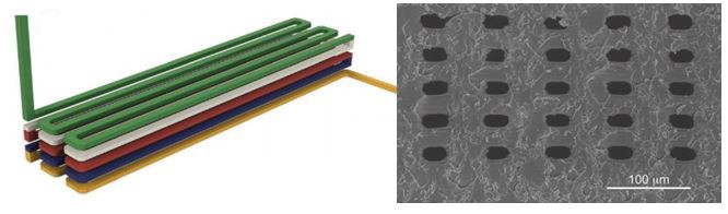
Left: Schematic illustration of 3D stacked serpentine channel design. Right: SEM image of 3D serpentine channel cross section.

Achieving true micro-scale printing resolution required innovation in both the 3D printer and the resin material. Gong et al. modified a digital light processing (DLP)-type 3D printer to include a high resolution light engine and a 385 nm UV LED light source. DLP printers project a pattern of light over the entire plane, crosslinking an entire layer in a single step. The light engine enables resolutions in the x-y plane of 7.6 µm and their choice of a 385 nm light source opened a wide range of possible UV-absorbers to them. Twenty different UV-absorbers were evaluated and 2-nitrophenyl phenyl sulfide (NPS) was chosen as having all the desired properties, with the main criterion of enabling smaller void sizes in the z-dimension. The final composition of the resin was a mixture of poly(ethylene glycol) diacrylate (PEGDA), NPS, and Irgacure 819. This mixture created structures that have low non-specific adsorption and had good resolution in the z-axis. Additionally, the structures could be photocured with a broad-spectrum UV lamp after printing.

Despite high-resolution printers and materials, a new printing regime also had to be developed to achieve the desired channel width. Because of scattering, the area of crosslinked resin tended to be smaller than the actual pixel size. This meant that channels, which are essentially voids of non-exposed space, came out wider than designed. The solution that Gong et al. devised was to perform two print patterns for each exposed layer. The first print pattern exposes the entire area, save for the channel void and the second exposure only projects a pattern along the channel walls. This method enabled reproducible printing of channels with a ~20 µm width.

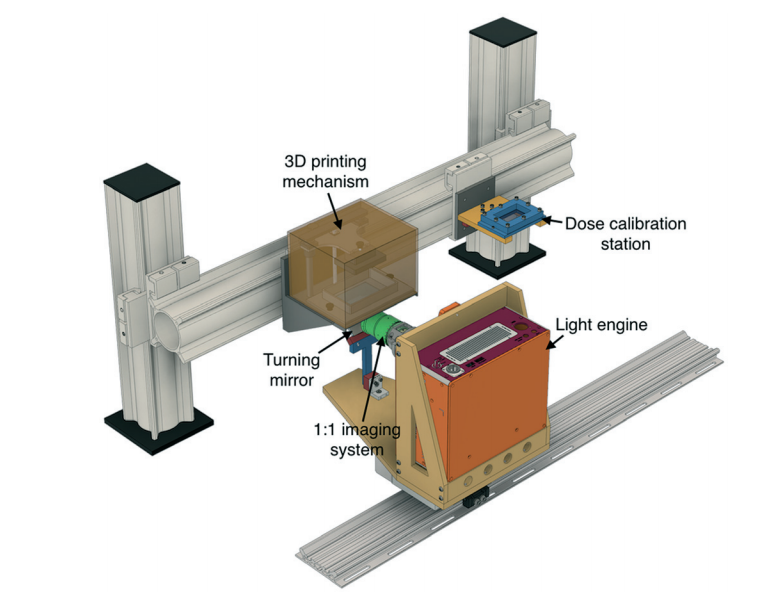
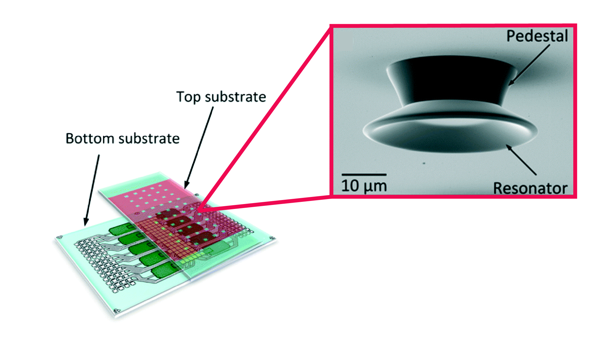
3D CAD model of the custom 3D printer by Gong et al.


So how soon can you expect to get your hands on a 3D printer capable of printing true microchannels? While this report has been very promising, it is currently the only printer and only resin formulation with these capabilities. According to Greg Nordin, the corresponding author, the main challenge is to figure out “how to get this into people’s hands in a convenient way so they don’t have to mess around much to get it to work.” Like any new technology, for it to be truly useful, users want to spend more time making and less time fiddling. Fortunately, there already are markets for printing small, intricate parts, such as jewelry and dentistry, so it’s only a matter of time before manufacturers catch on to the market for 3D printed microfluidics.


To download the full article for free* click the link below:

Custom 3D printer and resin for 18 μm × 20 μm microfluidic flow channels
Hua Gong, Bryce P. Bickham, Adam T. Woolley and Gregory P. Nordin
Lab Chip, 2017
DOI: 10.1039/C7LC00644F

*Free to access until 29th August 2017.


About the Webwriter

Darius Rackus is a postdoctoral researcher at the University of Toronto working in the Wheeler Lab. His research interests are in combining sensors with digital microfluidics for healthcare applications.”

Digg This
Reddit This
Stumble Now!
Share on Facebook
Bookmark this on Delicious
Share on LinkedIn
Bookmark this on Technorati
Post on Twitter
Google Buzz (aka. Google Reader)

How can biosensors provide real-time health monitoring?

Like a ripple spreading outwards on a pond’s surface, a plasmon is a collective oscillation of free electrons in a piece of metal. When the metal interacts with light (in this case k vector of light satisfies the momentum condition), the collective movement of oscillating electrons at the metal’s surface leads to the propagation of the light along the surface, also known as a surface plasmon. This simple physics rule is very helpful for sensor nanotechnology and optical signal processing applications since the offered advantages are significant, providing a smaller foot-print, lower limits of detection, and multiplexing opportunities. However, using sensors for biomolecule detection requires clever use of light. For example, surface plasmon resonance can be observed in nanohole arrays patterned on very thin gold films. Nanohole arrays can transmit light much more strongly than expected for the nanohole apertures at certain wavelengths. This phenomenon is called extraordinary optical transmission (EOT) and opens a new avenue for detection of minute amounts of biomolecules.

A nanoplasmonic biosensor was recently developed for real-time monitoring of proteins from live cells in a label-free configuration, thanks to extraordinary optical transmission. The biosensor is structured in a microfluidic device consisting of a cell module and the biosensor module. Microfluidic cell module design is based on a single zigzag channel for directly delivering the secreted proteins to the adjacent biosensor module via a tubing connection. The biosensor module consists of nanoholes fabricated on freestanding SiNx membranes. Antibodies specific to vascular endothelial growth factor (VEGF) are selectively immobilized on the gold nanohole arrays as biorecognition molecules. When the VEGF are captured on the sensor, a change of refractive index proximate to the surface is induced. This change results in a peak shift of the EOT spectrum (Figure 1). By tracing the spectral shifts continuously, the dynamics of cell secretion can be monitored. In this way, the secretion dynamics from live cancer cells are monitored and quantified for 10 hours. The proposed microfluidic device has unique capabilities for multiplexed and label-free detection in a compact footprint, which are promising for miniaturization and integration into lab-on-a-chip devices.

biosensor, microfluidics, extraordinary transmission phenomena, nanohole arrays

Design and working principle of microfluidic-integrated biosensor. Cancer cells grow in a zigzag channel, and secreted vascular endothelial growth factor are directly delivered to the adjacent detection module. Nanohole structures fabricated in the detection (biosensor) module are shown in SEM image with a scale bar of 1 μm (bottom left). The images on the right show the characteristic resonance peak (solid line) and EOT spectral shift of the peak upon binding (dashed line). Spectral displacements of the resonance peak based on molecular binding accumulation is shown in the sensorgram to reveal the real-time binding dynamics. Adapted from Li et al. (Lab Chip, 2017).

This technology will have a potentially significant impact on medicine. Most of the patients agree that preventive medicine is preferable to reactive medicine. However, preventive medicine requires frequent physical exams, which can only be maintained by spending substantial time and money at physicians’ offices. The need for frequent check-up exams could be reduced or eliminated by real-time monitoring of the health status by portable biosensors in the future. Developing biosensors that allow real-time monitoring of target biomolecules is a big step towards the addressing this future goal.


To download the full article for free* click the link below:

Plasmonic nanohole array biosensor for label-free and real-time analysis of live cell secretion

Xiaokang Li, Maria Soler, Cenk I. Özdemir, Alexander Belushkin, Filiz Yesilköy and Hatice Altug

Lab Chip, 2017

DOI: 10.1039/C7LC00277G


*Free to access until 7th August 2017.


About the Webwriter

Burcu Gumuscu is a postdoctoral fellow in Herr Lab at UC Berkeley in the United States. Her research interests include development of microfluidic devices for quantitative analysis of proteins from single-cells, next generation sequencing, compartmentalized organ-on-chip studies, and desalination of water on the microscale.

Digg This
Reddit This
Stumble Now!
Share on Facebook
Bookmark this on Delicious
Share on LinkedIn
Bookmark this on Technorati
Post on Twitter
Google Buzz (aka. Google Reader)

Freshwater from any water: a miniature hybrid water treatment and desalination system

With global freshwater supplies in decline, it is becoming more and more important to develop new technologies for water treatment. Given that much of the world is covered in saltwater, desalination is becoming an attractive option for generating fresh drinking water. However, the energy and capital costs of desalination can be prohibitive. Portable and scalable systems for water treatment and desalination would be useful in delivering freshwater to those who need it. Further, portable desalination systems would be particularly useful during humanitarian crises that arise due to intense weather events (e.g., hurricanes, tsunamis) in coastal regions.

Researchers from the Massachusetts Institute of Technology have recently developed a proof-of-concept microfluidic device for water treatment and desalination using electrochemical methods. The device uses two electrochemical techniques for water purification; electrocoagulation to remove particulate matter (including microorganisms) and ion concentration polarization to desalinate the water. In electrocoagulation, a sacrificial anode is oxidized to release metal coagulants that bind up and flocculate material in the water. Ion concentration polarization utilizes an electric field across an ion exchange membrane to generate an ion-rich and an ion-poor region, which can then be separated. The microfluidic device designed by Choi et al. uses one common pair of electrodes across several microchannels to achieve both electrocoagulation and ion concentration polarization. This has the advantage of minimizing power consumption as no extra power is needed to couple the two treatment methods. In their report, they demonstrated that the new hybrid device could remove nearly 90% of E. coli cells and approximately 95% of particulate matter as well as bring salt concentrations down from 20 mM NaCl to 8.6 mM NaCl (a drinkable level).

The work presented in this report lays the foundation for a truly universal and portable water treatment system. Someday you will be able to take water from any source—waste, seawater, or freshwater—and turn it into fresh clean drinking water. This will not only help those who do not have regular access to freshwater, but will be a great tool to have on hand in emergency situations.

To download the full article for free* click the link below:

Integrated pretreatment and desalination by electrocoagulation (EC)–ion concentration polarization (ICP) hybrid
Siwon Choi, Bumjoo Kim and Jongyoon Han
Lab Chip, 2017, 17, 2076-2084
DOI: 10.1039/C7LC00258K

*Free to access until 7th July 2017.


About the Webwriter

Darius Rackus is a postdoctoral researcher at the University of Toronto working in the Wheeler Lab. His research interests are in combining sensors with digital microfluidics for healthcare applications.”

Digg This
Reddit This
Stumble Now!
Share on Facebook
Bookmark this on Delicious
Share on LinkedIn
Bookmark this on Technorati
Post on Twitter
Google Buzz (aka. Google Reader)

The future of bioensors

St. Paul’s Cathedral in London has its own unique acoustics. The architecture of the dome allows a whisper to be heard from anywhere within the circular gallery, so-called the whispering gallery. The invention of whispering-gallery-mode (WGM) biosensors is indeed derived by this special gallery in St. Paul’s: just like a sound wave travelling within the dome, a light beam traveling within a glass sphere (in this case, a biosensor) circles multiple paths so that any molecule on the surface can be detected. Thanks to this powerful technique, interactions of unlabeled molecules can be analyzed with high sensitivity in real-time.

In early March of 2017, researchers from Max Planck Institute and University of Exeter published a comprehensive review paper in Lab on a Chip, explaining the advances of WGM sensors as scientific laboratory instruments, their development into lab on a chip devices, major challenges on the way towards real-world applications, and potential future applications.

WGM sensors probe the interaction between molecules and electromagnetic waves during a biomolecular reaction, and convert this information to a measurable signal. The probing is made possible thanks to the electromagnetic modes formed inside a resonator with axial symmetry. However, the electromagnetic waves slightly extend into the surrounding medium. Any changes in the surrounding medium, and therefore in the evanescent field, cause a shift of the resonance frequency—this is the basis of the sensing mechanism. WGMs are capable of sensing this shift in three ways: (1) Resonance frequency shift based sensing: measurable signal is the magnitude of the frequency shift, and the sensitivity of the sensor, which scale with the evanescent field strength at the distortion’s position, i.e. interaction of a single atomic ion with a plasmonic nanoparticle (Figure 1a). (2) Loss based sensing: it is based on the resonator’s energy loss per light wave oscillation, i.e. binding of polystyrene nanoparticles (Figure 1b). (3) Mode-splitting based sensing: a scattering molecule/particle couples clock-wise and counter clock-wise propagating WGMs, resulting in the formation of two different standing wave modes, i.e. deposition of multiple nanoparticles on a surface (Figure 1c).

Whishering gallery mode biosensors

Figure 1. Three different sensing mechanisms of whispering-gallery-mode biosensors. (a) Resonance frequency shift based sensing, (b) loss based sensing, (c) mode-splitting based sensing (from Kim et al., Lab Chip, 2017).

The review also focuses on several performance criteria of WGM sensors, such as single molecule sensitivity, time resolution, stability and specificity. Single molecule sensitivity of WGM sensors depends on the resonator’s size, the surrounding medium and excitation wavelength. Despite the fact that these parameters seem to limit the sensitivity, increasing the electric field inside a nanoscale volume significantly can circumvent this problem. Apart from that, WGM sensors can detect events happening in milliseconds to seconds whereas these detection speeds are mostly limited by the equipment, for example, the laser’s maximum scanning speed. When it comes to stability of WGM sensors, one common problem is reported to be the environmental noise sources, affecting the reliability of the measurements. A variety of methods to reduce those negative effects are further discussed in the review. One another notable functionality is that WGM sensors can be as specific as probing a surface-immobilized receptor molecule reacting with an analyte of interest.

microring resonator based on-chip sensor, pillar-supported high Q cavities

Figure 2. Lab on a chip WGMs. Left and middle images show a microring resonator based on-chip sensor with zoom-in images of different components, and right image shows a pillar-supported high Q cavities (from Kim et al., Lab Chip, 2017).

Lab on a chip applications of WGMs are discussed in two categories in the review (Figure 2): Planar resonators let the light to be coupled into multiple ring-resonators that are connected to channels containing different analytes. This type of resonators is low-cost and allows for in-parallel probing of samples. Pillar-supported high Q cavities is the second type, featuring a high Q factor owing to the air-gap between the substrate and the cavities. Pillar-supported resonators are high-cost due to several fabrication difficulties. Apart from those, droplet-based in vivo sensing via WGM sensors is also addressed as an alternative approach with the possibility of using the analyte medium itself as a resonator. Over the past decade, WGM sensors have been widely exploited to study molecular interactions with high sensitivity and seem to gain more and more attention.


To download the full article for free* click the link below:

Towards next-generation label-free biosensors: recent advances in whispering gallery mode sensors

Eugene Kim, Martin D. Baaske and Frank Vollmer

Lab Chip, 2017, Critical Review

DOI: 10.1039/C6LC01595F

*Free to access until 12th July 2017.


About the Webwriter

Burcu Gumuscu is a postdoctoral fellow in BIOS Lab on a Chip Group at University of Twente in The Netherlands. Her research interests include development of microfluidic devices for quantitative analysis of proteins of a single-cell, next generation sequencing, compartmentalized organ-on-chip studies, and desalination of water on the microscale.

Digg This
Reddit This
Stumble Now!
Share on Facebook
Bookmark this on Delicious
Share on LinkedIn
Bookmark this on Technorati
Post on Twitter
Google Buzz (aka. Google Reader)

Digital Whispers: Novel optical sensors enable label-free sensing with digital microfluidics

If you’ve ever visited St. Paul’s Cathedral in London or Grand Central Terminal in New York, you may be familiar with the interesting acoustic phenomenon termed a “whispering gallery”. The domed geometry of these structures allows sound to echo around the chambers such that a whisper spoken along the wall on one side can be clearly heard at the other end. This phenomenon can also apply to light, and microstructures tuned to a specific wavelength of light can be used as resonating sensors. Whispering-gallery mode (WGM) micro-goblet lasers use this phenomenon and can detect changes in the refractive index of the surrounding media as well as changes to the surface. This makes them ideal as label-free sensors that can detect changes to the surfaces of the microgoblets. When their surfaces are functionalized with capture moieties (e.g., antibodies, nucleic acids etc.) they can be used for sensitive label-free detection and would be a great tool to incorporate with microfluidics.

In their recent report, Wondimu et al. integrated arrays totaling 5,000 individually addressable sensors with a digital microfluidic (DMF) chip. DMF offers precise handling of nL-µL volume droplets in a compact format and with no moving parts. Typically, WGM sensors require coupling to fiber optics, but by doping the micro-goblets with organic dyes they can be operated as optically pumped lasers. This makes operating them less bulky and fits well with the streamlined philosophy behind DMF (i.e., no pumps, tubing, or connections). The fabrication of these large arrays is simple and relies on wet-etching and reflowing. Thus, scale-up is relatively straightforward. In their report, Wondimu et al. demonstrated the functionality of these sensors by testing liquids with different refractive indices as well as performing quantitative detection of streptavidin-biotin binding on the sensor surfaces. While these examples serve a demonstrative purpose, it will be possible to use these sensors for multiplexed affinity-based biosensing such as antibodies, nucleic acids, and aptamers. This will be a big leap for DMF as there haven’t been any examples of integrated multiplexed sensing on this scale before. One area where this could be applied to is the development of platforms to culture cells and perform multiplexed, label-free genetic analysis—a true micro total analysis system!

To download the full article for free* click the link below:


Integration of digital microfluidics with whispering-gallery mode sensors for label-free detection of biomolecules
Sentayehu F. Wondimu, Sebastian von der Ecken, Ralf Ahrens, Wolfgang Freude, Andreas E. Guber and Christian Koos
Lab Chip, 2017
DOI: 10.1039/C6LC01556E

*Free to access until 6th June 2017.


About the Webwriter

Darius Rackus is finishing his Ph.D. at the University of Toronto working in the Wheeler Lab. His research interests are in combining sensors with digital microfluidics for healthcare applications.

Digg This
Reddit This
Stumble Now!
Share on Facebook
Bookmark this on Delicious
Share on LinkedIn
Bookmark this on Technorati
Post on Twitter
Google Buzz (aka. Google Reader)

Game on!

Researchers at Standford University develop multi-level programming language for biotic games using swarms of microorganisms

Computer games are a ubiquitous pastime and a great example of how a single programming language give rise to a myriad of games. But what about biotic games? How could you program biological systems to function in an interactive way? Biotic games are interactive applications that interface biology and computer science for the promotion of science. The Riedel-Kruse Lab at Standford specialize in developing biotic games that use light to control swarms of Euglena gracilis—a phototaxic microorganism that avoids—and can direct, capture, and move whole swarms or individual organisms.

But programming swarms of microorganisms is no easy task. Swarms exhibit collective behaviour and therefore need to be controlled through local context rather than at the individual level. In their recent publication, the Riedel-Kruse Lab developed a set of hierarchical programming abstractions that allows swarms of Euglena within a biological processing unit (BPU; i.e., chip, microscope, and light stimuli) to be programmed in a single and efficient language at the stimulus, swarm, and system levels. At the lowest level, stimulus space programming (which the authors analogize to machine code) allows the programmer to have direct control over the various stimuli (e.g., turn left light on for 3 s), independent of the Euglena. Higher level programming at the swarm and system levels are more general and commands are given in terms of what the user wants the Euglena or system to do. For instance, swarm space commands direct the swarm in different operations such as move, split, and combine. System space commands incorporate conditional statements that can be used to confine a specific number of Euglena to a certain region or to clear Euglena from the field of view, for example.


While Lam et al. used this new language to program a biotic game, this new language and approach to swarm programming could be generalized for any type of swarm and stimuli. One application could be to program swarms to construct complex structures on the microscale. In future, by increasing access to BPUs through cloud computing and releasing this new programming language it will be possible for hobbyists and researchers alike to write new programs and applications. And maybe this is just the beginning of a revolution like the one ushered in by the release of the personal microcomputer.


To download the full article for free* click the link below:

Device and programming abstractions for spatiotemporal control of active micro-particle swarms

Amy T. Lam, Karina G. Samuel-Gama, Jonathan Griffin, Matthew Loeun, Lukas C. Gerber, Zahid Hossain, Nate J. Cira, Seung Ah Lee and Ingmar H. Riedel-Kruse

Lab Chip, 2017,17, 1442-1451

DOI: 10.1039/C7LC00131B


*Free to access until 24th May 2017.


About the Webwriter

Darius Rackus is finishing his Ph.D. at the University of Toronto working in the Wheeler Lab. His research interests are in combining sensors with digital microfluidics for healthcare applications.

Digg This
Reddit This
Stumble Now!
Share on Facebook
Bookmark this on Delicious
Share on LinkedIn
Bookmark this on Technorati
Post on Twitter
Google Buzz (aka. Google Reader)

Sample-in-answer-out

You have worked hard all year and wanted to treat yourself with something different for the summer. You decided to arrange a journey to South-Africa to enjoy the beautiful natural scenery. Discovering the range of wildlife in immense national parks, hiking in mountains, and meeting with warm locals made your journey unforgettable. You arrived at one of the best photography spots in a national park just before the sunset. While focussing on capturing the best image from the picturesque scenery, you got bitten by a Marsh mosquito, perhaps infected with a Plasmodium falciparum parasite. This parasite is known for causing malaria, the most significant parasitic disease of humans. You are not the only one: approximately 30,000 travellers from industrialized countries contract malaria each year. During the next 14 days, this parasite will differentiate and proliferate in the body. It will invade and destroy the red blood cells, eventually affecting the liver, spleen, and brain functionality. A few days after the bite, you found a small-scale laboratory for the malaria diagnosis test, but there was a problem: this laboratory can detect malaria only if you have 50-100 parasites per microliter of blood, occurring when the patient carries the parasite for weeks. You, then, had to find a larger laboratory equipped with a benchtop loop-mediated isothermal DNA amplification (LAMP) system, which can detect 1 malaria parasite per microlitre of blood. You wished there was a highly sensitive device for malaria diagnosis at the point of need. Well, we might have some good news for you.

Modern nucleic acid testing methods of malaria detection, such as LAMP, enable high sensitivity, high specificity, robust, and rapid analyses for asymptomatic infections. As performing these methods requires bulky and costly peripheral equipment and trained technicians, access to such equipment in rural areas is unlikely. Fortunately, researchers in Pennsylvania State University recently introduced a stand-alone, portable, and high sensitivity system that can perform “sample-in-answer-out” analyses. The system consists of a compact disc and a reader unit (Figure 1). The compact disc includes valves and microfluidic channels, where the blood sample is processed using magnetic beads. The reader unit can automatically perform all analysis steps including DNA purification, elution, amplification, and real-time detection. For a real demonstration of how the test is performed, the movie included below is well worth the watch. Test results can be displayed on a LCD screen or a smartphone within 40 minutes. The system can detect down to 0.6 parasites per microliter of blood. Each test costs around $1. With these specifications, this technology has the opportunity to create a new paradigm in molecular diagnosis at the point of care.

malaria detection test

Figure 1. Schematic view of an assembled compact disc made of PMMA; AnyMDX reading unit consisting of a magnet, heater plate, optical detection system, and LCD screen; and the illustration of integrated sample processing steps on the compact disc. The technique is based on DNA-carrying magnetic beads actuated against stationary reagent droplets.

To download the full article for free* click the link below:

A field-deployable mobile molecular diagnostic system for malaria at the point of need

Gihoon Choi, Daniel Song, Sony Shrestha, Jun Miao, Liwang Cuic and Weihua Guan

Lab Chip, 2016, Articles

DOI: 10.1039/C6LC01078D

About the Webwriter

Burcu Gumuscu is a postdoctoral fellow in BIOS Lab on a Chip Group at University of Twente in The Netherlands. Her research interests includedevelopment of microfluidic devices for next generation sequencing, compartmentalized organ-on-chip studies, and desalination of water on the microscale.

Digg This
Reddit This
Stumble Now!
Share on Facebook
Bookmark this on Delicious
Share on LinkedIn
Bookmark this on Technorati
Post on Twitter
Google Buzz (aka. Google Reader)

What can a magnet do?

The magic of magnetism can be shown through a simple classroom demonstration of bringing two magnetic pieces together and then trying to pull them apart. The attraction between the opposite poles of the magnets becomes very apparent as you struggle to tear them apart. This simple concept can be applied to lab-on-a-chip devices to eliminate the need for off-device hardware with power requirements, and therefore, enable the use of lab-on-a-chip technology in low-resource settings.

The majority of existing lab-on-a-chip systems use manual pipettes, syringe pumps, or pressure pump systems to manipulate the fluid flow. The dependence on off-chip hardware, however, makes the integration of these systems into low-cost environments rather challenging. Researchers in both, academia and industry think that this challenge can be addressed by “manually operated self-contained microfluidic devices”, which has gained significant attention over the past couple of years. In line with this objective, magnetically-adhesive based valves have for the first time shown to control fluid flow in a microfluidic device in a recent collaboration work by Sandia National Labs and Qorvo Inc. Here, the “magnetic-adhesive based valve” simply consists of a disk magnet seated on a thin ring of adhesive material.

In this study, a microfluidic device is designed to perform bioassays and contains a port connecting two chambers in different planes. The port is closed by an internal magnet located on a pressure-sensitive adhesive tape, and is opened by the help of an external magnet which displaces the internal magnet (figure 1). When the port is open, the reagents can flow in the microchannels, as shown in figure 2. The adhesive tape prevents any leakage within the microchip, while the magnet serves as an actuatable gate for reagents. The microfluidic device, therefore, allows for storage and on-demand transport of different types of reagents (both liquid, solid, and gas) to perform bioassays.

Magnetic-adhesive based valves are fabricated at the millimeter-scale, however, it is possible to manufacture micron-sized valves depending on the resolution of the laser ablation system used to cut the valve layout. Design considerations and characterization of magnetic-adhesive based valves are further addressed in the paper. Apart from this, the self-contained device is made of low-cost materials (such as PMMA and magnetic alloys), resulting in a fabrication cost as low as 0.2 dollars per chip. As portable and low-cost devices start to draw increasing attention in lab-on-a-chip technology, this work might be an important milestone for next generation micro total analysis systems.

Magnetic valves for lab on a chip sytems

Figure 1. Schematic and photos of magnetic-adhesive based valve working mechanism.

Magnetic valves for lab on a chip devices

Figure 2. Controlled transport and reaction of the stored components in a simple, power- and instrument-free manner in a three chambered microfluidic device.

This is a recently published Hot article and you can download it for free* by clicking the link below:

Magnetic-adhesive based valves for microfluidic devices used in low-resource settings

Jason C. Harper, Jenna M. Andrews, Candice Ben, Andrew C. Hunt, Jaclyn K. Murton, Bryan D. Carson, George D. Bachand, Julie A. Lovchik, William D. Arndt, Melissa R. Finley and Thayne L. Edwards

Lab Chip, 2016, Recent HOT Articles

DOI: 10.1039/C6LC00858E


About the Webwriter

Burcu Gumuscu is a postdoctoral fellow in BIOS Lab on a Chip Group at University of Twente in The Netherlands.

Her research interests include development of microfluidic devices for next generation sequencing, compartmentalized organ-on-chip studies, and desalination of water on the microscale .

Digg This
Reddit This
Stumble Now!
Share on Facebook
Bookmark this on Delicious
Share on LinkedIn
Bookmark this on Technorati
Post on Twitter
Google Buzz (aka. Google Reader)

Blood matters

Iron deficiency anemia (IDA) is not a trivial illness. Every individual in the world’s population has the potential to suffer from this nutritional disease. According to estimations, 900 million people worldwide are already afflicted with it. IDA is known to lower cognitive ability, work capacity, and future productivity of both children and adults. The situation appears to be grave when we consider the economic consequences of these problems.

The human body needs iron to produce red blood cells, and having low iron levels in the body leads to IDA. Diagnosis of IDA requires a complete blood count is performed by a bulky hematology analyzer. IDA has been a common disease for a really long time; however, its associated diagnosis costs are considerably high, and the diagnosis equipment is not available in many places of the world. Given the facts, IDA diagnosis actually deserves cheaper and easily accessible equipment, which has unfortunately remained elusive—up until now.

In last month’s issue of Lab on a Chip, Whitesides research group at Harvard University came up with a sound idea to diagnose IDA in a shorter and inexpensive way. They developed a low-cost and rapid-screening tool to diagnose IDA using aqueous multiphase systems containing layer of polymer-salt mixtures. These mixtures are loaded in a microhematocrit tube (depicted in the figure) together with a drop of blood from a fingerprick. Diagnosis results become available after a 2-minute low-cost centrifuging process.

The reported data suggest that diagnosis of IDA is improved by means of sensitivity and specificity when compared to the bulky hematology analyzer’s results. Several important red blood cell parameters, such as concentration of hemoglobin in a given volume of red blood cells, can be predicted. The technique’s ability to diagnose IDA was further improved using automated digital analysis. They also show that the tool is able to detect a wider range of anemia types including microcytic and hypochromic anemia. The portable and low cost screening tool could possibly find use in rural clinics where large fractions of the population at risk of IDA. Before entering the market, the performance of this technique will still have to be validated to demonstrate feasibility of using and interpreting the assay.

Design of the presented test loaded with blood before and after centrifugation for a representative IDA and Normal sample. Blood is loaded into the top of the tube, from a fingerprick, using capillary action provided by a hole in the side of the tube. Normal blood packs at the bottom of the tube, while less dense blood cells can be seen packing at the interfaces between the phases and inside the tube. Normal and IDA blood can be differentiated by eye after only 2 minutes of centrifugation. It is also possible to read the analysis results in an automated way. A commonly used software is used to convert the image to red intensity graphs.

This article was published in Lab on a Chip on 30th August 2016.

To download the full article for free* click the link below:

Diagnosis of iron deficiency anemia using density-based fractionation of red blood cells

Jonathan W. Hennek, Ashok A. Kumar, Alex B. Wiltschko, Matthew R. Patton, Si Yi Ryan Lee, Carlo Brugnara, Ryan P. Adams and George M. Whitesides
Lab Chip, 2016,16, 3929-3939
DOI: 10.1039/C6LC00875E

—————-

About the Webwriter


Burcu Gumuscu is a postdoctoral fellow in BIOS Lab on a Chip Group at University of Twente in The Netherlands. Her research interests include development of microfluidic devices for next generation sequencing, compartmentalized organ-on-chip studies, and desalination of water on the microscale.

—————-

*Access is free until 7th November 2016 through a registered RSC account – register here

Digg This
Reddit This
Stumble Now!
Share on Facebook
Bookmark this on Delicious
Share on LinkedIn
Bookmark this on Technorati
Post on Twitter
Google Buzz (aka. Google Reader)

Recent Advances in 3D Printing

Guest edited by Jennifer Lewis (Harvard University) and Howard Stone (Princeton University) this collection of papers showcases recent advances in the rapidly evolving field of 3D printing, with an emphasis on themes that impact lab-on-a-chip applications.

Free* Access: The upcoming 3D-printing revolution in microfluidics
Critical Review
Nirveek Bhattacharjee, Arturo Urrios, Shawn Kang and Albert Folch
Lab Chip, 2016,16, 1720-1742 DOI: 10.1039/C6LC00163G

Free* Access: High density 3D printed microfluidic valves, pumps and multiplexers
HOT Article
Hua Gong, Adam Trticle. Woolley and Gregory P. Nordin
Lab Chip, 2016,16, 2450-2458 DOI: 10.1039/C6LC00565A

Free* Access: Bioprinted Thrombosis-on-a-Chip
HOT Article
Rahmi Oklu et al.
Lab Chip, 2016, Accepted Manuscript, C6LC00380J

Open Access: 3D- printed microfluidic devices: enablers and barriers
Michael C. Breadmore., et al
Lab Chip, 2016,16, 1993-2013
DOI: 10.1039/C6LC00284F

This collection also features a video demonstration:

3D printing of liquid metals as fugitive inks for fabrication of 3D microfluidic channels
Dishit P. Parekh, Collin Ladd, Lazar Panich, Khalil Moussa and Michael D. Dickey
Lab Chip, 2016,16, 1812-1820 DOI: 10.1039/C6LC00198J

Browse our 3D Printing collection – we hope you enjoy the articles

*Access is free until 10th October via a registered RSC account.

Digg This
Reddit This
Stumble Now!
Share on Facebook
Bookmark this on Delicious
Share on LinkedIn
Bookmark this on Technorati
Post on Twitter
Google Buzz (aka. Google Reader)