Archive for the ‘Hot Articles’ Category

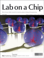
Hybrid opto-electric manipulation, droplet based microfluidics and digital PCR on the cover of issue 13!

The striking image on the outside front cover of this issue is courtesy of Aloke Kumar (Oak Ridge National Laboratory).  The Critical Review which it highlights discusses the fundamentals, applications and future of hybrid opto-electric manipulation techniques for microfluidics

Hybrid opto-electric manipulation in microfluidics—opportunities and challenges
Aloke Kumar, Stuart J. Williams, Han-Sheng Chuang, Nicolas G. Green and Steven T. Wereley
Lab Chip, 2011, 11, 2135-2148
DOI: 10.1039/C1LC20208A

The equally eye-catching image on the inside front cover is from Valérie Taly and Andrew D. Griffiths (ISIS, Strasbourg) et al., accompanying work on a droplet-based microfluidics method for digital PCR and a method for multiplexing quantitative digital PCR beyond the conventional limitations of color-encoded probes.
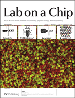
Quantitative and sensitive detection of rare mutations using droplet-based microfluidics
Deniz Pekin, Yousr Skhiri, Jean-Christophe Baret, Delphine Le Corre, Linas Mazutis, Chaouki Ben Salem, Florian Millot, Abdeslam El Harrak, J. Brian Hutchison, Jonathan W. Larson, Darren R. Link, Pierre Laurent-Puig, Andrew D. Griffiths and Valérie Taly
Lab Chip, 2011, 11, 2156-2166
DOI: 10.1039/C1LC20128J

Multiplex digital PCR: breaking the one target per color barrier of quantitative PCR
Qun Zhong, Smiti Bhattacharya, Steven Kotsopoulos, Jeff Olson, Valérie Taly, Andrew D. Griffiths, Darren R. Link and Jonathan W. Larson
Lab Chip, 2011, 11, 2167-2174
DOI: 10.1039/C1LC20126C

View the issue

Digg This
Reddit This
Stumble Now!
Share on Facebook
Bookmark this on Delicious
Share on LinkedIn
Bookmark this on Technorati
Post on Twitter
Google Buzz (aka. Google Reader)

HOT: continuous electrowetting, assaying sprouting angiogenesis and 3D patterns in a 2D platform

This Communication from Nathan B. Crane (University of South Florida) et al. describes new method of droplet transport, combining diode-like conduction and electrowetting on dielectric to achieve continuous electrowetting with a single electrode.
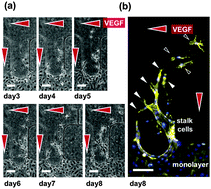
Continuous electrowetting via electrochemical diodes
Christopher W. Nelson, Corey M. Lynch and Nathan B. Crane
Lab Chip, 2011, 11, 2149-2152
DOI: 10.1039/C1LC20196D


In their paper Seok Chung (Korea University) et al. have developed a hydrogel incorporating a microfluidic platform which can mimic the 3D tissue microenvironment for the study of endothelial cell sprouting angiogenesis.  They are able to precisely control the gradient of soluble angiogenic factors, VEGF and ANG-1 and obtain a quantitative response to the assay.
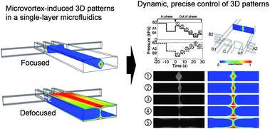
In vitro 3D collective sprouting angiogenesis under orchestrated ANG-1 and VEGF gradients
Yoojin Shin, Jessie S. Jeon, Sewoon Han, Gi-Seok Jung, Sehyun Shin, Sang-Hoon Lee, Ryo Sudo, Roger D. Kamm and Seok Chung
Lab Chip, 2011, 11, 2175-2181
DOI: 10.1039/C1LC20039A


William C. Messner (Carnegie Mellon University) and colleagues have also been working in 3D to achieve dynamic control of 3D chemical patterns in a single 2D microfluidic platform.  They are able to switch between ‘focused’ and ‘defocused’ 3D flow profiles, and to rapidly tune the patterns through feedback control of the inlet pressures.
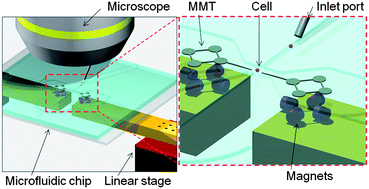
Dynamic control of 3D chemical profiles with a single 2D microfluidic platform
YongTae Kim, Sagar D. Joshi, Lance A. Davidson, Philip R. LeDuc and William C. Messner
Lab Chip, 2011, 11, 2182-2188
DOI: 10.1039/C1LC20077A

As with all our HOT papers, these are free to access for 4 weeks

Digg This
Reddit This
Stumble Now!
Share on Facebook
Bookmark this on Delicious
Share on LinkedIn
Bookmark this on Technorati
Post on Twitter
Google Buzz (aka. Google Reader)

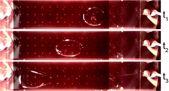
HOT: new driving force for high accuracy microrobot to manipulate single cells

Manipulating cells in microfluidic chips is often accomplished with a magnetically driven microtool (MMT), driven by a permanent magnet.  However MMTs driven by permanent magnets suffer from low positioning accuracy and response speed.  Here, Masaya Hagiwara (Nagoya University) and colleagues have devised a new way of driving MMTs – using a piezoelectric ceramic induce ultrasonic vibration and reduce the effective friction.  The result is a 1.1 mm positioning accuracy of the microrobot, which is 100 times higher than operating without vibration.

This HOT article is featured on the cover of Issue 12 and is free to access for 6 weeks:

On-chip magnetically actuated robot with ultrasonic vibration for single cell manipulations
Masaya Hagiwara, Tomohiro Kawahara, Yoko Yamanishi, Taisuke Masuda, Lin Feng and Fumihito Arai
Lab Chip, 2011, 11, 2049-2054
DOI: 10.1039/C1LC20164F

Digg This
Reddit This
Stumble Now!
Share on Facebook
Bookmark this on Delicious
Share on LinkedIn
Bookmark this on Technorati
Post on Twitter
Google Buzz (aka. Google Reader)

HOT: detecting CRP over six orders of magnitude using silicon photonic microring resonators

In their recent paper Ryan Bailey and colleagues at the University of Illinois at Urbana-Champaign address the challenge of creating a biomarker assay capable of spanning a clinically relevant dynamic range.  The ability to accurately detect protein biomarkers over a wide dynamic range is extremely important to determine the stage a disease is at, as well as monitoring the effects of any treatments.

They have developed a a silicon photonic microring resonator-based platform that can quantify the cardiovascular risk biomarker C-reactive protein over a dynamic range of six orders of magnitude.  The 3 step assay increases the dynamic range beyond that possible for a single-step assay and also reduces false positive results.

Download the article for the details – the article is currently free to access:

Sensitive on-chip detection of a protein biomarker in human serum and plasma over an extended dynamic range using silicon photonic microring resonators and sub-micron beads
Matthew S. Luchansky, Adam L. Washburn, Melinda S. McClellan and Ryan C. Bailey
Lab Chip, 2011, 11, 2042-2044
DOI: 10.1039/C1LC20231F

Digg This
Reddit This
Stumble Now!
Share on Facebook
Bookmark this on Delicious
Share on LinkedIn
Bookmark this on Technorati
Post on Twitter
Google Buzz (aka. Google Reader)

HOT: separating cells through their ability to engulf nanoparticles

Cells labelled with magnetic nanoparticles are useful for a number of applications, ranging from MRI to drug delivery and cancer therapy, and being able to sort said cells based on their magnetic nanoparticle loading is obviously necessary. Continuous flow methods currently exist for separating cells based on the extent of their magnetisation, but only for cells where the nanoparticles have been bound to them via specific surface markers.

Photograph of the microfluidic magnetophoresis chip

In this paper Claire Wilhelm (University of Paris Diderot) and Nicole Pamme (University of Hull) make use of the other mechanism of introducing nanoparticles to the cells  – exploiting the natural ability of monocytes and macrophages to engulf molecules. The team produced a free-flow microfluidic chip with an external magnet to separate the cells through five different exits, depending on the loading of magnetic nanoparticles. They demonstrated successful and efficient separation of the monocytes and macrophages, with monocytes displaying a much weaker endocytotic ability than macrophages, at rates of 10 to 100 cells per second.

For the full details download the article – it’s free to access for 4 weeks:

Cell sorting by endocytotic capacity in a microfluidic magnetophoresis device
Damien Robert, Nicole Pamme, Hélène Conjeaud, Florence Gazeau, Alexander Iles and Claire Wilhelm
Lab Chip, 2011, Advance Article
DOI: 10.1039/C0LC00656D

Digg This
Reddit This
Stumble Now!
Share on Facebook
Bookmark this on Delicious
Share on LinkedIn
Bookmark this on Technorati
Post on Twitter
Google Buzz (aka. Google Reader)

HOT article: webcam technology to identify cardiotoxicity

Identifying drugs that have the potential to cause heart dysfunction as early as possible is crucial for preventing the waste of time and money in drug discovery processes.

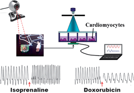
Cell-based biosensors using living cells have been applied in pharmacological screening, but current technologies encounter difficulties such as small measurement areas, complicated fabrication procedures and high costs.  However, the rapid development of digital consumer technologies has driven down the cost and created compact imaging sensor modules such as CMOS (complementary field oxide semiconductor) imaging senors.  

In this HOT article, Ali Khademhosseini (Harvard Medical School) and colleagues made use of this development, extracting the CMOS sensor from a webcam and using it with an image processing algorithm to measure cardiotoxicity in cardiomyocytes.  They were able to detect beat-to-beat variability in real-time after treatment with drugs iosprenaline and doxorubicin.

The authors hope this neat system may be useful for a range of cell-based biosensing applications.

Download the article for free for 4 weeks:

A cell-based biosensor for real-time detection of cardiotoxicity using lensfree imaging
Sang Bok Kim, Hojae Bae, Jae Min Cha, Sang Jun Moon, Mehmet R. Dokmeci, Donald M. Cropek and Ali Khademhosseini
Lab Chip, 2011, 11, 1801-1807
DOI: 10.1039/C1LC20098D, Paper

Digg This
Reddit This
Stumble Now!
Share on Facebook
Bookmark this on Delicious
Share on LinkedIn
Bookmark this on Technorati
Post on Twitter
Google Buzz (aka. Google Reader)

HOT article: use liquid metal to make simple microfluidic electrodes

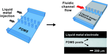
Ju-Hee So and Michael Dickey from North Carolina State University present an extremely simple fabrication route for microfluidic electrodes by using metal alloys with low melting point , such as eutectic gallium indium.

The liquid metal injected into the microchannels is inherently aligned and in direct contact with the fluid, but is neatly prevented from mixing with the fluid by posts with spacings that are too small to allow the metal to easily flow through.  The metal also maintains its shape despite being a liquid, due to the spontaneous formation of a thick oxide skin.  The mechanical stability of the electrodes was demonstrated in operating conditions commonly used in microfluidic applications, and as a proof-of-principle, used for electrohydrodynamic mixing, which requires extremely high electric fields.

The authors believe that this technique is significantly simpler and easier to implement than anything that has been published to date in the literature – why not download the article and see for yourself?  The article is free to access for 4 weeks!

Inherently aligned microfluidic electrodes composed of liquid metal
Ju-Hee So and Michael D. Dickey
Lab Chip, 2011, 11, 905-911
DOI: 10.1039/C0LC00501K

Digg This
Reddit This
Stumble Now!
Share on Facebook
Bookmark this on Delicious
Share on LinkedIn
Bookmark this on Technorati
Post on Twitter
Google Buzz (aka. Google Reader)

HOT article: phaseguides – primed and ready to revolutionise microfluidics

So you’ve designed this fantastic little lab-on-a-chip device that will revolutionise disease/virus/cancer detection or discover fantastic new drugs, but you can’t get your sample into all the complicated channels or bubbles are ruining your flow.  Sound familiar?

Well now Paul Vulto (Albert-Ludwigs-Universität Freiburg) and colleagues have developed a novel technique for priming microfluidic devices that could solve all these problems.  By using patterned ridges in their devices they are able to guide the liquid–air interfaceto fill complicated geometries – by forcing it to align with the ridge.     This allows them to fill complex microfluidic chambers and channels, independant of their shape, simply by the carefully patterning of these ridges.  The team have aptly coined the term  ‘phaseguides‘ to descibe their invention and hope that it ‘will prove a leap forward towards more simple, flexible and reliable microfluidic systems’.

This figure demonstrates the impressive use of phaseguides by the filling of a butterfly shaped chamber.  Inserts (a and b) demonstrate that when filling without a phaseguide pattern the liquid spreads radially leaving the structure largely un-filled.   Inserts (c–f) demonstrate that with phaseguides (visualized by dashed lines in (c)), the structure can be completely filled with liquid.

This paper comes highly recommended by our expert reviewers and is free to access for 6 weeks – why not download it today!

Phaseguides: a paradigm shift in microfluidic priming and emptying
Paul Vulto, Susann Podszun, Philipp Meyer, Carsten Hermann, Andreas Manz and Gerald A. Urban
Lab Chip, 2011, 11, 1596-1602
DOI: 10.1039/C0LC00643B

Digg This
Reddit This
Stumble Now!
Share on Facebook
Bookmark this on Delicious
Share on LinkedIn
Bookmark this on Technorati
Post on Twitter
Google Buzz (aka. Google Reader)

Free Lab on a Chip articles in Nature technology feature

Work published in Lab on a Chip features heavily in Nature’s latest Special Technology Feature ‘Tissue models: A living system on a chip‘.

The article, summarising the current state-of-the-art in creating living tissue models on chips, references work from LOC Editorial Board member Donald Ingber (Harvard Medical School) and LOC publications from Michael Shuler (Cornell),  John March (Cornell), Linda Griffith (MIT) and Axel Günther (University of Toronto).

We’ve made these great articles free to access for 2 weeks – why not take a look!

A microfluidic device for a pharmacokinetic–pharmacodynamic (PK–PD) model on a chip
Jong Hwan Sung, Carrie Kam and Michael L. Shuler
Lab Chip, 2010, 10, 446-455

Microscale 3-D hydrogel scaffold for biomimetic gastrointestinal (GI) tract model
Jong Hwan Sung, Jiajie Yu, Dan Luo, Michael L. Shuler and John C. March
Lab Chip, 2010, 11, 389-392

Perfused multiwell plate for 3D liver tissue engineering
Karel Domansky, Walker Inman, James Serdy, Ajit Dash, Matthew H. M. Lim and Linda G. Griffith
Lab Chip, 2010, 10, 51-58

A microfluidic platform for probing small artery structure and function

Axel Günther, Sanjesh Yasotharan, Andrei Vagaon, Conrad Lochovsky, Sascha Pinto, Jingli Yang, Calvin Lau, Julia Voigtlaender-Bolz and Steffen-Sebastian Bolz
Lab Chip, 2010, 10, 2341-2349
From our 2010 Emerging Investigators themed issue

Digg This
Reddit This
Stumble Now!
Share on Facebook
Bookmark this on Delicious
Share on LinkedIn
Bookmark this on Technorati
Post on Twitter
Google Buzz (aka. Google Reader)

HOT article: packed to precision – capillary-driven automatic packaging for microfluidic devices

The fabrication  of 3D microstructures for microfluidic devices continues to be a challenge as traditional microfabrication techniques are not suitable for the construction of these devices.  For example, PDMS (polydimethylsiloxane) is one of the most common substrate materials for microfluidic devices, but standard packaging techniques are not able to bond multiple substrate layers with the high precision required.

Tingrui Pan (University of California, Davis) et al. have now come up with an easy and robust technique which they hope will make this problem a thing of the past.  Their new technique, CAP (capillary-driven automatic packaging) uses the interactions between a liquid capillary bridge and the top and bottom substrates to align multiple substrate layers with high precision, and has a bonding strength comparable to standard oxygen plasma processes.  The technique is also transferable to other materials, requires no thermal or mechanical treatment, nor any specialist equipment.

Illustration of the CAP-enabled microdevice fabrication process, including (a) micropatterning of a shadow mask made of dry film, (b) PDMS replica molding, (c) selective oxygen plasma treatment through the shadow mask, (d) DI water loading in the defined hydrophilic regions, and finally (e) self-alignment and self-engagement steps between two chips with identical capillary alignment patterns.

Pan et al. believe that this technique has the ability to be employed in microdevices for point-of-care diagnosis, controlled drug delivery, and combinatorial biological screening – why not take a look and see for yourself – the article’s free to access for four weeks!

Capillary-driven automatic packaging
Yuzhe Ding, Lingfei Hong, Baoqing Nie, Kit S. Lam and Tingrui Pan
Lab Chip, 2011, 11, 1464-1469
DOI: 10.1039/C0LC00710B

Digg This
Reddit This
Stumble Now!
Share on Facebook
Bookmark this on Delicious
Share on LinkedIn
Bookmark this on Technorati
Post on Twitter
Google Buzz (aka. Google Reader)