Author Archive

A New Method of Droplet Handling for Ultra-Low Volume Kinetics Studies

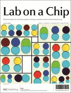
In a recent paper in Lab on a Chip, which featured as the Cover Article of Issue 22, Vol 13, researchers from the Ecole Polytechnique, Palaiseau, France describe a micro-droplet based method for measuring reaction kinetics using tiny (20 nL) sample volumes.


When working with precious reagents such as enzymes, minimizing sample volume is of the utmost importance.  Droplet-based microfluidic methods can greatly reduce sample volumes, but still waste considerable amounts of sample in fluid handling processes (e.g. pushing the sample through a channel).  A team led by Marten Vos and Charles Baroud has utilized a unique “rails and anchors” method1 of forming and controlling the movement of droplets to observe the kinetics of a chemical reaction using only 20 nL of each reagent solution.

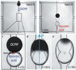
In this scheme, the flow chamber geometry is carefully designed so that when an aqueous solution is injected into the oil-filled chamber, droplets of a reproducible size are released and constrained between the top and bottom surfaces of the chamber.  Then, widening grooves (“rails”) in the bottom of the chamber guide the droplets via surface tension to “anchors” in the center.  (See figure a and b.)  In this way, no additional solution is wasted pushing droplets around.

Droplet-based scheme for observing reaction kinetics (Fig 2 from paper)

The authors used the “rails and anchors” to place two droplets, each containing a different reagent, in proximity, and then used an IR laser pulse to break the surface tension between them (figure c). The interface between the two solutions (in this case, DCPIP and ascorbic acid) can clearly be seen in figure d below.  By tracking the front of the color change that moves across the droplet as the reaction proceeds (figure e), it is possible to calculate the reaction rate (accounting also for the diffusion rates of the reactants).

The researchers also demonstrated the potential of multiplexing their technique by monitoring six different reactions in parallel droplets. They were able to obtain results in good agreement with a commercial stopped-flow spectrometer with greatly reduced cost and time.

Read this HOT article in Lab on a Chip today!

Parallel Measurements of Reaction Kinetics using Ultralow Volumes, Etienne Fradet, Paul Abbyad, Marten H. Vos, and Charles N. Baroud, DOI: 0.1039/c3lc50768h

References:

  1. P. Abbyad et al., Lab on a Chip 2011, 11, 813-821.
Digg This
Reddit This
Stumble Now!
Share on Facebook
Bookmark this on Delicious
Share on LinkedIn
Bookmark this on Technorati
Post on Twitter
Google Buzz (aka. Google Reader)

Capillarics – microfluidic circuits

The term ‘microfluidics’ immediately invokes the image of elegant, tiny devices with a small footprint in which fluids are manipulated with ease. It is an image that is most definitely correct. Still, every researcher in the field of microfluidics is aware of ‘the dirty little secret’ of these devices. Namely, that they often require bulky pumps and lots of tubing to be operated.

The requirement for macroscopic equipment to control microfluidic devices is a serious problem. It hinders the development of complex and integrated microfluidic circuits and it prevents the integration of the devices in small portable equipment.

Many researchers in the field are actively developing methods to get rid of all macroscopic equipment to control microfluidic devices. One such method is to build ‘autonomous’ devices, in which the flow is driven only by capillary forces and not by external pumps.

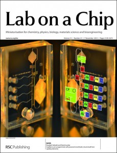
In a recent paper, which featured as the Cover article for Issue 21 of Lab on a Chip, Safavieh and Juncker give a solid overview of the various elements that can be used to autonomously control flow, such as pumps, vents and valves. Moreover, they propose a method called ‘capillarics’ in which autonomous fluidic circuits are designed in a way that is similar to electronic circuits. Finally, they demonstrate the power of their approach by rationally designing a ‘capillaric circuit’ and using it to perform a biochemical assay.

To learn more, read the full HOT article: Capillarics: pre-programmed, self-powered microfluidics circuits built from capillary elements, Roozbeh Safavieh and David Juncker, DOI: 10.1039/C3LC50691F

Digg This
Reddit This
Stumble Now!
Share on Facebook
Bookmark this on Delicious
Share on LinkedIn
Bookmark this on Technorati
Post on Twitter
Google Buzz (aka. Google Reader)

Follow Your Nose: A Micro-aquarium for Probing Bacterial Chemotaxis in Gas Gradients

Many organisms can direct their movements according to their chemical environment. In bacteria, this chemical-directed migration (i.e. chemotaxis) is enabled by receptor-mediated signaling to the bacterial flagellar motors.1 Equipped with this remarkable sensory-motor system, bacteria can measure chemical gradients and swim towards nutritious substances (positive chemotaxis) or flee from cytotoxic chemicals (negative chemotaxis).

Bacterial chemotaxis is critical for various processes including biofilm formation, biomediation, and diseases pathogenesis.2 For example, at the onset of various digestive diseases, bacteria rely on chemotaxis to find a suitable colonization site in the human gastrointestinal tract. Thus, there is great interest in studying the rates and molecular mechanisms in these chemotactic processes.

In the past 10 years, microfluidic gradient-generators have improved the way scientists study bacterial chemotaxis;3 however, these studies have not addressed gas gradients (e.g. O2 and CO2), which are known to be important in bacterial motility.4 Microfluidics devices have enabled the generation of well-controlled chemical gradients and the measurement of bacterial response at high spatiotemporal resolution. Gradients are typically formed by flowing a sample liquid (e.g. aspartate in buffer) and a reference liquid (e.g. buffer) into the device.

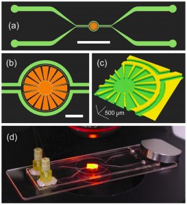
A team of researchers at the RIKEN Advanced Science Institute, Japan and Hanyang University, Korea carried out the world’s first microfluidic study of bacterial chemotaxis in gas gradients.5 The microfluidic device (see picture) comprises an isolated chamber (micro-aquarium) for bacterial culture and two bypass channels for sample and reference inputs. In this design, the sample molecules in the channel permeate through the PDMS walls (150 µm thick) and diffuse into the liquid of the micro-aquarium, facilitating the generation of a stable, shear-free gradient. The bacterial response can be monitored optically and quantified via image processing.

Using this device, the research team studied the chemotactic behaviour of Euglena gracilis microbial cells in the presence of CO2 gradients. These studies revealed that the chemotaxis of Euglena cells is rapid and is dependent on CO2 concentration. In particular, negative chemotaxis was observed at high concentrations (>15%) and positive chemotaxis was observed at low concentrations (<15%)— the maximum positive chemotaxis was observed at ~5% CO2, corresponding to the most favourable condition for photosynthesis. Interestingly, the team observed evidence of chemotactic “adaption” when they consecutively switched the sample and reference inputs.

The microfluidic device is also compatible with small molecule liquid samples such as ethanol, H2O2, and culture media. By continuously perfusing culture medium in the bypass channels, the viability of the Euglena cells in the micro-aquarium can be maintained for more than 2 months.

In summary, the research team created a microfluidic gradient-generator that is versatile and straightforward to use. The device is useful for bacterial chemotaxis studies in chemical gradients formed from gas or liquid samples. Furthermore, the device may potentially be useful for other applications including drug screening, toxicity assays, and environmental monitoring.

  1. H. C. Berg, Annual Review of Biochemistry, 2003, 72, 19-54.
  2. T. Ahmed, T. S. Shimizu and R. Stocker, Integrative Biology, 2010, 2, 604-629.
  3. J. Wu, X. Wu and F. Lin, Lab on a Chip, 2013, 13, 2484-2499.
  4. C. Douarche, A. Buguin, H. Salman and A. Libchaber, Physical Review Letters, 2009, 102, 198101.
  5. K. Ozasa, J. Lee, S. Song, M. Hara and M. Maeda, Gas/liquid sensing via chemotaxis of Euglena cells confined in an isolated micro-aquarium, Lab on a Chip, 2013, 13, 4033-4039
Digg This
Reddit This
Stumble Now!
Share on Facebook
Bookmark this on Delicious
Share on LinkedIn
Bookmark this on Technorati
Post on Twitter
Google Buzz (aka. Google Reader)

High-throughput chip for drug screening in 3D

A simple micro-array chip developed by scientists in China could sharpen the search for new drugs by enabling the high-throughput screening of drug candidates against cells cultured in three dimensions.

Developing new drugs for the treatment of complex diseases such as cancer is an expensive and time consuming process. Often, the first step in designing a new drug is to use high-throughput screening of large chemical libraries to identify compounds that have the desired medicinal effect.

Most cell-based drug screening technologies currently rely on cell cultures grown in two dimensions (i.e. as a flat layer of cells) and require sophisticated robotic systems to seed the cells and administer the drugs. However, Yanan Du and colleagues from Tsinghua University may have revolutionised the process by developing a micro-array chip that can be used to culture three dimensional balls of cells, called spheroids, and administer candidate drugs on a high-throughput scale.

Du’s chip consists of two plates containing arrays of micro-wells. The micro-wells of the first plate contain porous gelatin sponges. Once cells and culture medium are added, these sponges act as a rudimentary extracellular matrix enabling the cells to form spheroids. Meanwhile, the micro-wells of the second plate are filled with potential drug candidates. Pushing the two plates together brings the drug into contact with the spheroids so the effect of the drug on the cells can be observed.

The technique should be accessible to any laboratory with basic cell culture facilities

There are two key advantages to this system. Firstly, cells that are cultured in 3D more closely mimic the tissues in the body than cells grown in 2D. Consequently, the effect that the drug has on these cells is likely to be more representative of the effect that the drug would actually have in the body, making the results of the screening process more reliable. Cells cultured in 3D are also more likely to develop drug resistance so these compounds can be weeded out at an earlier stage in the drug development process. The second advantage is that the technique doesn’t require any expensive or sophisticated equipment, and so should be accessible to any laboratory with basic cell culture facilities.

‘By using microwells they can scale-down the number of cells and the amount of drug they use whilst increasing throughput by having a device compatible with standard laboratory instrumentation,’ says Tony Cass from Imperial College London, UK, who also develops devices for high throughput analysis.

Mario Cabodi, an expert in microfabrication at Boston University in Massachusetts, US, says the chip potentially has a promising future: ‘3D cell culture systems, like the one described here, might be helpful in bridging the gap between in vitro tests and in vivo results.’

Du’s team are now in the process of standardising the chip and are collaborating with drug development companies to perform cell-based tests of potential anti-cancer drugs.

To see the original Chemistry World article, and many others, click here.

Alternatively, you can download the full paper here.

Digg This
Reddit This
Stumble Now!
Share on Facebook
Bookmark this on Delicious
Share on LinkedIn
Bookmark this on Technorati
Post on Twitter
Google Buzz (aka. Google Reader)

Molecular Force Assay on a Microfluidic Chip

Researchers in Munich have adapted their molecular force assay technique to a microfluidic system, allowing the strength of protein-DNA interactions to be measured in a chip format.

The measurement of intermolecular forces is a challenging and important task. Two well-known methods, atomic force microscopy and optical trapping, are capable of probing these forces1. However, these instrumentation-intensive techniques are not suitable for every application. A team from the Ludwig-Maximilians-Universität, Germany, headed by Hermann Gaub, a leader in the field of force spectroscopy, has now developed a technique, termed molecular force assay (MFA), to probe the strength of DNA hybridization and the effects of DNA-binding proteins2.

Schematic of the molecular force assay (adapted from Figure 1 of paper)

In MFA, a surface is functionalized with a carefully designed DNA construct, as seen in the left panel of the figure. This surface is brought into contact with another surface which is coated with neutravidin (center). The neutravidin binds the biotin at the end of the DNA construct, so when the two surfaces are separated, the DNA construct rips apart (dehybridization, right). The breakage can happen in one of two ways—if the hybridization of the “probe strand” is stronger than that of the “reference strand”, the reference strand will dehybridize, leaving behind a Cy3 tag. If the hybridization of the reference strand is stronger, the probe strand will dehybridize, leaving both a Cy3 tag and a Cy5 tag behind. Via the resulting fluorescence, it is possible to compare the hybridization strength of the probe strand to that of the known reference. DNA binding proteins which modify the hybridization strength of the probe strand can then be introduced.

In the original formulation of MFA, the surfaces were brought into contact using a piezoelectric device; however, in a recent paper in Lab on a Chip, the researchers have translated their assay into a microfluidic format. A microfluidic chip was used in which the lower layer contains sample wells and the upper layer contains control valves. By pressurizing button valves above each well, the top and bottom surfaces can be brought into contact and then separated. The authors demonstrated the effect of EcoRI binding on probe strand hybridization strength as a proof-of-concept, “paving the way for studies of currently unknown protein-DNA interactions, including those of transcription factors.”

Read this HOT article in Lab on a Chip today!

Protein–DNA force assay in a microfluidic format, Marcus Otten, Philip Wolf, and Hermann E. Gaub. DOI: 10.1039/C3LC50830G

References:

1. K. C. Neuman and A. Nagy, Nature Methods, 2008, 5, 491-505.

2. P. M. D. Severin et al., Lab on a Chip, 2011, 11, 856-862.

Digg This
Reddit This
Stumble Now!
Share on Facebook
Bookmark this on Delicious
Share on LinkedIn
Bookmark this on Technorati
Post on Twitter
Google Buzz (aka. Google Reader)

Paper device spots antibiotic-resistant bacteria

Scientists in Canada have developed a paper-based device that checks if bacteria are resistant to certain antibiotics. The simple system could help users in remote areas pick the most appropriate treatment for bacterial infections.

Testing bacteria to see which antibiotics will be effective against them is vital for properly medicating patients. Methods exist to assess bacteria but the equipment needed is often expensive and requires highly trained operators and laboratory conditions. Now, Ratmir Derda and colleagues at the University of Alberta have designed a portable device made from paper and other low-cost materials. The team sought the help of high school students to make and test the devices to show how easy they are to construct and use.

Derda’s device consists of a paper support with a clear plastic window over an area of nutritious media permeated into a sheet of thick blotting paper. This culture media has a uniform pattern of hydrophobic spots to ensure samples disperse evenly across it. Two zones of antibiotics are added onto the media before the device is sterilised in an autoclave. After autoclaving, a cell viability dye is dropped on the culture media and the bacterial sample is then added on top of the dye. Finally, the device is sealed and incubated overnight.

In areas with no bacterial growth the dye remains blue and in areas where bacteria do grown the dye turns pink. The colours are easy to see through the device’s plastic window – there is no need to open the device. A blue area around the antibiotic zones indicates that the bacteria are susceptible to the antibiotics. The hydrophobic spots on the culture media aid quantification of the blue area to give an idea of how sensitive the bacteria are to the antibiotics.

Preparing and using the device is simple. (M: culture media; A: antibiotic zone; C: culture zone)

‘This work shows that living microorganisms can be grown and screened for antibiotic resistance using paper devices that are small and light enough to store in a person’s pocket,’ says Marya Lieberman, an expert in paper-based sensors at the University of Notre Dame in Indiana, US.

The team also found that the device could easily be stored long-term. After assembling to the point that it contained the culture media and antibiotics, it could be left for up to 70 days in a sealed bag. This storage ability along with the device’s cheap components and ease of use make it very promising for use in remote areas.

Visit Chemistry World to read this article and others by clicking here.

Or read the full paper: Antimicrobial susceptibility assays in paper-based portable culture devices, DOI: 10.1039/C3LC50887K

Digg This
Reddit This
Stumble Now!
Share on Facebook
Bookmark this on Delicious
Share on LinkedIn
Bookmark this on Technorati
Post on Twitter
Google Buzz (aka. Google Reader)

Insect-powered tweezers

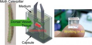
Scientists in Japan have developed the first biohybrid microdevice that can function in air. The microtweezers powered by insect muscle tissue could be used to handle cells and other fragile objects as part of a microelectromechanical system (MEMS).

cyborg jellyfish driven by rat heart cells is part of a recent flurry of research into devices that use mammalian muscle cells to provide motor power. Higher energy efficiency and the ability to self-repair are just some of the benefits of using cells in tiny devices. However, biohybrid devices don’t typically work outside cell culture medium. Now, a team lead by Keisuke Morishima at Osaka University has used insect muscle tissue to fabricate a microtweezer device that can operate in the open air.

Morishima‘s team assembled the microtweezers from polydimethylsiloxane and muscle cells taken from moth larvae and packaged them into a capsule containing a small amount of cell medium. The capsule’s clever design exploits a surface tension effect to stop medium from spilling even if the device is flipped upside down.

‘These results indicate that it will be possible to utilise insect muscle tissue and cells as microactuators both in a wet environment, such as in a microchannel, and on a dry substrate, such as on a silicon MEMS structure,’ says Morishima. When the team layered the medium surface with paraffin to slow evaporation from the opening of the device the microtweezers were functional for up to five days.


Shuichi Takayama a microfluidics and nanotechnology specialist at the University of Michigan in the US says that ‘while a variety of muscle-based actuators have been reported, this device is interesting as it is capable of operating in harsh environments. It is a great idea to combine robust muscle tissue from insects with clever packaging.’

The group now hopes to integrate muscle tissue that functions in both wet and dry environments into devices to expand their work into the nanorobotics field.

This article was taken from Chemistry World. To read this one, and many more, click here!

Digg This
Reddit This
Stumble Now!
Share on Facebook
Bookmark this on Delicious
Share on LinkedIn
Bookmark this on Technorati
Post on Twitter
Google Buzz (aka. Google Reader)

Lab on a Chip Co-hosts EU-Korea Microfluidics Workshop

We are very pleased to announce that Lab on a Chip will once again Co-host the third EU-Korea Workshop on microfluidics, focusing on “Emerging Microfluidic Platform Technologies: From Biosciences to Applications”.

Please come along and see us at the meeting, which will be held in Postech International Centre, Pohang, Korea. The workshop takes place on October 3rd to 5th, 2013.

Meet the Editor and International speakers:

Jean-Louis Viovy, Institute Curie, France
Andreas Manz, KIST, Europe
Dongpyo Kim, Pohang, Koreas
Chris Abell, Cambridge, UK
Noo Li Jeon, Seoul, Korea
Sabeth Verpoorte, Groningen, Netherlands
Hywel Morgan, Southampton, UK
Petra Dittrich, ETH Zurich, Switzerland
Sanghyun Lee, FEMTOLAB, Korea
Samuel Sanchez, Max-Planck, Germany
Yoon Kyoung Cho, UNIST, Korea
Francois Leblanc, CEO Fluigent

Digg This
Reddit This
Stumble Now!
Share on Facebook
Bookmark this on Delicious
Share on LinkedIn
Bookmark this on Technorati
Post on Twitter
Google Buzz (aka. Google Reader)

Personal kidney disease monitoring on your phone

Angharad Rosser-James, Publishing Editor in the Lab on a Chip editorial production team, recently wrote this fantastic article for Chemistry World. It focuses on a recent Lab on a Chip paper, and shows how the miniaturisation field can have a huge impact on our daily lives: 

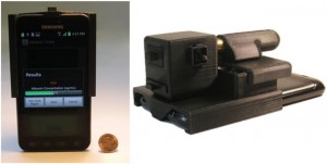
A smart phone attachment and accompanying app that could be used by people in their own home to monitor the health of their kidneys have been developed by scientists in the US. The lightweight and cost-effective device contains a fluorescent assay which works with the phone’s existing camera to provide results within minutes. 

The lightweight and compact attachment is installed on the existing camera unit of a smart-phone

Millions of people die each year from chronic kidney disease with 11% of US adults thought to have some form of kidney-related problem. Early detection and treatment is the key to prevent or control kidney damage. Routine screening for kidney damage checks albumin levels in urine with high levels of the protein indicating a potential problem. These tests are currently carried out using bench-top urine analysers and require patients to make regular trips to a clinic or hospital. 

The Albumin Tester, a digital fluorescent tube reader accompanied by an android smart phone app devised by Aydogan Ozcan and colleagues at the University of California in Los Angeles could save patients from having to make so many of these trips. Weighing only 148 g, a similar weight to the smart phone itself, the whole device can be attached to the back of a smart phone. Urine is added to fluorescent assays confined within disposable test tubes and the smart phone’s camera collects images of the assays via an external plastic lens. The app converts the fluorescence signals into an albumin concentration value within 1 second. Its detection limit of 5–10 µg ml-1 is more than 3 times lower that the clinically accepted healthy threshold. 

The user-friendly app converts fluorescence signals into albumin concentrations within 1 s and can give daily or weekly reports

Ozcan envisions the device’s application in ‘the early diagnosis of kidney disease or for routine monitoring of high-risk patients, especially those suffering from chronic conditions such as diabetes, hypertension, and/or cardiovascular diseases.’ Govind Kaigala, who develops microsystems for biomolecule analysis at IBM Research in Switzerland agrees and says ‘the albumin tester is a gadget which holds the promise of a simple, rapid and low-cost test for regular use by the patient.’ 

‘This technology has the potential to make widespread impact on health care in developing as well as developed countries,’ says Olav Solgaard, an expert in optical microelectromechanical systems at Stanford University in the US. 

Ozcan anticipates that their next step is to make it possible to measure other kidney disease biomarkers, such as creatinine, using the same smart phone attachment. 

View this article on the Chemistry World website, or access the full paper: A F Coskun et alLab Chip, 2013, DOI: 10.1039/c3lc50785h

Digg This
Reddit This
Stumble Now!
Share on Facebook
Bookmark this on Delicious
Share on LinkedIn
Bookmark this on Technorati
Post on Twitter
Google Buzz (aka. Google Reader)

Making cilia without the bunny suit

Cilia are microscale ‘eyelash-like’ extensions of eukaryotic cells found in epithelial linings throughout the body. In the fallopian tubes, windpipe, and lungs, motile cilia beat rhythmically to move objects within the viscous liquid above. Non-motile cilia in the inner ear transduce mechanical vibrations to electrical signals to ultimately excite auditory nerves.

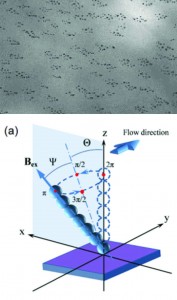
Previously, these appendages have been built using advanced microfabrication techniques. Now, for the first time, researchers at Eindhoven University of Technology in the Netherlands present a simple bench-top fabrication method for self-assembly of artificial cilia using magnetic beads and latex particles


Jaap den Toonder and his team created the artificial cilia sans cleanroom by coating magnetic beads with latex particles in a fluid cell using a magnetic field to control bead orientation. Latex particles were attracted to the beads by electrostatic forces and the whole structure was bonded together using a heating cycle. The completed artificial cilia were 3 μm in diameter and could be made into lengths of up to 33 μm by optimizing the magnetic field strength and protocol duration. The cilia were actuated by oscillating magnetic fields after fabrication to produce flow velocities of 3 μm/s.

Microfluidic devices operate under low Reynolds numbers where inertia is negligible, presenting a significant challenge to efficient mixing and moving of objects. Cilia and flagella evolved in organisms living in low Reynolds numbers to enable swimming by generating fluid flow using nonreciprocal (nonreversible) motions of beating and twisting.1, 2 The fabrication method presented in this work powerfully enables artificial cilia to be fabricated in situ in assembled platforms and “ship-in-a-bottle” constructed devices, thus facilitating practical applications for these structures in existing microfluidic platforms for bio-inspired fluid manipulation at the microscale.

Out of the cleanroom, self-assembled magnetic artificial cilia, Ye Wang, Yang Gao, Hans Wyss, Patrick Anderson, and Japp den Toonder, Lab Chip, 2013, 13, 3360-3366. DOI: 10.1039/C3LC50458A


References:
1. E. M. Purcell, AIP Conference Proceedings, 1976, 28, 49.
2. S. Khaderi, J. den Toonder and P. Onck, Biomicrofluidics, 2012, 6, 014106.

Digg This
Reddit This
Stumble Now!
Share on Facebook
Bookmark this on Delicious
Share on LinkedIn
Bookmark this on Technorati
Post on Twitter
Google Buzz (aka. Google Reader)