Author Archive

New YouTube Videos


Monitoring CO2 invasion processes at pore scale using Geological Labs on Chip

Transitioning from multi-phase to single-phase microfluidics for long-term culture and treatment of multicellular spheroids

Magnetic force-assisted self-locking metallic bead array for fabrication of diverse concave microwell geometries


Digg This
Reddit This
Stumble Now!
Share on Facebook
Bookmark this on Delicious
Share on LinkedIn
Bookmark this on Technorati
Post on Twitter
Google Buzz (aka. Google Reader)

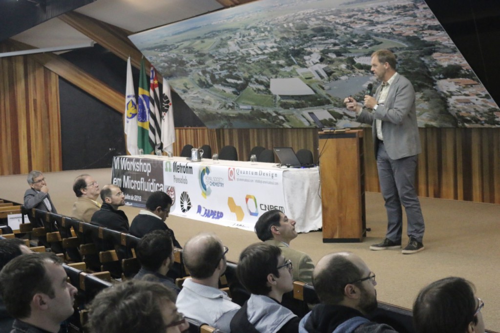
6th Workshop on Microfluidics, State University of Campinas, Brazil

Invited keynote talk “Lab, Cells and Organ on a Chip” by Dr. Albert van den Berg (University of Twente, The Netherlands)

The 6th Workshop on Microfluidics was held at the Convention Center of UNICAMP (State University of Campinas) in Campinas, Sao Paulo (Brazil), from 20-22 July, 2016. Since 2011, the workshop has brought together young students, researchers, and companies from different states of Brazil to discuss topics related to fundamentals, fabrication technologies, innovations, and applications in microfluidic science. This field has been spread out around different regions of Brazil presenting outstanding contributions for microfabrication and microfluidic technologies.

The event was supported by the Royal Society of Chemistry for the fourth time. Prof Dosil P. de Jesus (UNICAMP), member of the 6th Workshop on Microfluidics scientific board, presented the poster competition awards, sponsored by the Royal Society of Chemistry’s Lab on a Chip and Analytical Methods journals.


The winner of the Analytical Methods Poster Competition was Gabriela B. Almeida, from State University of Campinas, for her work “Microfluidic Devices Combining Dielectrophoresis Trapping and Surface Enhanced Raman Spectroscopy”.

Gabriela F. Giordano, from The Brazilian Nanotechnology National Laboratory (LNNano), Campinas, was the winner of the Lab on a Chip Poster Competition for her work “Gravity-Assisted Distillation on a Chip: a Novel Concept for Sample Preparation in Microfluidics”.

Gabriela Brito Almeida (centre), winner of the Analytical Methods Competition, with Prof. Dosil P. de Jesus (UNICAMP) (right) and Dr Elizabeth Magalhaes (RSC Manager, Brazil) (left)

Gabriela F. Giordano (centre), winner of the Lab on a Chip Competition, with Prof. Dosil P. de Jesus (UNICAMP) (right) and Dr Elizabeth Magalhaes (RSC Manager, Brazil) (left)

Digg This
Reddit This
Stumble Now!
Share on Facebook
Bookmark this on Delicious
Share on LinkedIn
Bookmark this on Technorati
Post on Twitter
Google Buzz (aka. Google Reader)

Chlorophyll lasers

From space shuttles to military equipment, or even the kitchenware that we use on daily basis, lasers have found use in more places than we often realise. Interestingly, the solid-state lasers were believed to be a solution to an unknown problem after their invention in 1950s. In that time nobody—including their developer Charles Townes—noticed that the lasers were one of the game changer inventions in the world’s history. After tens of years, in 1964, the laser technology was awarded with a Nobel prize when its potential for diverse applications were realized.

So far we have only discussed solid-state lasers, but there is definitely capacity in the laser world to improve the performance and versatility with the help of little twists, such as optofluidic lasers. “Optofluidics” is a synergic combination of optical systems and microfluidics. In other words, optical systems are built or synthesized from liquids, aiming to serve as good as their solid-state equivalents. For example, two immiscible liquids form a smooth surface in their interface, leading to a laser cavity or an optical resonator with a very high Q-factor that allows for operating at low energy levels. This emerging field gains importance when considering most of the biochemical reactions that occur in aqueous environments. Optofluidic laser systems are flexible to change their optical properties by just replacing the liquid media; and with this twist, lasers have new application areas including diagnosis of genetic disorders at the cellular level and in vivo biosensing.

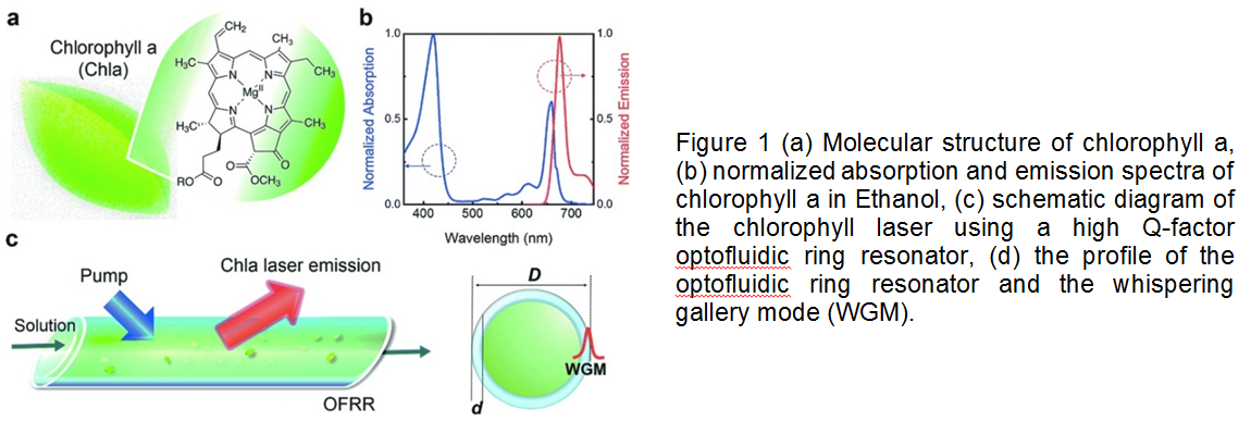
What is more exciting about the optofluidic lasers is that they can be biodegradable and easily tunable in microenvironments. Researchers in University of Michigan recently showed that one of the most abundant pigments on earth, chlorophylls, can maintain both biodegradability and tunability in optical systems owing to their fluorescence capabilities. Chlorophylls have a high Q-factor, dual-absorption bands in the visible spectrum, and a large shift between absorption and emission bands, suggesting that chlorophylls can be used as donors in fluorescence resonance energy transfer (FRET) laser (Figure 1). In this study, chlorophyll a was isolated from spinach leaf and used as the gain medium and the donor to develop a novel optofluidic laser. Two lasing bands of chlorophyll a was investigated by both theoretical and experimental means. Concentration-dependent studies enabled more insight for the mechanism determining when, where, and why the laser emission band appears. This new technique seems to gain increasing attention for applications in in vivo and in vitro biosensing, solar lighting and energy harvesting.

This article, published on 12th May 2016, is included in the Lab on a Chip Recent HOT Articles themed collection.

To download the full article for free* click the link below:

Optofluidic chlorophyll lasers
Yu-Cheng Chen, Qiushu Chena, Xudong Fan
Lab Chip, 2016, 16, 2228-2235
DOI: 10.1039/C6LC00512H

About the Webwriter

Burcu Gumuscu is a PhD researcher in BIOS Lab on a Chip Group at University of Twente in The Netherlands. Her research interests include development of microfluidic devices for second generation sequencing, organ-on-chip development, and desalination of water on the micron-scale.

*Access is free until 23/09/2016 through a registered RSC account.

Digg This
Reddit This
Stumble Now!
Share on Facebook
Bookmark this on Delicious
Share on LinkedIn
Bookmark this on Technorati
Post on Twitter
Google Buzz (aka. Google Reader)

New YouTube videos

Optofluidic holographic microscopy with custom Field of View by a linear array detector


Active pneumatic control of centrifugal microfluidic flows for lab-on-a-chip applications

Moving droplets between closed and open microfluidic systems

Optofluidic Guiding, Valving, Switching and Mixing based on Plasmonic Heating in Random Gold Nanoisland Substrate

Digg This
Reddit This
Stumble Now!
Share on Facebook
Bookmark this on Delicious
Share on LinkedIn
Bookmark this on Technorati
Post on Twitter
Google Buzz (aka. Google Reader)

New YouTube videos

Fabrication and Characterization of Optogenetic, Multi-Strip Cardiac Muscles

Direct detection and drug-resistance profiling of bacteremias using inertial microfluidics

Droplet-in-oil array for picoliter-scale analysis based on sequential-inkjet printing

The microenvironment of double emulsions in rectangular microchannels

Digg This
Reddit This
Stumble Now!
Share on Facebook
Bookmark this on Delicious
Share on LinkedIn
Bookmark this on Technorati
Post on Twitter
Google Buzz (aka. Google Reader)

New YouTube videos

A versatile technology for droplet-based microfluidics: thermomechanical actuation

Design and fabrication of magnetically functionalized flexible micropillar arrays for rapid and controllable microfluidic mixing

Active pneumatic control of centrifugal microfluidic flows for lab-on-a-chip applications

Digg This
Reddit This
Stumble Now!
Share on Facebook
Bookmark this on Delicious
Share on LinkedIn
Bookmark this on Technorati
Post on Twitter
Google Buzz (aka. Google Reader)

When microfluidics meets inkjet printing

Since its inception over 50 years ago, inkjet printing has become the most widely adopted method for the reproduction of images and text on paper. Inherently a microfluidic technology, inkjet printing is a process in which individual ink droplets, between 10 to 100 µm diameters, are placed at software-configurable locations on a substrate. The resulting dots can combine to create photo-quality images with resolutions of up to several thousand dots per inch. Such exquisite precision and flexibility make inkjet printing particularly attractive for the scalable fabrication of fine features. Indeed, in addition to coloured inks, this technology has also been adapted to deposit a wide range of materials for manufacturing, including metals, ceramics, polymers, and even biological cells.1

Not surprisingly, the lab on a chip community is also beginning to leverage printing technologies for the fabrication of microfluidic components.2 However, this is not straightforward because the quality of the printed features often depends on the interaction of the ink droplets with the substrate material. Since microfluidic devices are predominantly prototyped using polydimethylsiloxane (PDMS),3 recent research has focused on the development of PDMS-compatible methods for inkjet printing.

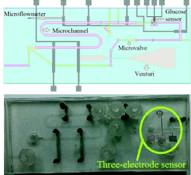
In one example, Profs Dachao Li at the Tianjin University, China and Robert C. Roberts at the University of Hong Kong used inkjet printing to form robust silver microelectrodes on PDMS-based microfluidics for glucose sensing.4 In forming these electrodes, the PDMS material posed two challenges: 1) poor adhesion of silver to PDMS inhibited robust electrode formation and, 2) the inherent hydrophobicity of PDMS promoted coalescence of neighboring ink droplets, which impaired the precision of printed features.

To overcome these challenges, the researchers modified the substrate using (3-Mercaptopropyl)trimethoxysilane, a coupling reagent that presents thiol groups on the PDMS surface. The thiol groups not only served as covalent anchoring sites for the inkjet-printed silver but also increased the wettability of the ink droplet, which improved the precision of the printed features.

Remarkably, the printed electrodes formed on these substrates tolerated very harsh stress tests, including immersion in water, exposure to high pressure air stream, and even ultrasonication. This new fabrication strategy enabled the team to create an all-in-one PDMS system with fluid handling and a three-electrode electrochemical cell for transdermal detection of glucose (Picture 1).
In another example, Profs Yanlin Song and Fengyu Li at the Chinese Academy of Sciences, China developed an elegant inkjet-printed method 5 for the fabrication of enclosed PDMS microchannels. This process conventionally requires at least 4 steps: 1) create a solid template via photolithography, 2) cast PDMS prepolymer mixture in the template, 3) cure and separate the PDMS from the template, and 4) bond the molded PDMS to a substrate.

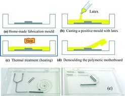
To simplify this procedure, the researchers implemented a liquid template formed by inkjet-printing (Picture 2). Here, an immiscible liquid pattern is printed directly on a pool of PDMS prepolymer solution. Within seconds, the liquid spontaneously submerges below the prepolymer solution and acts as a template. Upon heating, the polymer solidifies while the liquid template evaporates, leaving behind an enclosed PDMS microchannel device.
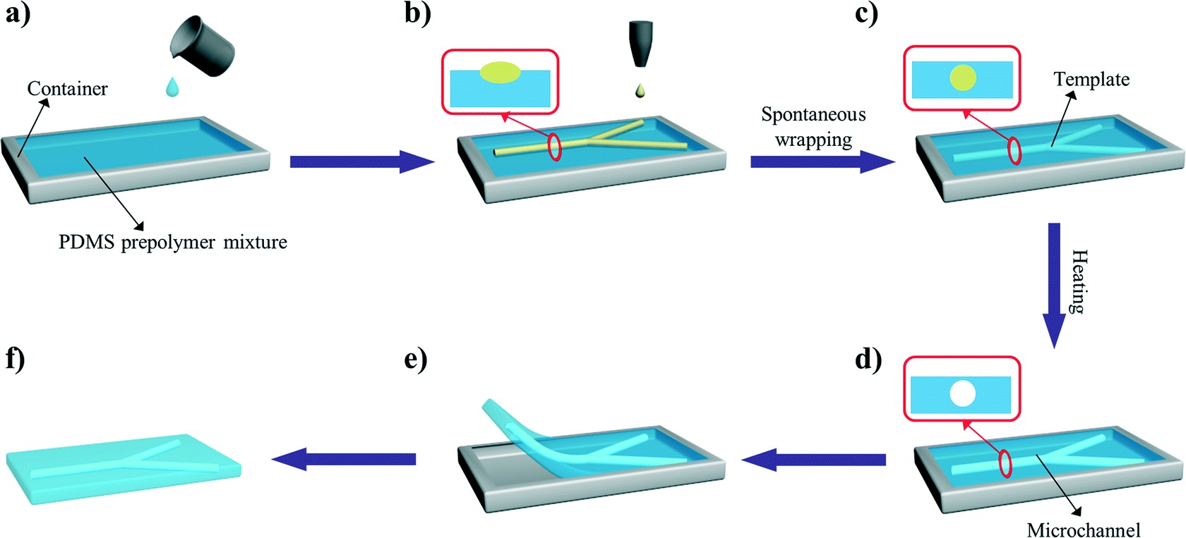
In the development of this method, one major challenge was maintaining the stability of the liquid template in the prepolymer solution. Due to surface tension, printed liquid templates tend to break up and relax into discontinuous spherical droplets.  Although this instability can be inhibited by increasing the liquid viscosity, high viscosities are not compatible with inkjet printing. To address this challenge, the researchers used an ink whose viscosity is tunable by temperature. At room temperature, the ink has relatively low viscosity (10 centipoise), which is compatible with inkjet printing. When the ink is printed on a cooled (3-4oC) pool of prepolymer solution, the viscosity of the liquid increases dramatically (>40 centipoise), which enabled the formation of a stable liquid template with no breakages.

This inkjet-printed approach enabled the formation of remarkably versatile PDMS microchannels. The diameters of the fabricated channels can range from 100 to 900 µm just by changing the template design. In addition, the interior surface of the channel can be modified by including vinyl-terminated functional molecules in the ink composition, which covalently incorporates in the PDMS matrix during thermal curing. Applying this technique with vinyl-terminated poly(ethylene glycol)methacrylate molecules, the researchers effortlessly imbue their devices with protein fouling resistance.

In summary, the lab on a chip community is beginning to leverage the benefits of inkjet printing in the fabrication of microfluidic components. By engineering the interaction of the ink droplets with the substrate material, researchers are devising innovative ways to fabricate robust electrodes and versatile microchannels without the need for cleanroom facilities and complicated procedures.

1.    I. M. Hutchings and G. D. Martin, Inkjet technology for digital fabrication, John Wiley & Sons, 2012.
2.    P. Tseng, C. Murray, D. Kim and D. Di Carlo, Lab on a Chip, 2014, 14, 1491-1495.
3.    E. Berthier, E. W. K. Young and D. Beebe, Lab on a Chip, 2012, 12, 1224-1237.
4.    J. Wu, R. Wang, H. Yu, G. Li, K. Xu, N. C. Tien, R. C. Roberts and D. Li, Lab on a Chip, 2015, 15, 690-695.
5.    Y. Guo, L. Li, F. Li, H. Zhou and Y. Song, Lab on a Chip, 2015, 15, 1759-1764.

Digg This
Reddit This
Stumble Now!
Share on Facebook
Bookmark this on Delicious
Share on LinkedIn
Bookmark this on Technorati
Post on Twitter
Google Buzz (aka. Google Reader)

Free access to February’s HOT Articles

These HOT articles published in February 2015 were recommended by our referees and are free* to access for 4 weeks


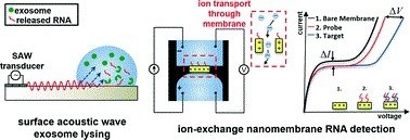
On-chip surface acoustic wave lysis and ion-exchange nanomembrane detection of exosomal RNA for pancreatic cancer study and diagnosis
Daniel Taller, Katherine Richards, Zdenek Slouka, Satyajyoti Senapati, Reginald Hill, David B. Go and Hsueh-Chia Chang
Lab Chip, 2015,15, 1656-1666
DOI: 10.1039/C5LC00036J, Paper


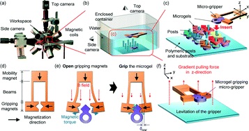
Three-dimensional heterogeneous assembly of coded microgels using an untethered mobile microgripper
Su Eun Chung, Xiaoguang Dong and Metin Sitti
Lab Chip, 2015,15, 1667-1676
DOI: 10.1039/C5LC00009B, Paper

A flexible lab-on-a-chip for the synthesis and magnetic separation of magnetite decorated with gold nanoparticles
Flávio C. Cabrera, Antonio F. A. A. Melo, João C. P. de Souza, Aldo E. Job and Frank N. Crespilho
Lab Chip, 2015, Advance Article
DOI: 10.1039/C4LC01483A, Paper

Take a look at our Lab on a Chip 2015 HOT Articles Collection!

*Access is free until 30.04.15 through a publishing personal account. It’s quick, easy and free to register!


Digg This
Reddit This
Stumble Now!
Share on Facebook
Bookmark this on Delicious
Share on LinkedIn
Bookmark this on Technorati
Post on Twitter
Google Buzz (aka. Google Reader)

New YouTube Videos

Towards microfluidic-based depletion of stiff and fragile human red cells that accumulate during blood storage 


Development of the centrifugal multiplex RT-LAMP—ICS microdevice for influenza A virus subtyping 
   

Microfluidic Continuous Flow Digital Loop-Mediated Isothermal Amplification (LAMP) 

Digg This
Reddit This
Stumble Now!
Share on Facebook
Bookmark this on Delicious
Share on LinkedIn
Bookmark this on Technorati
Post on Twitter
Google Buzz (aka. Google Reader)

LOC issue 2 – sorting stem cell-derived cardiomyocyte clusters, cross talk between cancer and immune cells and Genome Sequence Scanning

Welcome to Issue 2 of Lab on a Chip! This issue features the winner of the Lab on a Chip Pioneers of Miniaturisation Lectureship, Andrew deMello, on the back cover. Read more about this award and others given at MicroTAS here.

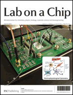
On the front cover of issue 2 is featured work from the group of Luke Lee at the University of California, Berkeley, in conjunction with colleagues at Stanford University.

In the paper, the authors use a ‘non-genetic, label-free cell cytometry method based on electrophysiological response to stimulus’ to sort undifferentiated stem cells from heterogeneous stem cell progeny.

The cell cytometer can identify human induced pluripotent stem cell-derived cardiomyocyte clusters from their extracellular field potential signals – these stem cells can then be used for various stem cell therapies.

Label-free electrophysiological cytometry for stem cell-derived cardiomyocyte clusters
Frank B. Myers, Christopher K. Zarins, Oscar J. Abilez and Luke P. Lee
DOI: 10.1039/C2LC40905D

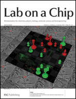
Work from the Italian National Research Council – Institute for Photonics and Nanotechnologies and the Istituto Superiore di Sanità is featured on the inside front cover.

The groups, led by Fabrizio Mattei and Luca Businaro, have used an on-chip co-culture system to investigate interactions between cancer cells and a host’s immune system.

Cross talk between cancer and immune cells: exploring complex dynamics in a microfluidic environment
Luca Businaro, Adele De Ninno, Giovanna Schiavoni, Valeria Lucarini, Gabriele Ciasca, Annamaria Gerardino, Filippo Belardelli, Lucia Gabriele and Fabrizio Mattei
DOI: 10.1039/C2LC40887B

On the inside back cover, work from Robert Meltzer and co-workers at Pathogenetix, Inc. is featured.

In their paper, they present a novel compound funnel design for use with Genome Sequence Scanning (GSS) technology, which improves molecule throughput.

High-throughput genome scanning in constant tension fluidic funnels
Joshua W. Griffis, Ekaterina Protozanova, Douglas B. Cameron and Robert H. Meltzer
DOI: 10.1039/C2LC40943G

As with all our cover articles these are free to access for 6 weeks (following a simple registration for an RSC Publishing account).

Other HOT articles featured in the issue include:

Benchtop fabrication of microfluidic systems based on curable polymers with improved solvent compatibility
Michinao Hashimoto, Robert Langer and Daniel S. Kohane
DOI: 10.1039/C2LC40888K

Microfluidic assisted self-assembly of chitosan based nanoparticles as drug delivery agents
Fatemeh Sadat Majedi, Mohammad Mahdi Hasani-Sadrabadi, Shahriar Hojjati Emami, Mohammad Ali Shokrgozar, Jules John VanDersarl, Erfan Dashtimoghadam, Arnaud Bertsch and Philippe Renaud
DOI: 10.1039/C2LC41045A

Microfluidic devices for X-ray studies on hydrated cells
Britta Weinhausen and Sarah Köster
DOI: 10.1039/C2LC41014A

For even more exciting microfluidics research, read the full issue here.

Digg This
Reddit This
Stumble Now!
Share on Facebook
Bookmark this on Delicious
Share on LinkedIn
Bookmark this on Technorati
Post on Twitter
Google Buzz (aka. Google Reader)