Author Archive

Microrobot gets to grips with bubbles

A remotely controlled micro-robot uses air bubbles to grip on to and assemble sub-millimetre sized components.

The microgripper lifts a poly(ethylene glycol) diacrylate hydrogel disc (pink) and stacks it on top of a another hydrogel disc (blue).

Source: Royal Society of Chemistry


Gripping and manipulating sub-millimetre objects is a tricky problem in microrobotics. Mechanical grasping is difficult at micro level, and techniques such as micro-machined grippers, although very accurate, need to be tethered to a control system and so are unsuitable for work in confined spaces.

To overcome these problems, Metin Sitti and his team from the Max Planck Institute in Germany have developed an untethered microgripper, which uses surface tension, rather than mechanical means, to grab small parts, such as a hair, a cloth fibre or a piece of muscle tissue. The gripper works in an aqueous environment, can grab both hydrophilic and hydrophobic items and, unlike other systems, does not need to be customised to fit to the item.

Read the full article in Chemistry World.


Programmable assembly of heterogeneous microparts by an untethered mobile capillary microgripper

Joshua Giltinan, Eric Diller and Metin Sitti

Lab Chip, 2016, 16, 4445-4457

DOI: 10.1039/C6LC00981F, Paper

Digg This
Reddit This
Stumble Now!
Share on Facebook
Bookmark this on Delicious
Share on LinkedIn
Bookmark this on Technorati
Post on Twitter
Google Buzz (aka. Google Reader)

Chips out of the Lab – Outreach at MicroTAS 2016

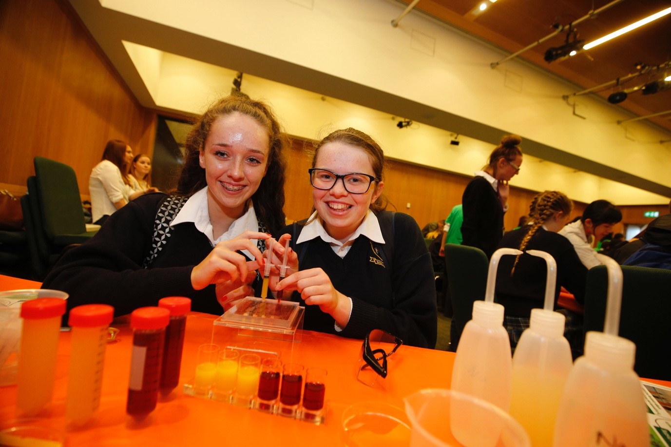
At the Tuesday lunch break of this year’s conference for Miniaturized Systems for Chemistry and Life Sciences (MicroTAS) in Dublin, you may have thought the conference audience was getting significantly younger, despite being in its 20th year. Pupils from local schools filled lecture halls along with members of the conference delegation. Conference chairs Nicole Pamme and Jens Ducrée invited attendees to join 500 local pupils for a science outreach event sharing the capabilities of microfluidic technologies. Pupils attended a lecture by Professor Sabeth Verpoorte explaining what microfluidics and lab-on-a-chip technologies are and had the chance to participate in a number of hands-on activities. The event served to increase student interest in STEM as well as a way for researchers to share activities they use for science outreach.

Pupils had a chance to use real microfluidic chips taken straight from the lab.

Researchers regularly participate in outreach events to promote an interest in STEM subjects. This is often through universities and community science festivals open to the public. Conferences serve as a great opportunity for international researchers to connect and share their work with other like-minded scientists, but in the past have rarely been opportunities to engage with the general public. For the first time in its history, MicroTAS included public engagement in its 2016 programme.

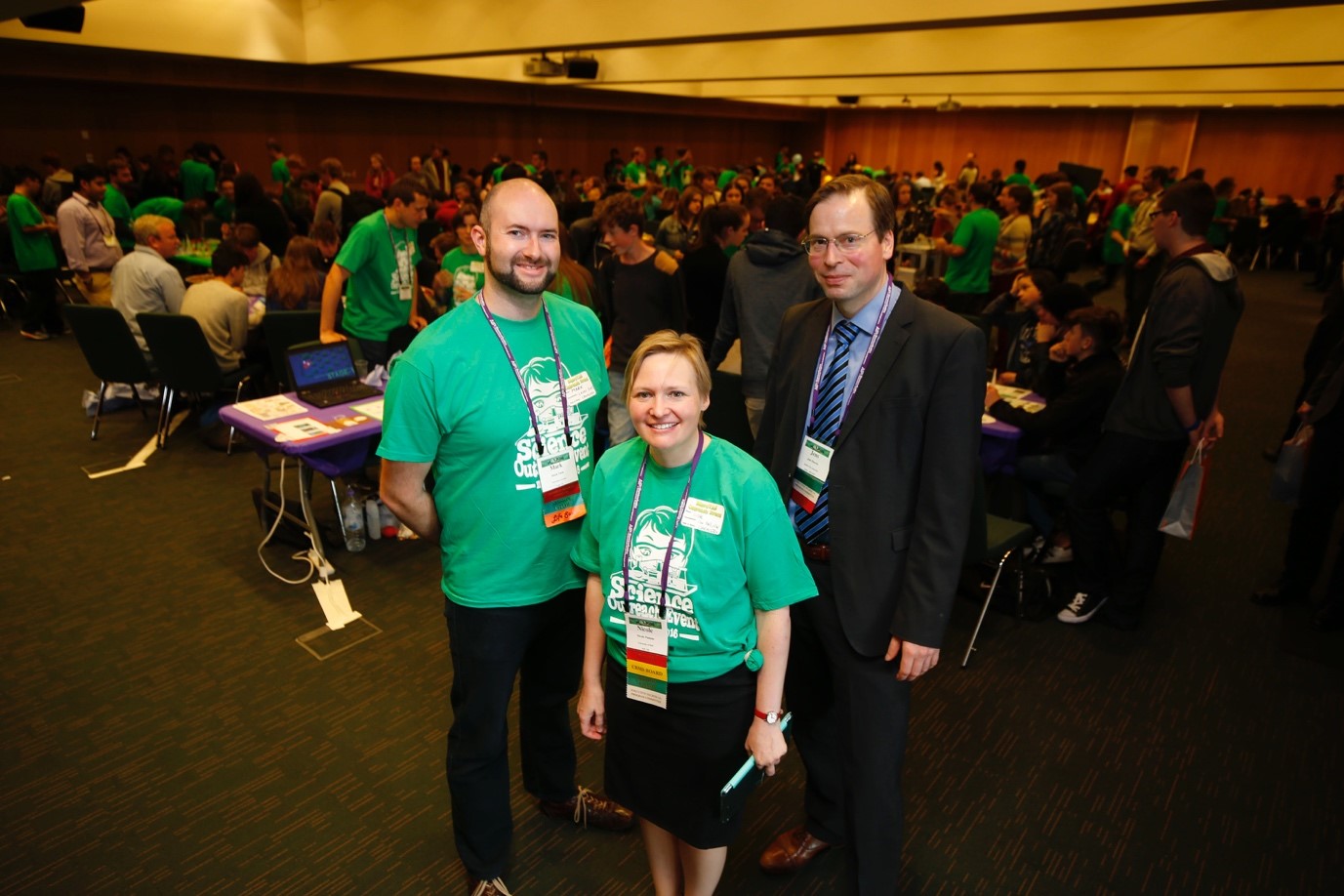
Pupils from eight local schools participated in a number of hands-on activities highlighting lab-on-a-chip technologies. Such activities ranged from interacting with large-scale versions of chips to trap ping-pong balls (cells); applying fabrication technology like injection molding to produce chocolate treats; to using microfluidic chips that volunteers brought and demonstrated. The outreach event was made possible through financial support from the RSC Outreach Fund, the Institute of Physics (Ireland), and the Analytical Chemistry Trust Fund. Eilish McLoughlin, Director of the Centre for the Advancement of STEM Teaching and Learning (CASTeL) at Dublin City University connected the conference with local schools, and Mark Tarn (Leeds University) coordinated all the conference volunteers. Volunteers represented 20 different universities and companies, highlighting a range of possibilities for careers in STEM.

Pupils file in for the event in Dublin. (Photo credit Tae Jae Lee).

Professor Pamme, who spearheaded the event, wanted to create an opportunity for researchers to share the different types of educational activities they have developed using microfluidics. Publications using microfluidics as a teaching tool do crop up in the literature (for example: Lab on a Chip, 12, 696-701; Lab on a Chip, 15, 947-957), but many microfluidic outreach and teaching activities do not get published. Professor Pamme hoped that there would be cross-pollination of ideas by having volunteers help out with peers at different institutions. A poster exhibit highlighting all the different activities was on display for the duration of the conference and these will be available from the Chemical and Biological Microsystems Society website (the host organization for MicroTAS).

This year’s outreach event may well serve as a template for future MicroTAS conferences as well as for other disciplines including outreach as a part of their international meetings.

Outreach organizers (from left to right) Mark Tarn, Nicole Pamme, and Jens Ducree.

Digg This
Reddit This
Stumble Now!
Share on Facebook
Bookmark this on Delicious
Share on LinkedIn
Bookmark this on Technorati
Post on Twitter
Google Buzz (aka. Google Reader)

New YouTube Videos

The importance of travelling wave components in standing surface acoustic wave (SSAW) systems

Multiple actuation microvalves in wax microfluidics

Multiple actuation microvalves in wax microfluidics

Digg This
Reddit This
Stumble Now!
Share on Facebook
Bookmark this on Delicious
Share on LinkedIn
Bookmark this on Technorati
Post on Twitter
Google Buzz (aka. Google Reader)

Microfluidics made easy

Automated design helps researchers find the right chip for the job

Scientists in the US have devised an algorithmic process to speed up the design of microfluidic chips, generating a library containing thousands of different chip designs that researchers can search by functionality.

Microfluidic chips, which are widely used in areas such as disease diagnostics and DNA sequencing, consist of tiny channels etched into a glass or plastic. These microchannels are connected to achieve a specific function, for example mixing fluids. The design process, however, has remained relatively unchanged since their emergence as William Grover from the University of California, who led the new study, explains: ’We design them by hand and we test them – if they work great, but more often than not, they don’t, so then I have to start all over again. That process is so slow and inefficient.’

With a new online database created by Grover and his team, even researchers with no microfluidics experience can find the perfect chip to suit their needs. Grover’s team created a computer program that generates thousands of random microfluidic chip designs and simulates their behaviour. The database collects these simulated designs, and users can query it to find chips suitable for given tasks.

Read the full article in Chemistry World.


Random design of microfluidics

Junchao Wang, Philip Brisk and William H. Grover

Lab Chip, 2016, Advance Article

DOI: 10.1039/C6LC00758A, Paper

Digg This
Reddit This
Stumble Now!
Share on Facebook
Bookmark this on Delicious
Share on LinkedIn
Bookmark this on Technorati
Post on Twitter
Google Buzz (aka. Google Reader)

New YouTube Videos

Microfluidic partition with in situ biofabricated semipermeable biopolymer membranes for static gradient generation


Dielectric elastomer actuator for mechanical loading of 2D cell cultures


A one-step strategy for ultra-fast and low-cost mass production of plastic membrane microfluidic chips

Digg This
Reddit This
Stumble Now!
Share on Facebook
Bookmark this on Delicious
Share on LinkedIn
Bookmark this on Technorati
Post on Twitter
Google Buzz (aka. Google Reader)

Sepsis’ sticking point in for a shock

Simple microfluidic method helps detect life-threatening condition earlier

A new method for monitoring the onset of sepsis touted by US researchers may assist clinicians in helping more of their patients survive this potentially fatal condition.

Sepsis, often referred to as blood poisoning, can be the result of an innocuous infection or injury. Without treatment, the immune system becomes overwhelmed, leading to organ shutdown. In the UK alone, 30,000 people die as a direct result of the condition. Sepsis needs to be caught early to maximise the chances of a full recovery. However, existing diagnostics rely on antibody labelling, which requires samples to be prepared and tested by specialist laboratory staff. This requires time that a patient might not be able to spare. ‘Existing methods are too cumbersome,’ explains Joel Voldman, heading up the new study at Massachusetts Institute of Technology.

detecting sepsis with electrochemical monitoring

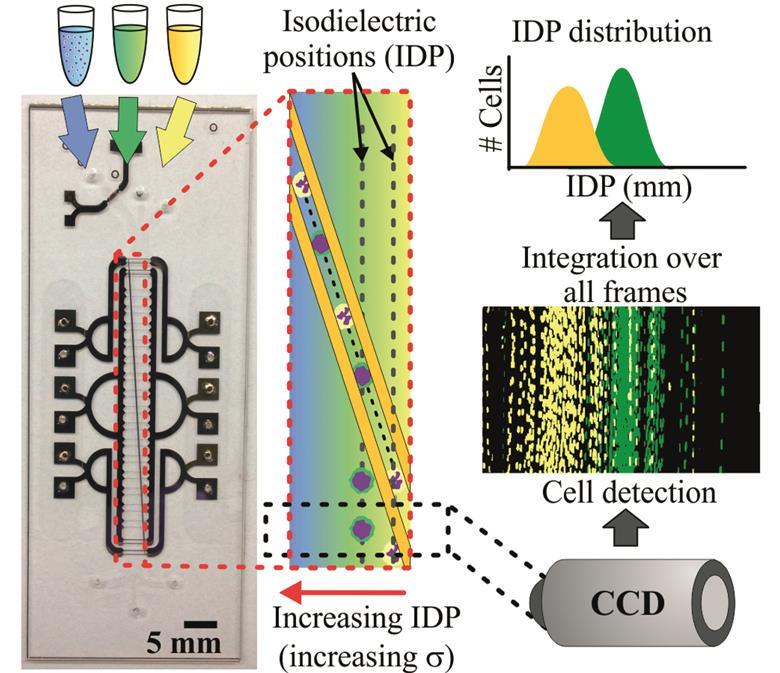
The tiny device can process large amounts of blood and detect activated white blood cells that are an indicator for sepsis. Source: © Royal Society of Chemistry

Voldman’s group devised a microfluidic method that takes advantage of the white blood cells’ electrical properties, which makes it possible to detect sepsis quickly in its earliest stages. Sepsis triggers white blood cells to become activated and circulate in the blood. The number of these activated cells indicates the disease’s progression. Applying an electric field then separates activated and non-activated cells based on subtle differences in their electrical properties.

Read the full article in Chemistry World.


Monitoring sepsis using electrical cell profiling

Javier L. Prieto, Hao-Wei Su, Han Wei Hou, Miguel Pinilla Vera, Bruce D. Levy, Rebecca M. Baron, Jongyoon Han and Joel Voldman

Lab Chip, 2016, Advance Article

DOI: 10.1039/C6LC00940A, Paper

Digg This
Reddit This
Stumble Now!
Share on Facebook
Bookmark this on Delicious
Share on LinkedIn
Bookmark this on Technorati
Post on Twitter
Google Buzz (aka. Google Reader)

Blood matters

Iron deficiency anemia (IDA) is not a trivial illness. Every individual in the world’s population has the potential to suffer from this nutritional disease. According to estimations, 900 million people worldwide are already afflicted with it. IDA is known to lower cognitive ability, work capacity, and future productivity of both children and adults. The situation appears to be grave when we consider the economic consequences of these problems.

The human body needs iron to produce red blood cells, and having low iron levels in the body leads to IDA. Diagnosis of IDA requires a complete blood count is performed by a bulky hematology analyzer. IDA has been a common disease for a really long time; however, its associated diagnosis costs are considerably high, and the diagnosis equipment is not available in many places of the world. Given the facts, IDA diagnosis actually deserves cheaper and easily accessible equipment, which has unfortunately remained elusive—up until now.

In last month’s issue of Lab on a Chip, Whitesides research group at Harvard University came up with a sound idea to diagnose IDA in a shorter and inexpensive way. They developed a low-cost and rapid-screening tool to diagnose IDA using aqueous multiphase systems containing layer of polymer-salt mixtures. These mixtures are loaded in a microhematocrit tube (depicted in the figure) together with a drop of blood from a fingerprick. Diagnosis results become available after a 2-minute low-cost centrifuging process.

The reported data suggest that diagnosis of IDA is improved by means of sensitivity and specificity when compared to the bulky hematology analyzer’s results. Several important red blood cell parameters, such as concentration of hemoglobin in a given volume of red blood cells, can be predicted. The technique’s ability to diagnose IDA was further improved using automated digital analysis. They also show that the tool is able to detect a wider range of anemia types including microcytic and hypochromic anemia. The portable and low cost screening tool could possibly find use in rural clinics where large fractions of the population at risk of IDA. Before entering the market, the performance of this technique will still have to be validated to demonstrate feasibility of using and interpreting the assay.

Design of the presented test loaded with blood before and after centrifugation for a representative IDA and Normal sample. Blood is loaded into the top of the tube, from a fingerprick, using capillary action provided by a hole in the side of the tube. Normal blood packs at the bottom of the tube, while less dense blood cells can be seen packing at the interfaces between the phases and inside the tube. Normal and IDA blood can be differentiated by eye after only 2 minutes of centrifugation. It is also possible to read the analysis results in an automated way. A commonly used software is used to convert the image to red intensity graphs.

This article was published in Lab on a Chip on 30th August 2016.

To download the full article for free* click the link below:

Diagnosis of iron deficiency anemia using density-based fractionation of red blood cells

Jonathan W. Hennek, Ashok A. Kumar, Alex B. Wiltschko, Matthew R. Patton, Si Yi Ryan Lee, Carlo Brugnara, Ryan P. Adams and George M. Whitesides
Lab Chip, 2016,16, 3929-3939
DOI: 10.1039/C6LC00875E

—————-

About the Webwriter


Burcu Gumuscu is a postdoctoral fellow in BIOS Lab on a Chip Group at University of Twente in The Netherlands. Her research interests include development of microfluidic devices for next generation sequencing, compartmentalized organ-on-chip studies, and desalination of water on the microscale.

—————-

*Access is free until 7th November 2016 through a registered RSC account – register here

Digg This
Reddit This
Stumble Now!
Share on Facebook
Bookmark this on Delicious
Share on LinkedIn
Bookmark this on Technorati
Post on Twitter
Google Buzz (aka. Google Reader)

New YouTube Videos

Autonomous microfluidic capillaric circuits replicated from 3D-printed molds

The intercell dynamics of T cells and dendritic cells in a lymph node-on-a-chip flow device

A reconfigurable stick-n-play modular microfluidic system using magnetic interconnects
Digg This
Reddit This
Stumble Now!
Share on Facebook
Bookmark this on Delicious
Share on LinkedIn
Bookmark this on Technorati
Post on Twitter
Google Buzz (aka. Google Reader)

New YouTube Videos

A conductive liquid-based surface acoustic wave device

Highly efficient sample stacking by enhanced field amplification on a simple paper device

A lab-on-a-disc with reversible and thermally stable diaphragm valves


Digg This
Reddit This
Stumble Now!
Share on Facebook
Bookmark this on Delicious
Share on LinkedIn
Bookmark this on Technorati
Post on Twitter
Google Buzz (aka. Google Reader)

New YouTube Videos

An In-line Spectrophotometer on Centrifugal Microfluidics Platform for Real-time Protein Determination

and Calibration

Hydrodynamic trapping for rapid assembly and in situ electrical characterization of droplet interface bilayer arrays

A fully automated microfluidic micellar electrokinetic chromatography analyzer for organic compound detection

Digg This
Reddit This
Stumble Now!
Share on Facebook
Bookmark this on Delicious
Share on LinkedIn
Bookmark this on Technorati
Post on Twitter
Google Buzz (aka. Google Reader)