Archive for July, 2019

We are delighted to announce that Hang Lu is the 2019 winner of the “Pioneers of Miniaturization” Lectureship!

The 14th “Pioneers of Miniaturization” Lectureship, sponsored by Dolomite and Lab on a Chip , is for early to mid-career scientists who have made extraordinary or outstanding contributions to the understanding or development of miniaturised systems.

The 2019 “Pioneers of Miniaturization” Lectureship will be presented to Professor Lu at the µTAS 2019 Conference in Basel, Switzerland, being held on 27-31 October 2019. Professor Lu will receive a certificate, a monetary award and will give a short lecture during the conference.

Many congratulations to Professor Hang Lu on this achievement from the Lab on a Chip Team!

About the Winner

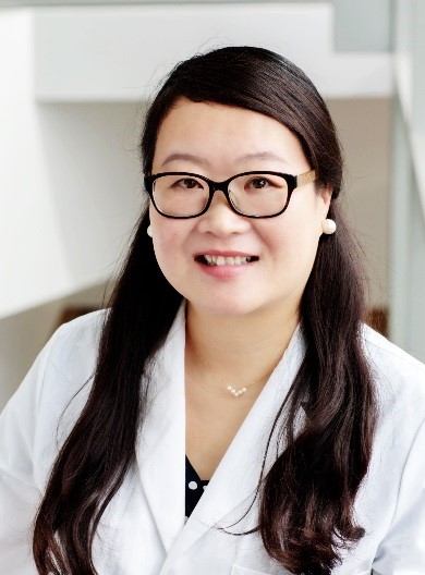
Professor Hang Lu is the 
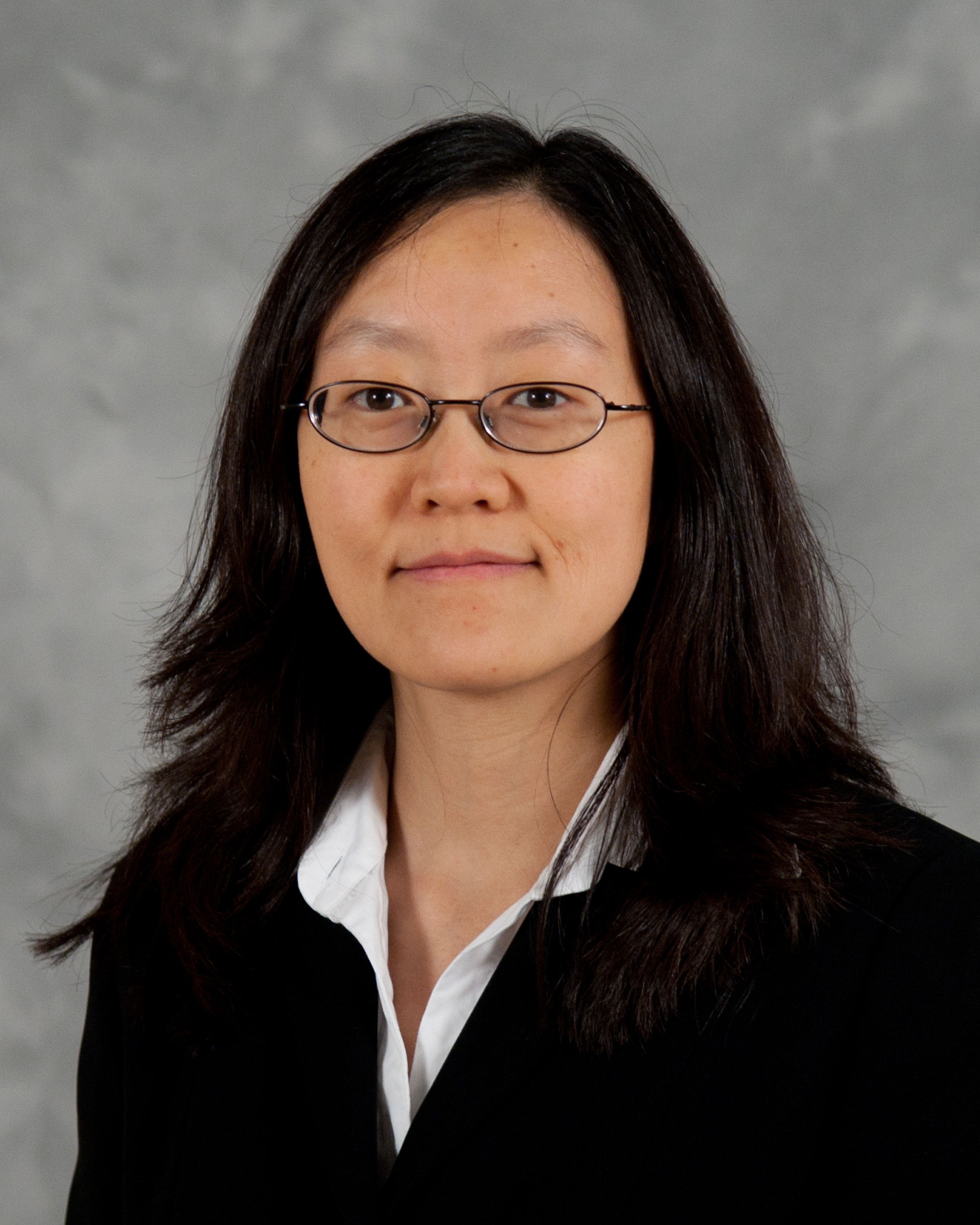
Love Family Professor, School of Chemical and Biomolecular Engineering, Georgia Institute of Technology, USA.

Professor Lu earned her PhD in Chemical Engineering from Massachusetts Institute of Technology, USA in 2003. After a postdoctoral fellowship with Professor Cornelia I. Bargmann, at University of California San Francisco and the Rockefeller University, she was appointed as an Assistant Professor at School of Chemical and Biomolecular Engineering, Georgia Institute of Technology.

In recognition of her outstanding achievements, Professor Lu has received numerous awards and international recognition, including being invited to join Board of Directors, Chemical and Biological Microsystems Society, invited to present at the Nobel Symposium on Microfluidics (2017) and the National Academy of Sciences’ Kavli Frontiers of Science Symposia (2014, 2012, 2009), awarded the ACS Analytical Chemistry Young Innovator Award, Chemical and Biological Microsystems Society (2013), Council of Systems Biology in Boston (CSB2) Prize in Systems Biology (2011), a National Science Foundation CAREER award (2010), an Alfred P. Sloan Foundation Research Fellowship (2009), a DARPA Young Faculty Award (2007), a DuPont Young Professor Award (2006), the Saville Lectureship of Princeton University (2013), the H. C. Van Ness Award Lectures of Rensselaer Polytechnic Institute (2011), and is a fellow of the American Institute for Medical and Biological Engineering (AIMBE) and  a fellow of the American Association for the Advancement of Science (AAAS). She has authored more than 140 peer-reviewed publications and has served on the Editorial Board of Lab on a Chip as Associate Editor since 2017. She is currently the director of the Interdisciplinary Bioengineering Program, and the associate director of the NSF-Simons Foundation supported Southeast Center for Mathematics and Biology, Georgia Institute of Technology.

Professor Lu has pioneered the use of microfluidic systems for imaging and performing genetic studies with small organisms, primarily the nematode C. elegans. In a series of studies published since 2008 she established a set of technologies to streamline imaging, phenotyping, and sorting of C. elegans based on features that are difficult to distinguish and discern by human eyes. The throughput of these technologies were often 1,000 times that of conventional approaches. Professor Lu’s technology has enable faster and more accurate experiments and revolutionized how genetic screens and high-content imaging experiments are done currently in other scientists’ labs. In parallel, her lab has also engineered micro systems for high-content experiments with cells, aggregates, organoids, and embryos to extract high-dimensional information for systems biology studies.

The Lu group performs research at the interface of engineering and biology. They engineer automated microfluidic systems, microscopy tools, and image imformatic technologies to address questions in neuroscience, cell biology, and biotechnology that are difficult to answer using conventional techniques. Applied to the study of fundamental biological questions, these new techniques allow the Lu group to gather large-scale quantitative data about complex systems.

Learn about the Lu group online

Digg This
Reddit This
Stumble Now!
Share on Facebook
Bookmark this on Delicious
Share on LinkedIn
Bookmark this on Technorati
Post on Twitter
Google Buzz (aka. Google Reader)

Emerging Investigator Series – Joo H. Kang

Dr. Joo H. Kang is currently an Assistant Professor of the Department of Biomedical Engineering, School of Life Sciences at Ulsan National Institute of Science and Technology (UNIST), Korea. He received double Bachelor’s degrees in Chemical Engineering and Life Science from Sogang University in 2002 and his M.S. and Ph.D. in Bioengineering from Korea Advanced Institute of Science and Technology (KAIST) in 2004 and 2008, respectively. He joined Children’s Hospital Boston, Harvard Medical School as a research fellow in 2009, and he continued his work at the Wyss Institute, Harvard University as a Wyss Technology Development Fellow from 2012-2016. He received several awards in his early career, including Postdoctoral Award for Professional Development from Harvard University, Wyss Technology Development Fellowship from Harvard University, Baxter Young Investigator Award from Baxter Inc., and Young Frontiers in Bio and Braining Engineering from KAIST. His research interests include multiscale biofluidic approaches for tackling infectious diseases and cancer, and miniaturized organ-mimicking microsystems.

Read Joo H. Kang’s Emerging Investigator article “Measurement of the magnetic susceptibility of subtle paramagnetic solutions using the diamagnetic repulsion of polymer microparticles” and find out more about him in the interview below:


Your recent Emerging Investigator Series paper focuses on measuring magnetic susceptibility of subtle paramagnetic solution using diamagnetic repulsion of polymer microparticles. How has your research evolved from your first article to this most recent article?

One of the research topics that interested me was to discriminate the subtle differences in the magnetic susceptibility of materials in a microfluidic regime. The first paper I published in regards to this (Kang,JH, et al., JACS, 2009) demonstrated the capability of discriminating the magnetic susceptibility of “solid microparticles” where they are diamagnetically forced to be located at a quasi-isomagnetic position in a microfluidic channel (a position where the differences of the magnetic susceptibility between the solid particles and surrounding media become nearly zero). When I was invited to make contribution to the Emerging Investigator Series of Lab on a Chip last year, I wanted to revisit this, and this time I aimed to assess the subtle magnetic susceptibility of “surrounding paramagnetic solutions”.

What aspect of your work are you most excited about at the moment?

As for the paper, I was surprised of the sensitivity of the device that can discriminate the magnetic susceptibility. We compared our results with those assessed by a conventional superconducting quantum interference device (SQUID), and found that our approach is even more sensitive than the conventional one. Likewise, we can unveil various scientific approaches when exploring fluidic regimes at the micro and nanoscale, and this is the most exciting aspect as being a part of the research community in this field.

In your opinion, what applications can your current approach be used for?

Because this is a platform technology, various applications are possible where we need to measure the magnetic susceptibility of paramagnetic solutions. Assessment of residual magnetic nanoparticles in biological samples, for examples, would be one of the potential uses. We could also use this platform to evaluate metal contamination of drinking water, such as chromium or iron oxide, which alters the magnetic susceptibility of water.

What do you find most challenging about your research?

Taking research from the “bench to products”. Since I started my independent research career, I realized that I have to make considerable efforts to get this happen while playing multiple roles at the same time. But I am enjoying it.

In which upcoming conferences or events may our readers meet you?

I am planning to attend microTAS 2019 that will be held in Basel, Switzerland this year.

How do you spend your spare time?

I am spending my time with my family, hiking, swimming, and playing soccer or games with my little son and daughter.

Which profession would you choose if you were not a scientist?

Probably an architect. This was one of the paths I was thinking of when I was a high school student.

Can you share one piece of career-related advice or wisdom with other early career scientists?

A clear vision on your own research and collaborators who you can share your vision with.

Digg This
Reddit This
Stumble Now!
Share on Facebook
Bookmark this on Delicious
Share on LinkedIn
Bookmark this on Technorati
Post on Twitter
Google Buzz (aka. Google Reader)

Emerging Investigator Series – Mei He

Dr. He is a tenure-track assistant professor at the University of Kansas. She received her PhD degree from the University of Alberta with professor Jed Harrison, and postdoctoral training from the University of California, Berkeley with professor Amy Herr. She is the vice chair of the ASABE Biosensor program and the Councilor of the American Electrophoresis Society. Dr. He is also the founder of Clara Biotech Inc. and the founder committee for the MidWest 3D technology society. Dr. He Received NIH Maximizing Investgator’s Research Award for Early Stage Investigators in 2019. She also received the Lab on Chip Outstanding Reviewer for the year of 2018. One of her publications also received the 2018 SLAS Technology Readers Choice Award. Her research interests include biomedical microfluidic devices and sensing approaches, 3D biomaterials, and nanodelivery, employed in programming and monitoring biomimetic immunity associated with extracellular vesicles.

Read Mei He’s Emerging Investigator article “3D-printing enabled micro-assembly of a microfluidic electroporation system for 3D tissue engineering” and find out more about her in the interview below:


Your recent Emerging Investigator Series paper focuses on “3D-printing Enabled Micro-assembly of Microfluidic Electroporation System for 3D Tissue Engineering”. How has your research evolved from your first article to this most recent article?

My first article observed the microscale evolution of porous polymer materials in the microfluidic channel when I was a PhD student. I found very interesting phenomena in the microfluidic device which actually inspired me to explore more surrounding dimensions, surface chemistry, and scales. Till to my recent article focusing on 3D geometric influence on cellular behavior and their extracellular vesicles secretion dynamics, the 3D dimension in microscale is intriguing in the biological system.

What aspect of your work are you most excited about at the moment?

I am very excited to take the microfluidic technology and phenomena into the biology world, as it will bring new investigation and discovery. Biology is still in its infancy stage, I am very excited to see how microfluidic technology could advance this growth.

In your opinion, what is the biggest impact your microfluidic electroporation system will have in tissue engineering?

Intracellular delivery of regulatory or therapeutic targets into the cell is very crucial in the field of tissue engineering and regenerative medicine. Current existing electro-transfection systems, including microfluidic platforms and commercial benchtop systems, are only able to study monolayer cell suspensions in vitro, which is incapable of clinical translation within in vivo tissue microenvironment. So developing a 3D, in vivo like tissue microenvironment with effective electro-transfection is very important to move to the clinical study in the future. We actually are more interested in downstream, precise control and manipulation of cellular machinery for secreting exosomes and extracellular vesicles under the transfection-induced stimulus, such technology is not existing yet but very important for understanding the interconnection of cargo internalization with cellular level responses elicited by exosomes delivery pathway.

What do you find most challenging about your research?

Building up an in vivo like tissue system with precise control is not straightforward. The environment in a controlled lab setting is totally different than in an in vivo biological system. So the analyzed information actually is not representative of the real situation in the human in vivo system. There are huge heterogeneities present in the cell population as well as human individuals, which poses the challenges for correctly understanding cellular system regulation, such as immunity, in our human body. Mimicking in vivo living system is very challenging, but crucial for understanding quite a few of mechanism and disease pathogenesis. Our research introduces new microfluidic technology and material solutions to solve such challenges.

In which upcoming conferences or events may our readers meet you?

I will attend next year Gorden Research Conference in Bioanalytical Sensors as well as the MicroTAS annual meeting.

How do you spend your spare time?

I have a 7-year-old boy and expecting a new baby girl this year. My spare time definitely is occupied by kids and watching them growing.

Which profession would you choose if you were not a scientist?

I always like to discover new things since I was a child. If I am not a scientist, I would like to be a greeting card designer or paleontologist.

Can you share one piece of career-related advice or wisdom with other early career scientists?

Being a life-long learner and always keeping strong scientific curiosity will definitely help with your research development. Get good mentors around you and you will appreciate their advice.

Digg This
Reddit This
Stumble Now!
Share on Facebook
Bookmark this on Delicious
Share on LinkedIn
Bookmark this on Technorati
Post on Twitter
Google Buzz (aka. Google Reader)