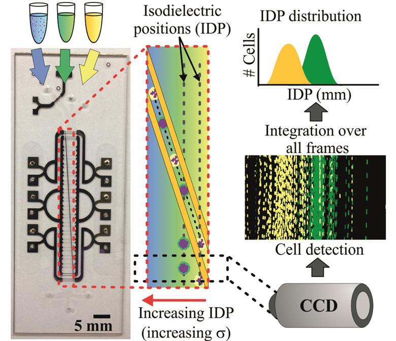
| The importance of travelling wave components in standing surface acoustic wave (SSAW) systems
|
| Multiple actuation microvalves in wax microfluidics
|
| Multiple actuation microvalves in wax microfluidics |


")
The µTAS 2016 Conference was held during 9-13th October in Dublin, Ireland. Sam Keltie and Maria Southall, Executive and Deputy Editors of Lab on a Chip, attended this conference and announced the prestigious Lab on a Chip awards which include the Pioneers of Miniaturization Lectureship (in partnership with Corning Inc), the Widmer Young Researcher Poster Prize, the Art in Science competition (in partnership with NIST) and the µTAS video competition (in partnership with Dolomite Microfluidics). The competition was tough but we are pleased to announce this year’s Prize Winners below.
“Pioneers of Miniaturization” Lectureship

Congratulations to all of our Prize Winners: Dr Daniel Irimia (top left), Vaibhav Jain (top right), Enrica Rollo (bottom right) and Adam Churchman (bottom right).
Dr Daniel Irimia (Massachusets General Hospital) was announced as the winner of the 11th “Pioneers of Miniaturization” Lectureship, sponsored by Lab on a Chip and Corning Inc and supported by the Chemical and Biological Microsystems Society (CBMS). The “Pioneers of Miniaturization” Lectureship rewards early to mid-career scientists who have made extraordinary or outstanding contributions to the understanding or development of miniaturised systems. Dr Daniel Irimia received a certificate and a monetary award from Po Ki Yuen (Corning Inc), and delivered a short lecture titled “The amazing neutrophil: unexpected insights from tiny devices” at the conference.
Art in Science Competition
Lab on a Chip and Darwin Reyes from the National Institute of Standards Technology (NIST) presented the Art in Science award to Vaibhav Jain, Purdue University. The award aims to highlight the aesthetic value in scientific illustrations while still conveying scientific merit. You can see his winning photograph “The Rising Sun” along with the runner ups on our Art in Science blog post.
µTAS Video Competition
Widmer Young Researcher Poster Prize
The Widmer Young Researcher Poster Prize was awarded to Adam Churchman, PhD student at Leeds University. His poster highlighted his research on the formation of oil layer inside microbubbles through single step microfluidics.
Also of interest: Browse through our collection of archived µTAS Abstracts online!
Congratulations to all the winners at the conference! We look forward to seeing you at µTAS 2017 in Savannah, Georgia.
Automated design helps researchers find the right chip for the job
Scientists in the US have devised an algorithmic process to speed up the design of microfluidic chips, generating a library containing thousands of different chip designs that researchers can search by functionality.
Microfluidic chips, which are widely used in areas such as disease diagnostics and DNA sequencing, consist of tiny channels etched into a glass or plastic. These microchannels are connected to achieve a specific function, for example mixing fluids. The design process, however, has remained relatively unchanged since their emergence as William Grover from the University of California, who led the new study, explains: ’We design them by hand and we test them – if they work great, but more often than not, they don’t, so then I have to start all over again. That process is so slow and inefficient.’
With a new online database created by Grover and his team, even researchers with no microfluidics experience can find the perfect chip to suit their needs. Grover’s team created a computer program that generates thousands of random microfluidic chip designs and simulates their behaviour. The database collects these simulated designs, and users can query it to find chips suitable for given tasks.
Read the full article in Chemistry World.
Random design of microfluidics
Junchao Wang, Philip Brisk and William H. Grover
Lab Chip, 2016, Advance Article
DOI: 10.1039/C6LC00758A, Paper

We are both proud and very pleased to introduce the 2016 edition of our Emerging Investigators issue, which celebrates the most promising and brightest amongst early career miniaturisation scientists around the world.
Guest editors Charles M. Schroeder, Sarah Köster and Yanyi Huang introduce the issue in their Editorial.
Emerging Investigators 2016: discovery science meets technology
DOI: 10.1039/C6LC90076C
Read the full collection online today: http://rsc.li/loc-emginv-16
Free* Access: AC electric field induced droplet deformation in a microfluidic T-junction
Communication
Heng-Dong Xi, Wei Guo, Michael Leniart, Zhuang Zhi Chong and Say Hwa Tan
Lab Chip, 2016,16, 2982-2986 DOI: 10.1039/C6LC00448B
Open Access: Arrayed water-in-oil droplet bilayers for membrance transport analysis
R. Watanabe, N. Soga, M. Hara and H. Noji
Lab Chip, 2016,16, 3043-3048
DOI: 10.1039/C6LC00155F
Free* Access: Cell-on-hydrogel platform made of agar and alginate for rapid, low-cost, multidimensional test of antimicrobial susceptibility
Han Sun, Zhengzhi Liu, Chong Hu and Kangning Ren
Lab Chip, 2016,16, 3130-3138
DOI: 10.1039/C6LC00417B
Free Access*: One-step immunoassay of C-reactive protein using droplet microfluidics
Matthew Y. H. Tang and Ho Cheung Shum
Lab Chip, 2016, Advance Article
DOI: 10.1039/C6LC01121G
*Access is free until 11 November 2016 via a registered RSC account.
Lab on a Chip and the National Institute of Standards Technology (NIST) were pleased to present the Art in Science award at the µTAS 2016 Conference on 13 October 2016. The award highlights the aesthetic value in scientific illustrations while still conveying scientific merit. Many fantastic submissions were received this year with the winner selected by Sam Keltie, Lab on a Chip Executive Editor, Darwin Reyes, NIST and Dino Di Carlo, Lab on a Chip Editorial Board member.
And the winner is…
Vaibhav Jain, Purdue University
“The Rising Sun”

Dark field micrograph of sweat advancing in a paper channel dyed with Cobalt chloride dihydrate (Bluish), changing it to cobalt chloride hexahydrate (Reddish orange)
And the runners up are…
Gokce Ozkazanc, Bilkent University
“Composition of Particles in a Droplet”

Visualization of Particle Composition inside a Water Droplet
Susanna Lladó Maldonado, TU Braunschweig
“Wellness microresort for cells“

Mixing performance in a microbubble column-bioreactor
A big thank you to all the contributors this year!
Simple microfluidic method helps detect life-threatening condition earlier
A new method for monitoring the onset of sepsis touted by US researchers may assist clinicians in helping more of their patients survive this potentially fatal condition.
Sepsis, often referred to as blood poisoning, can be the result of an innocuous infection or injury. Without treatment, the immune system becomes overwhelmed, leading to organ shutdown. In the UK alone, 30,000 people die as a direct result of the condition. Sepsis needs to be caught early to maximise the chances of a full recovery. However, existing diagnostics rely on antibody labelling, which requires samples to be prepared and tested by specialist laboratory staff. This requires time that a patient might not be able to spare. ‘Existing methods are too cumbersome,’ explains Joel Voldman, heading up the new study at Massachusetts Institute of Technology.
The tiny device can process large amounts of blood and detect activated white blood cells that are an indicator for sepsis. Source: © Royal Society of Chemistry
Voldman’s group devised a microfluidic method that takes advantage of the white blood cells’ electrical properties, which makes it possible to detect sepsis quickly in its earliest stages. Sepsis triggers white blood cells to become activated and circulate in the blood. The number of these activated cells indicates the disease’s progression. Applying an electric field then separates activated and non-activated cells based on subtle differences in their electrical properties.
Read the full article in Chemistry World.
Monitoring sepsis using electrical cell profiling
Javier L. Prieto, Hao-Wei Su, Han Wei Hou, Miguel Pinilla Vera, Bruce D. Levy, Rebecca M. Baron, Jongyoon Han and Joel Voldman
Lab Chip, 2016, Advance Article
DOI: 10.1039/C6LC00940A, Paper
Iron deficiency anemia (IDA) is not a trivial illness. Every individual in the world’s population has the potential to suffer from this nutritional disease. According to estimations, 900 million people worldwide are already afflicted with it. IDA is known to lower cognitive ability, work capacity, and future productivity of both children and adults. The situation appears to be grave when we consider the economic consequences of these problems.
The human body needs iron to produce red blood cells, and having low iron levels in the body leads to IDA. Diagnosis of IDA requires a complete blood count is performed by a bulky hematology analyzer. IDA has been a common disease for a really long time; however, its associated diagnosis costs are considerably high, and the diagnosis equipment is not available in many places of the world. Given the facts, IDA diagnosis actually deserves cheaper and easily accessible equipment, which has unfortunately remained elusive—up until now.
In last month’s issue of Lab on a Chip, Whitesides research group at Harvard University came up with a sound idea to diagnose IDA in a shorter and inexpensive way. They developed a low-cost and rapid-screening tool to diagnose IDA using aqueous multiphase systems containing layer of polymer-salt mixtures. These mixtures are loaded in a microhematocrit tube (depicted in the figure) together with a drop of blood from a fingerprick. Diagnosis results become available after a 2-minute low-cost centrifuging process.
The reported data suggest that diagnosis of IDA is improved by means of sensitivity and specificity when compared to the bulky hematology analyzer’s results. Several important red blood cell parameters, such as concentration of hemoglobin in a given volume of red blood cells, can be predicted. The technique’s ability to diagnose IDA was further improved using automated digital analysis. They also show that the tool is able to detect a wider range of anemia types including microcytic and hypochromic anemia. The portable and low cost screening tool could possibly find use in rural clinics where large fractions of the population at risk of IDA. Before entering the market, the performance of this technique will still have to be validated to demonstrate feasibility of using and interpreting the assay.
 |
|
This article was published in Lab on a Chip on 30th August 2016.
To download the full article for free* click the link below:
Diagnosis of iron deficiency anemia using density-based fractionation of red blood cells
Jonathan W. Hennek, Ashok A. Kumar, Alex B. Wiltschko, Matthew R. Patton, Si Yi Ryan Lee, Carlo Brugnara, Ryan P. Adams and George M. Whitesides
Lab Chip, 2016,16, 3929-3939
DOI: 10.1039/C6LC00875E
—————-
About the Webwriter

—————-
*Access is free until 7th November 2016 through a registered RSC account – register here
We are delighted to welcome our new Advisory Board members!
 Yoshinobu Baba – Nagoya University
Yoshinobu is a Professor in the Department of Advanced Medical Science Graduate School of Medicine at Nagoya University. His major area of interest is nanobiosicence and nanobiotechnology for omics, systems biology, cancer diagnosis, cancer therapy regenerative medicine, and molecular in vivo imaging.
 Jean-Christophe Baret – University of Bordeaux, France
Jean-Christophe is a Professor at the University of Bordeaux. His research group focuses on the fundamental study of interfaces in liquid systems through the dynamics of droplets, bubbles and emulsions.

Anja Boisen – Technical University of Denmark, Denmark
Anja is a Professor in the Department of Micro- and Nanotechnology at the Technical University of Denmark. Her research group focuses on development and application of micro and nano mechanical sensors and microfabricated solutions for oral drug delivery. The group also explores integration of micro and nano sensors onto centrifugal microfluidic platforms.
 Qun Fang – Zhejiang University
Qun is a Qiushi Distinguished Professor in the Department of Chemistry at Zhejiang University, and the Director of Institute of Microanalytical Systems at Zhejiang University. His research interests include microfluidic analysis, capillary electrophoresis, flow injection analysis, and miniaturization of analytical instruments, especially the development of automated and high-throughput droplet-based microfluidic analysis and screening techniques.
 Martin A. M Gijs – EPFL, Switzerland
Martin is Professor in the School of Engineering at EPFL and Head of the Laboratory of Microsystems. His present interests are in developing technologies for novel magnetic devices, new microfabrication technologies for microsystems fabrication in general and the development and use of microsystems technologies for microfluidic and biomedical applications in particular.
Noo Li Jeon – Seoul National University, South Korea
Noo Li is a Professor at the School of Mechanical and Aerospace Engineering, Seoul National University. His research group use cell culture and microfluidic techniques to investigate biological processes.
 Gwo-Bin Lee – National Tsing Hua University
Gwo-Bin is a Professor at the National Tsing Hua University. His research interests are on nano-biotechnology, micro/nanofluidics and their biomedical applications. He is currently developing integrated micro/nano systems incorporated with nano/biotechnology for a variety of applications, including fast diagnosis of infectious diseases, screening of biomarkers for cancer and cardiovascular diseases, and optofluidics.
 Hang Lu – Georgia Institute of Technology, USA
Hang is a Professor at the School of Chemical & Biomolecular Engineering, Georgia Institute of Technology. Her research group work at the interface of engineering and biology. They engineer BioMEMS and microfluidic devices to address questions across a variety of disciplines.
 Adrian Neild – Monash University, Australia
Adrian is an Associate Professor and Director of Research in the Department of Mechanical and Aerospace Engineering Department at Monash University. His research interest are focused on non-linear ultrasound including acoustic radiation forces and acoustic streaming as well as viscosity, microfluidic systems, micron-scale particle and biological cell handling, air-coupled ultrasound, transducer design, non-destructive testing, and finite-element analysis.
 Nicole Pamme – University of Hull, UK
Nicole is a Professor in Analytical Chemistry at the University of Hull. Following her PhD, where she worked on single particle analysis in microfluidic chips, Nicole spent a year in Japan, working as an independent research fellow in the International Centre for Young Scientists (ICYS) at the National Institute for Materials Science. She has been at the University of Hull since 2005 and is currently Director of Research.
 Sámuel Sánchez – Institute of Bioengineering of Catalonia, Spain
Sámuel leads the lab-in-a-tube and nanorobotic biosensors research group at the Institute of Bioengineering of Catalonia. His research focuses on the design of miniaturized devices that bridge multidisciplinary fields from material science, chemistry and biology. His research group studies a broad range of phenomena occurring at the interface between materials and biology, from fundamental studies to applications.
 Anderson Shum – University of Hong Kong, China
Anderson is an Associate Professor in the Department of Mechanical Engineering and the University of Hong Kong. His research area covers microscaled fluid dynamics, biomedical applications of microfluidics, eye-on-a-chip, and all-aqueous microfluidics; and his main area of expertise include droplet microfluidics, emulsion-templated materials and microscaled interfacial phenomena.