Archive for the ‘Hot Article’ Category

Treating cancer with nanoparticles

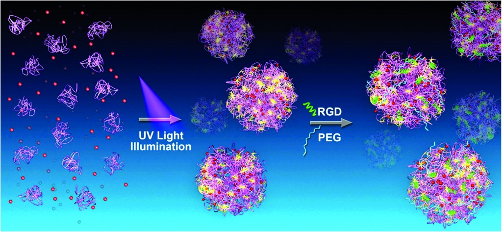
Cancer therapeutics is a burgeoning field of research, particularly because chemotherapy has serious side effects that include the devastation of healthy tissue and the possibility that some cancerous tissue survives the radiation. Research is therefore underway to specifically target cancerous tissue by using targeted drug delivery systems.   We know that nanoparticles can accumulate in cancer tissue, and Wei Wang’s group takes advantage of this fact to create a cancer-specific nanoparticle-based drug delivery system. 

The researchers produce nanoparticles that contain UV-sensitive proteins.  When exposed to UV light, the nanoparticle proteins assemble with a cancer drug and a cancer cell-targeting protein fragment.  The researchers aim to create a nanoparticle-drug complex that specifically attaches to cancerous cells.   When fluorescent nanoparticles were injected into cancerous mice, the researchers found that the particles specifically accumulated in the cancerous tissue.  Further, these nanoparticles were able to effectively kill cancer cells in a dish.  Finally, the researchers showed that these nanoparticles could decrease the size of a tumor in cancerous mice.  This research demonstrates an important breakthrough in cancer treatment. 

Photo synthesis of protein-based drug-delivery nanoparticles for active tumor targeting
Jinbing Xie, Ying Li, Yi Cao, Chun Xu, Mao Xia, Meng Qin, Jiwu Wei and Wei Wang
Biomater. Sci., 2013, Advance Article DOI: 10.1039/C3BM60174A

Debanti Sengupta recently completed her PhD in Chemistry from Stanford University.  She is currently a Siebel postdoctoral scholar at the University of California, Berkeley.  

Follow the latest journal news on Twitter @BioMaterSci or go to our Facebook page.

To keep up-to-date with all the latest research, sign-up to our RSS feed or Table of contents alert.

Digg This
Reddit This
Stumble Now!
Share on Facebook
Bookmark this on Delicious
Share on LinkedIn
Bookmark this on Technorati
Post on Twitter
Google Buzz (aka. Google Reader)

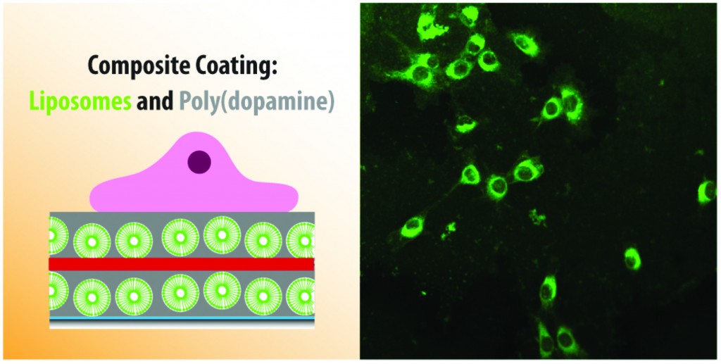
Cargo delivery to adhering myoblast cells from liposome-containing poly(dopamine) composite coatings

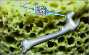
Most of us are accustomed to thinking of medicinal drugs as something we ingest or inject into our bodies.  However, if the drug can be introduced at the particular part of the body where it is needed the most, it will be more effective. Further, if we can slow down the release of the drug, we may be able to use less drugs for the same purpose.  With these long-term goals in mind, Brigitte Städler’s group has recently published a paper where they investigate the release of drugs from a biological coating.  This work can one day be used to modify the surface of implants in our body (commonly used examples include bone implants or pacemakers).  Modifying the surface of these implants would allow us to use them as drug depots in our body.  These implants can then release drugs more slowly into the bloodstream as opposed to all at once, thus making the drugs more effective.

The researchers used a structure known as a liposome – particles made out of fat that can potentially trap a drug in their centers – and surrounded them with a material that can be used to coat implant surfaces. They found that muscle cells could grow on these surfaces without dying, thus proving that the surfaces are not toxic. The liposomes were made out of a fat that fluoresces (emits light under certain conditions).  The muscle cells could absorb these fluorescent fats, which made the cells themselves fluorescent and allowed the cells to be tracked. Next, the researchers trapped a toxic drug in the liposomes, and found that the presence of the drug meant that they could control whether the cells survived or died.  While any kind of drug could in theory be trapped in the coating, a toxic drug could specifically be useful if the coating was near harmful or cancerous cells.

This work shows a great deal of promise in improving the way we deliver drugs to patients.

Cargo delivery to adhering myoblast cells from liposome-containing poly(dopamine) composite coatings
Martin E. Lynge, Boon M. Teo, Marie Baekgaard Laursen, Yan Zhang and Brigitte Städler 
Biomater. Sci., 2013, Advance Article DOI: 10.1039/C3BM60107B

Debanti Sengupta recently completed her PhD in Chemistry from Stanford University.  She is currently a Siebel postdoctoral scholar at the University of California, Berkeley.  

Follow the latest journal news on Twitter @BioMaterSci or go to our Facebook page.

To keep up-to-date with all the latest research, sign-up to our RSS feed or Table of contents alert.
Digg This
Reddit This
Stumble Now!
Share on Facebook
Bookmark this on Delicious
Share on LinkedIn
Bookmark this on Technorati
Post on Twitter
Google Buzz (aka. Google Reader)

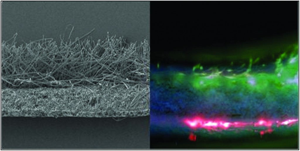
Nanofibrous/microfibrous biodegradable scaffolds for the regeneration and segregation of multiple tissue types

The advent of regenerative medicine has begun to expand the treatment options available to patients who require tissue reconstruction due to birth defects or injuries.  However, it still offers limited solutions for those patients whose wounds span multiple layers and types of tissues.  This is in part due to the shortcomings of the barrier membranes that are typically implanted in order to maintain tissue layer separation.  In this article, researchers from the University of Sheffield propose novel biodegradable bilayer and trilayer fibrous scaffolds that can effectively segregate tissue layers and maintain proper tissue structure during healing.

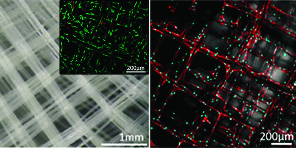
Electrospinning was used to create monolayers of microfibrous polylactic acid (PLA), microfibrous poly ε-caprolactone (PCL), and nanofibrous polyhydroxy-butyrate-co-hydroxyvalerate (PHBV).  Initially, fluorescently labeled fibroblasts and mesenchymal progenitor cells (hESMP) were co-cultured on separate sides of each of the monolayer scaffolds.  After seven days, cell mixing was observed on both the PLA and PCL scaffolds.  However, cell segregation was still clearly evident on the PHBV scaffold, indicating that its nanofibrous structure was acting as a barrier to cell penetration. 

The researchers then created bilayer membranes of PHBV-PLA and PHBV-PCL that exhibited similar barrier properties and maintained high levels of cell viability.   These bilayer scaffolds were designed such that the different degradation rates of the composite polymers were comparable to the different growth rates of hard and soft tissue, thus making them better candidates for the treatment of conditions, such as cleft palate, which require bone and soft tissue segregation.  Finally, a PLA-PHBV-PLA trilayer membrane was designed with the goal of supporting two different types of soft tissue growth.  Subsequent results confirmed that the nanofibrous PHBV layer effectively separated the fibroblasts and keratinocytes that were cultured in the two PLA layers.

Ultimately the researchers were able to demonstrate that nanofibrous scaffolds are capable of promoting cell viability while maintaining separation between different cell types.  The methodology used is simple and reproducible and the resulting scaffold is biocompatible and biodegradable.

Development of bilayer and trilayer nanofibrous/microfibrous scaffolds for regenerative medicine
Frazer J. Bye, Julio Bissoli, Leanne Black, Anthony J. Bullock, Sasima Puwanun, Keyvan Moharamzadeh, Gwendolen C. Reilly, Anthony J. Ryan and Sheila MacNeil 
Biomater. Sci., 2013, 1, 942-951 DOI: 10.1039/C3BM60074B

Ellen Tworkoski is a guest web-writer for Biomaterials Science.  She is currently a second year Ph.D. student in the biomedical engineering department at Northwestern University (Evanston, IL, USA).

Follow the latest journal news on Twitter @BioMaterSci or go to our Facebook page.

To keep up-to-date with all the latest research, sign-up to our RSS feed or Table of contents alert.

Digg This
Reddit This
Stumble Now!
Share on Facebook
Bookmark this on Delicious
Share on LinkedIn
Bookmark this on Technorati
Post on Twitter
Google Buzz (aka. Google Reader)

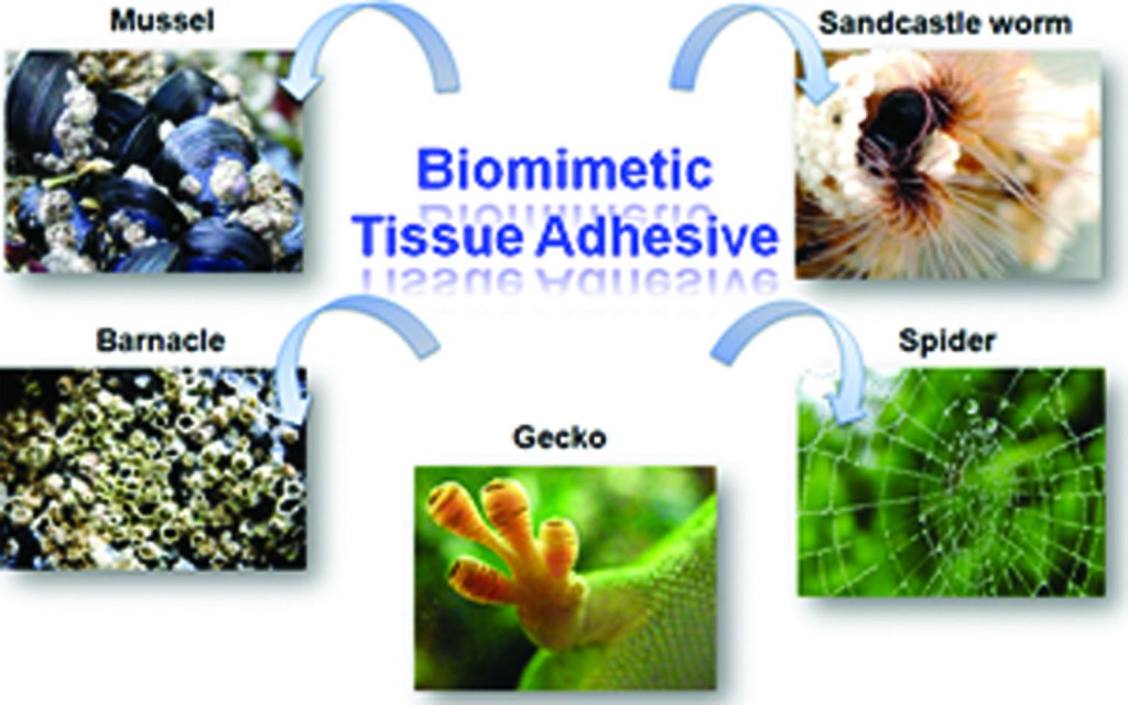
Hot paper: Review of tissue adhesives

Wound closure has traditionally been performed using surgical sutures, staples or clips. These products are not always the most desirable solution especially for suturing friable tissues such as internal organs. Tissues adhesives appear to be an ideal alternative to improve wound healing and overcome fluid leakage and the possibility of additional trauma to the wound sometimes caused by these traditional products.

This Review by Pêgo, Wang and colleagues, covers the recent progress in synthetic/semi-synthetic adhesives and biomimetic adhesives that could be useful for future biomedical applications. They discuss the mechanisms behind the adhesive performances as well as their advantages and disadvantages.

Taking tissue adhesives to the future: from traditional synthetic to new biomimetic approaches
Biomater. Sci., 2013, 1, 239-253.  DOI: 10.1039/c2bm00121g

Follow the latest journal news on Twitter @BioMaterSci or go to our Facebook page.

To keep up-to-date with all the latest research, sign-up to our RSS feed or Table of contents alert.

Digg This
Reddit This
Stumble Now!
Share on Facebook
Bookmark this on Delicious
Share on LinkedIn
Bookmark this on Technorati
Post on Twitter
Google Buzz (aka. Google Reader)

Hot paper: Review of DNA origami technology

DNA nanotechnology allows the construction of self-assembled scaffolds for use in the arrangement of functional molecules and nanomaterials. These can be used to create complex molecular devices. DNA origami is a new programmed DNA assembly system that enables the design of 2D nanostructures which can be functionalised with molecules and nanoparticles.

This Review by Endo, Yang and Sugiyama covers the rapidly moving field of DNA materials science. The review describes the state of current DNA origami research and describes its applications in biomaterials such as the selective functionalisation and single molecule imaging of biomolecules; cell-targeting and as a basis for molecular machines.

DNA origami technology for biomaterials applications
Biomater. Sci., 2012, Advance Article.  DOI: 10.1039/c2bm00154c

Follow the latest journal news on Twitter @BioMaterSci or go to our Facebook page.

To keep up-to-date with all the latest research, sign-up to our RSS feed or Table of contents alert.

Digg This
Reddit This
Stumble Now!
Share on Facebook
Bookmark this on Delicious
Share on LinkedIn
Bookmark this on Technorati
Post on Twitter
Google Buzz (aka. Google Reader)

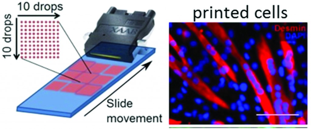
Hot paper: Bio-ink for printing living cells on demand

A bio-ink to print living cells onto a surface using a commercial printer has been developed by Dr Marc in het Panhuis and colleagues at the University of Wollongong, Australia. Bioprinting can be used for tissue engineering and analytical applications. The bio-ink consists of a gel – gellan gum – that’s used in food additives. The gel makes sure that the cells in the bio-ink remain suspended with no sign of aggregation, which was the problem with previous inks. Aggregation means an uneven distribution of cells being printed out onto a surface.

Bio-ink for printing living cells on demand

Biomater. Sci., 2012, Advance Article.  DOI: 10.1039/c2bm00114d

Follow the latest journal news on Twitter @BioMaterSci or go to our Facebook page.

To keep up-to-date with all the latest research, sign-up to our RSS feed or Table of contents alert.

Digg This
Reddit This
Stumble Now!
Share on Facebook
Bookmark this on Delicious
Share on LinkedIn
Bookmark this on Technorati
Post on Twitter
Google Buzz (aka. Google Reader)

Fabrication of scaffolds for regenerative medicine from electrospinning and additive manufacturing minireview

Producing scaffolds for use in tissue engineering is a major focus in the field. Electrospinning and additive manufacturing are two of the methods in which scaffolds can be fabricated. Additive manufacturing is broadly defined as the construction of complex structures in a layer-by-layer fashion using computer aided design.

This minireview by Dalton, Hutmacher and colleagues from the Institute for Health and Biomedical Innovation at Queensland University of Technology describes solution and melt electrospinning use in conjunction with additive manufacturing for tissue engineering scaffolds. The minireview describes the emerging areas of biomodal and multiphasic scaffolds, and scaffolds published using melt electrospinning writing as an additive manufacturing technique.

Electrospinning and additive manufacturing: converging technologies
Biomater. Sci., 2012, Advance Article.  DOI: 10.1039/c2bm00039c

Follow the latest journal news on Twitter @BioMaterSci or go to our Facebook page.

To keep up-to-date with all the latest research, sign-up to our RSS feed or Table of contents alert.

Digg This
Reddit This
Stumble Now!
Share on Facebook
Bookmark this on Delicious
Share on LinkedIn
Bookmark this on Technorati
Post on Twitter
Google Buzz (aka. Google Reader)

More advance articles published

A tissue engineering approach based on the use of bioceramics for bone repair

A tissue engineering approach based on the use of bioceramics for bone repair

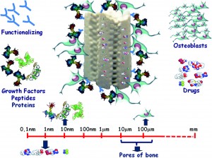
Mesoporous bioactive ceramics that form scaffolds by prototyping are excellent candidates for bone regeneration. This review by Salinas, Esbrit and Vallet-Regí describes the use of bioceramics such as those based on oxides, phosphates, carbonates, nitrides, carbides, carbons and glasses for bone repair.

(Biomater. Sci., 2012, DOI: 10.1039/c2bm00071g, Advance article)


Progress and perspectives in developing polymeric vectors for in vitro gene deliveryProgress and perspectives in developing polymeric vectors for in vitro gene delivery

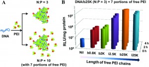
The problems associated with gene-delivery vectors for human gene therapy as discussed in this Review by Yue and Wu. The Review focuses on how polymeric vectors navigate each intracellular obstacle or “slit”. Intracellular trafficking mechanisms of DNA-polymer complexes are particularly focussed upon.

(Biomater. Sci., 2012, DOI: 10.1039/ c2bm00030j, Advance article)

Effects of zinc and strontium substitution in tricalcium phosphate on osteoclast differentiation and resorptionEffects of zinc and strontium substitution in tricalcium phosphate on osteoclast differentiation and resorption

Biomaterials for bone replacement must be able to regulate osteoblast and osteoclast functions to maintain the dynamics of bone remodelling. In this paper, Bose and co-workers report osteoclast-like cell differentiation and resorption activity in the presence of β-tricalcium phosphate and Zn-and Sr-doped tricalcium phosphate materials in an in vitro study. The presence of Zn was found to reduce activity in all cultures, but the osteoclast-like cellular resorption process was not affected.

(Biomater. Sci., 2012, DOI: 10.1039/ c2bm00012a, Advance article)

Don’t forget to keep up-to-date with all the latest research you can sign-up for the Biomaterials Science RSS feed or Table of contents alert.

To keep up with the journal news you can Like us on Facebook or Follow us on Twitter.

Digg This
Reddit This
Stumble Now!
Share on Facebook
Bookmark this on Delicious
Share on LinkedIn
Bookmark this on Technorati
Post on Twitter
Google Buzz (aka. Google Reader)

Biomaterials Science – new Advanced Articles

Understanding the role of nano-topography on the surface of a bone-implant

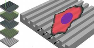
This review by Walboomers and co-authors discusses the interaction of cells with the nano-topographical features of bone implants. The review also details the characterisation of implant surfaces and their manufacture.

(Biomater. Sci., 2012, DOI: 10.1039/c2bm00032f, Advance article)


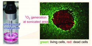
Titanium dioxide nanoparticle-entrapped polyion complex micelles generate singlet oxygen in the cells by ultrasound irradiation for sonodynamic therapy

The main cytotoxic agent in photodynamic therapy is believed to be the reactive oxygen species 1O2 which is used to treat cancerous diseases.  In this paper, Harada and co-workers investigate the generation of reactive oxygen species using sonication of TiO2 nanoparticles. They first synthesised TiO2 nanoparticle-entrapped micelles with a core-shell structure. The nanoparticles were able to generate reactive oxygen species by sonication when inside the micelles. The frequently of sonication was appropriate for clinical situations, thus they has the potential to be used in sonodynamic therapy.

(Biomater. Sci., 2012, DOI: 10.1039/c2bm00066k, Advance article)

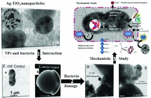
A progressive approach on inactivation of bacteria using silver–titania nanoparticles

The antibacterial properties of silver compounds have long been known. In this paper by Li, Luo and Bashir, Ag-decorated TiO2 nanoparticles were prepared by a colloidal chemistry method. The nanoparticles were coincubated with model microbes, that are found in water, to investigate their biocidal effectiveness. The bactericidal mechanism was also investigated with a focus on the role of the microbial outer membrane.

(Biomater. Sci., 2012, DOI: 10.1039/c2bm00010e, Advance article)

Don’t forget to keep up-to-date with all the latest research you can sign-up for the Biomaterials Science RSS feed or Table of contents alert.

To keep up with the journal news you can Like us on Facebook or Follow us on Twitter.


Digg This
Reddit This
Stumble Now!
Share on Facebook
Bookmark this on Delicious
Share on LinkedIn
Bookmark this on Technorati
Post on Twitter
Google Buzz (aka. Google Reader)

The first advance articles for Biomaterials Science have been published!

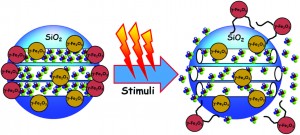
Mesoporous silica nanoparticles for the design of smart delivery nanodevices

The nanomedical use of mesoporous silica nanoparticles has the potential to be revolutionary. This review by María Vallet-Regí and co-workers focuses on the design of mesoporous silica nanoparticles as smart drug delivery systems. The review also covers stimuli-responsive nanocaps and magnetic nanoparticles.

(Biomater. Sci., 2012, DOI: 10.1039/c2bm00085g, Advance article)


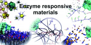
Enzyme responsive materials: design strategies and future developments

Enzyme responsive materials are discussed in this review by Rein V. Ulijn and co-authors. Several strategies for the development of the materials are described such as polymer hydrogels, supramolecular materials and quantum dot particles. The review also discusses the challenges in the development of these materials for specific applications.

(Biomater. Sci., 2012, DOI: 10.1039/c2bm00041e, Advance article)


Citrate-based biodegradable injectable hydrogel composites for orthopaedic applications

Bone biomaterials and scaffolds that have excellent in vivo responses and properties have been a much researched topic over the last 20 years. In this paper by Jian Yang and co-workers, a new bioactive citrate-based injectable biodegradable composite is developed. The composite consists of poly (ethylene glycol) (a FDA approved polymer) and hydroxyapatite which is used for delivering cells into irregular bony defects.

(Biomater. Sci., 2012, DOI: 10.1039/c2bm00026a, Advance article)


Don’t forget to keep up-to-date with all the latest research you can sign-up for the Biomaterials Science RSS feed or Table of contents alert.

To keep up with the journal news you can Like us on Facebook or Follow us on Twitter.

Digg This
Reddit This
Stumble Now!
Share on Facebook
Bookmark this on Delicious
Share on LinkedIn
Bookmark this on Technorati
Post on Twitter
Google Buzz (aka. Google Reader)