Archive for the ‘Hot Article’ Category

Facile Synthesis of Antibacterial Graphene Films Doped with Silver Nanoparticles

The development of antibacterial materials that can guard against microbial infections during medical procedures has been a topic of increasing interest. In this paper, a team of researchers from Beijing University of Chemical Technology and the University of Bremen describe a facile synthesis method for a hybrid nanomaterial that has previously demonstrated excellent antibacterial activity: reduced graphene oxide films decorated with silver nanoparticles. The efficient, dimensionally scalable, and environmentally friendly nature of this method make it a potentially valuable tool for other groups interested in fabricating these hybrid films for applications ranging from medical biomaterials, to cell culture scaffolds, to drug delivery platforms, to environmental remediation.

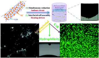
Simultaneous reduction and thermal evaporation-driven self assembly fabrication method used to create RGO/AgNP hybrid film
Schematic model for fabricating RGO/AgNP hybrid film

Zhang et al. created these hybrid films by using sodium citrate to simultaneously reduce aqueous graphene oxide (GO) and aqueous silver nitrate to reduced graphene oxide (RGO) and silver nanoparticles (AgNPs), respectively. This was followed by a prolonged incubation at 80°C to induce thermal evaporation-driven self-assembly of the hybrid RGO/AgNP films. The mass ratio of silver nitrate to GO in the initial solution could be adjusted to produce films with varying densities of AgNPs. In addition, this mass ratio as well the thermal evaporation duration provided some control over the thickness of the resulting hybrid film. The film’s size could be readily changed via adjustments to the dimensions of the reaction container and the film could be later transferred to other substrates without any additional post processing steps.

Subsequent characterisation of the film confirmed the successful reduction of GO as well as the formation of primarily spherical AgNPs between 12 and 30 nm in size on the surface of the film. The resulting material was found to be very hydrophilic and supported the adhesion and proliferation of mouse osteoblasts. An antibacterial assay measuring E. coli adhesion on the surface of silicon wafers coated with these RGO/AgNP films found that the few bacteria that did manage to adhere to the film’s surface exhibited damaged cell walls. A subsequent E. coli colony viability assay proved that no living bacterial colonies were present on the surface of the hybrid film, a result that could not be obtained when using either an uncoated silicon wafer or a pure RGO film.

The biocompatibility, hydrophilicity, durability, and antibacterial activity of these RGO/AgNP hybrid films make them intriguing candidates for a number of medical and environmental applications. The simple, dimension-scalable, and environmental friendly synthesis method presented by Zhang et al. in this paper opens up new avenues for the creation and widespread use of these versatile hybrid films.

Check out the full article:
Graphene film doped with silver nanoparticles: self-assembly formation, structural characterizations, antibacterial ability, and biocompatibility by Panpan Zhang, Haixia Wang, Xiaoyuan Zhang, Wei Xu, Yang Li, Qing Li, Gang Wei, and Zhiqiang Su

Ellen Tworkoski is a Web Writer for Biomaterials Science and is currently a graduate student in the biomedical engineering department at Northwestern University (Evanston, IL, US).

Follow the latest journal news on Twitter @BioMaterSci or go to our Facebook page.

Digg This
Reddit This
Stumble Now!
Share on Facebook
Bookmark this on Delicious
Share on LinkedIn
Bookmark this on Technorati
Post on Twitter
Google Buzz (aka. Google Reader)

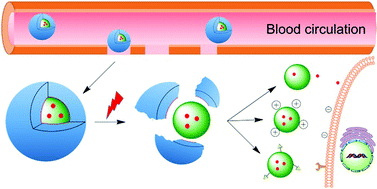
Nanocarriers for cancer drug delivery

Chen et al. discuss the emerging antitumor applications of extracellularly reengineered polymeric nanocarriers.


Chen et al. write an informative and interesting review on the methods by which nanoparticle drug delivery vehicles are engineered using diverse triggers that result in drug release.

In the field of drug delivery, particularly to sites of tumour, there are many different considerations – the drug must be delivered to the site of the tumour, it must be intact when delivered, and it must act to destroy cancerous tissue while remaining as nontoxic as possible to healthy tissue. As a result, much research has been devoted to the development of core-shell drug delivery structures that consist of the drug in the nanoparticle core surrounded by a protective shell. This protective shell may be removed using both internal and external triggers. Many nanocarriers use PEG (polyethylene glycol)-based shells for ease of solubility and in order to prevent proteins from being absorbed onto the surface of the shell. However, additional materials are also increasingly used for the development of these materials.

The authors review the materials as well as common strategies used to remove the shell. Specifically, they summarise literature that exploits changes in pH, since the acidity of the tumour microenvironment differs from its healthy surroundings. Charge-reversal nanocarriers with a positively charged core and a negatively charged shell are also used. In addition, enzymes can degrade the external shell. An enzyme family known as matrix metalloproteinases, or MMPs, is commonly used for this purpose, but other enzymes are also beginning to be explored. Finally, these nanocarriers can also be assembled or de-assembled using interactions between the nanocarrier and the host body.

Emerging antitumor applications of extracellularly reengineered polymeric nanocarriers by Jinjin Chen, Jianxun Ding, Chunsheng Xiao, Xiuli Zhuang and Xuesi Chen

Debanti Sengupta


Debanti Sengupta completed her PhD in Chemistry in 2012 from Stanford University.  She was previously a Siebel postdoctoral scholar at the University of California, Berkeley, and is currently a postdoctoral scholar in Radiation Oncology at Stanford University. Follow her on Twitter @debantisengupta.

Follow the latest journal news on Twitter @BioMaterSci or go to our Facebook page.

Digg This
Reddit This
Stumble Now!
Share on Facebook
Bookmark this on Delicious
Share on LinkedIn
Bookmark this on Technorati
Post on Twitter
Google Buzz (aka. Google Reader)

To crosslink or not to crosslink? – How polymeric micelles can target tumors effectively

Although big strides have been made in the development of anti-cancer drugs, a major hindrance in their development is the lack of appropriate models to test their efficacy. 2D models are poor representations for real-life situations, while animal models present ethical issues. 3D cell-based tumour models would be a major improvement, yet transport modes in 3D models are poorly understood, impeding targeted strategies. A new study published in Biomaterials Science by Lu et al. elucidates how polymeric micelles, a main cancer-treatment platform, are taken up and transported, allowing for improved development of strategies to effectively deliver drug payloads to tumours.

Despite the plethora of anti-cancer drugs developed in the past decades, testing efficacy prior to human trials remains suboptimal at best. 2D cell models poorly reflect the real-life situation, with different pharmacokinetics and nutrient availability, leading to misleading observations and faulty conclusions. Better representations are animal tumour models, yet – separate from obvious ethical concerns – animal metabolism is not necessarily comparable to humans. The development of a more representative 3D multicellular tumour spheroid (MCTS) model to investigate treatment modalities would be a big step towards alleviating those issues, yet the mode of drug carrier transport in 3D models is poorly understood, impeding targeted strategies.

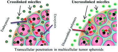
To address the current caveats in MCTS knowledge, Dr. Martina Stenzel’s research group at the University of New South Wales investigated how polymeric micelles are taken up and transported through the outer layers of MCTS’s. Her group created both crosslinked and uncrosslinked polymeric micelles (called CKM and UCM, respectively) and delivered them to a pancreatic MCTS’s. The penetration was monitored using a fluorescent payload, Nile Red, and various modes of endocytosis, as well as exocytosis, were blocked. Their results indicate that it is both caveolae-mediated endocytosis and exocytosis mechanisms are required for good penetration depth of micelles into MCTS’s. Taken together, this evidence points toward transcellular mechanisms as the primary mode of transport for drug-loaded polymeric micelles.

Dr. Stenzel’s group further shows that UCM micelles could not penetrate as far as CKM micelles. The rapid release of their toxic payload doxorubicin creates an apoptotic peripheral cell layer, leading to cessation of additional transcellular transport. Perhaps the most captivating aspect of her research, though understated in the main article, is that the mode of micellar transport seems to be identical in other tumour models.

How polymer micelles are transported in tumor models


The primary mechanism for micellar penetration in MCTS models shown in this article creates important guidance to other researchers investigating anti-cancer drug delivery to tumours. An essential insight is that micelles need to be capable of retaining their structural integrity long enough to prevent payload-induced penetration limitations. Intriguingly, there are indications that the shown penetration mechanisms are extrapolatable to other tumours as well. This study therefore represents a great step forward towards creating better utilization of in vitro tumour models.

Check out the full article:
H. Lu, R.H. Utama, U. Kitiyotsawat, K. Babiuch, Y. Jiang and M.H. Stenzel
Biomater. Sci., 2014, Advance Article, DOI: 10.1039/C4BM00323C


Biomaterials Science web writer Robert van Lith

Robert van Lith (@RvLith) is currently a Post-Doc in the Biomedical Engineering department at Northwestern University, developing intrinsically antioxidant  biomaterials. He recently received his Ph.D. from Northwestern University for his work on citrate-based antioxidant polyesters, receiving an American Heart Association Fellowship and Society for Biomaterials award for his work. He was trained in the Netherlands, holding an M.S. degree in Biomedical Engineering from Eindhoven University of Technology. Read more about Robert’s research publications here.


Follow the latest journal news on Twitter @BioMaterSci or go to our Facebook page.
Digg This
Reddit This
Stumble Now!
Share on Facebook
Bookmark this on Delicious
Share on LinkedIn
Bookmark this on Technorati
Post on Twitter
Google Buzz (aka. Google Reader)

Size of Internalized Calcium Phosphate Particles Plays a Critical Role in Cell Fate

Calcium phosphate (CaP) based materials have long been popular choices for a range of medical applications including bone replacement and drug delivery.  However, recent studies indicate a need for a closer look at how cells react to small, degraded CaP particles that find their way into the cell’s interior.  In a recent study, a research team from the University of Birmingham demonstrated that when CaP particles with a diameter larger than 1.5μm penetrated the interior of the cell but were not sequestered by the cell’s lysosomes, a series of events eventually leading to cell death could be observed.

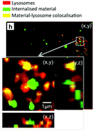
Within the past few years, an increased focus on the end results of CaP degradation have shown that the eventual cytotoxicity of these materials is heavily dependent on the total volume of internalized material.  A study by Motskin et al. in 2009 illustrated that many of these degraded particles are localized to the lysosomes, cellular structures whose acidic environments are responsible for the degradation of waste products which, in the case of CaP, results in the generation of calcium ions.  It was suggested that the generation of a surplus of these ions could interfere with the cell’s signalling pathways, potentially triggering apoptosis.

Intracellular Distribution of CaP showing co-localization with lysosomes

In a study published recently in Biomaterials Science, Williams et al. quantified the volume and size distribution of CaP material within cells by chemically grafting a fluorescent probe to the surface of silicon-substituted hydroxyapatite (SiHA), and then exposing a mouse osteoblast precursor cell line to the labelled particles.  Following exposure, the group observed changes in cell behaviour that were indicative of the onset of cell death including alterations in cell morphology, an increase in the number of lysosomes, and cellular detachment from the underlying substrate.  As the cells transitioned from the early stages of cell death (morphology changes) to later stages (detachment from the substrate), there was a marked increase in the amount of SiHA within the cytoplasm but outside of the lysosomes.  Moreover, the onset of cell death was correlated with SiHA aggregates within the cytoplasm that were 1.5μm in diameter or larger.

The group hypothesized that this may have been due to a destabilization of the lysosome membrane which prevented undissolved CaP from remaining within the lysosomes or, alternatively, the destabilization of endosomes which were responsible for delivering particles to the lysosomes.  Regardless, this study proves a need for additional research into the size-dependent effects of CaP particles on cell health and suggests a new design concern for CaP based medical materials.

Check out the full article here:
Quantification of volume and size distribution of internalised calcium phosphate particles and their influence on cell fate
by Richard L. Williams, Isaac Vizcaíno-Castón, and Liam M. Grover


Ellen Tworkoski is a Web Writer for Biomaterials Science and a graduate student in the department of Biomedical Engineering at Northwestern University.

Digg This
Reddit This
Stumble Now!
Share on Facebook
Bookmark this on Delicious
Share on LinkedIn
Bookmark this on Technorati
Post on Twitter
Google Buzz (aka. Google Reader)

Making it personal – Engineered tissues for neural regeneration

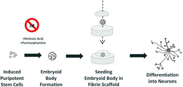
The “holy grail” of neural tissue engineering is to develop functional tissue constructs in an effort to reconnect nerves that have been previously disconnected from disease or injury (i.e. spinal cord injury).  One technique for developing personalized regenerative cell therapies is to use adult somatic cells from a patient (such as skin cells) and re-program them to an induced pluripotent stem cell (iPSC) state. After isolating iPSCs, patient-specific neurons may be generated and transplanted back into the patient, with the hope that the newly formed neurons will form connections in the tissue and restore function.

At the University of Victoria, Montgomery et al. have developed a method to differentiate mouse iPSCs into neurons within a fibrin gel. Fibrin has been regarded as a traditional biomaterial to effectively differentiate embryonic stem cells into neurons. In this study, fibrin was utilized to evaluate the efficiency of two iPS differentiation protocols. The first protocol tested was the traditional “4-/4+” method, involving no treatment to iPS embryoid bodies for 4 days of culture, then adding retinoic acid (RA) for the subsequent 4 days of culture. The second protocol tested was the newer “2-/4+” method, involving no treatment to iPS embryoid bodies for 2 days of culture, then adding RA and purmorphamine for the subsequent 4 days of culture. Individual embryoid bodies were then isolated and placed within a 3D fibrin gel to observe differentiation of neurons.


Interestingly, the overall differentiation efficiency was increased using the 6 day 2-/4+ method compared to using the traditional 4-/4+ method after seeding the treated iPS cells in 3D fibrin gels. Phenotypes were characterized using immunofluorescence staining and RT-PCR, with increases in expression of the neuronal markers TUJ1 and nestin at Day 14 of culture using the 2-/4+ method compared to the 4-/4+ method. The SOX2 pluripotent marker remained low at Day 14, indicating cells were differentiating to a neuronal state.

Taken together, this study demonstrates the ability to differentiate neurons from mouse iPSCs using simple differentiation and encapsulation protocols. Future work will need to be conducted to see if these protocols can be implemented to achieve neuronal differentiation using human iPSCs.

Check out the full article:
Engineering personalized neural tissue by combining induced pluripotent stem cells with fibrin scaffolds, by Amy Montgomery, Alix Wong, Nicole Gabers and Stephanie M. Willerth


Brian Aguado

Brian Aguado (@BrianAguado) is currently a Ph.D. Candidate and NSF Fellow in the Biomedical Engineering department at Northwestern University. He holds a B.S. degree in Biomechanical Engineering from Stanford University and a M.S. degree in Biomedical Engineering from Northwestern University. Read more about Brian’s research publications here.


Follow the latest journal news on Twitter @BioMaterSci or go to our Facebook page.

Digg This
Reddit This
Stumble Now!
Share on Facebook
Bookmark this on Delicious
Share on LinkedIn
Bookmark this on Technorati
Post on Twitter
Google Buzz (aka. Google Reader)

Growing replacement bones – is biomaterial geometry important?

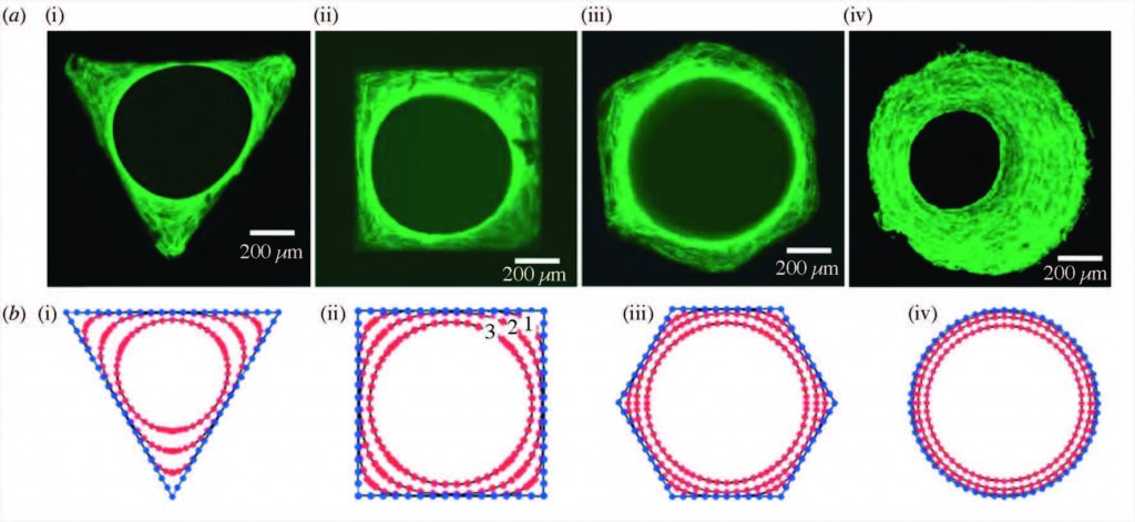
Staining of actin stress fibers

Staining of actin stress fibers to visualize the tissue formed in vitro and to study the effects of curvature (a). The predicted tissue regeneration based on a linear curvature-dependent theoretical model is depicted in subfigure (b). Theoretical predictions match these in vitro experimental observations.

In this paper, Professor Amir Zadpoor reviews the role of biomaterial scaffold geometries on regenerating bone tissue.  Scaffold curvature, pore size, and pore shape are all shown to be important in stimulating more bone growth.

In the case of large bone injuries, the role of scaffolds in regenerating bones becomes increasingly critical.  The ideal bone scaffolds need to be biocompatible, mechanically strong, and contain pores that allow the transport of nutrients and new cell growth.

The geometry of the scaffold plays a very important role in regenerating bone tissue, and is explored in this paper.  Broadly speaking, the curvature of the scaffold, the shape, and the size of the pores are all key components that influence how well the bone is structured and grows.

Strikingly, it has been found that curvature of the surface proportionally impacts the rate of tissue regeneration. Curvature of the surface can be much smaller than, on the scale of, or much larger than cells that grow on the surface. Smaller curvatures can impact individual cell focal adhesions, through which cells can both sense the underlying surface and apply force to it. Larger curvatures can impact cell stress fibers as well as the overall forces on the growing bone tissue.

Pore size can also dramatically impact bone regeneration.  It is already known that a limited amount of inflammation is actually good for bone growth.  Pore size can affect inflammation – for example, larger pores and wider scaffold geometry angles can increase local inflammation.  Pore size can also dictate the amount of oxygen and nutrients reaching the cells, and whether cartilage or bone is formed first.

Pore shape is also extremely important – for example, it has been shown that when cells are grown in scaffolds shaped like parallelograms, alkaline phosphatase activity is increased. Since alkaline phosphatase is a byproduct of bone growth, this data suggests a role for the shape of scaffold pores in bone generation.

However, there are significant caveats to be considered when studying bone regeneration in the lab. Since bone grows in stages, a short in vitro study may not capture all nuances of bone growth that occur in vivo. It is important to study in vitro and in vivo bone generation in parallel to reconcile any contradictory data. Further, it must be noted that it is often difficult to isolate the independent impact of one specific property of a scaffold without affecting another property.  For example, changing pore size can also change the overall mechanical properties of the scaffold.

Despite these limitation, it is clear that the geometrical properties of the scaffold can significantly impact bone growth.  Computational studies and modeling are also now being used to optimize many scaffold properties, and have the potential to drive further research to elucidate the role of scaffold geometry in clinically valuable bone growth.

Check out the full article:

Bone tissue regeneration: the role of scaffold geometry by Amir A. Zadpoor


Web writer Debanti Sengupta

Web writer Debanti Sengupta


Debanti Sengupta completed her PhD in Chemistry in 2012 from Stanford University.  She was previously a Siebel postdoctoral scholar at the University of California, Berkeley, and is currently a postdoctoral scholar in Radiation Oncology at Stanford University. Follow her on Twitter @yoginiscientist.
Follow the latest journal news on Twitter @BioMaterSci or go to our Facebook page.

Digg This
Reddit This
Stumble Now!
Share on Facebook
Bookmark this on Delicious
Share on LinkedIn
Bookmark this on Technorati
Post on Twitter
Google Buzz (aka. Google Reader)

Bioprinting vascular networks for tissue-engineered organs

Ex vivo engineering of 3D organs for transplantation purposes has made tremendous strides in recent years, yet complex tissues remain challenging due to their need for vascularization. A new study takes great steps towards solving this issue using a new, simple bioprinting process to create branched vascular networks capable of effective media exchange. The introduced methodology provides a pathway towards successful incorporation of a branched vasculature into tissue-engineered organs.


In vitro study of directly bioprinted perfusable vasculature conduits

The concept of tissue engineering – combining cells with biomaterials to create living, functional tissues – to provide a solution to the lack of suitable organs for transplantation has been tremendously popular for several decades now, and for good reason. It harnesses the potential to not only tailor organ characteristics to individual patients and reduce transplant rejection risks, but also to virtually erase waiting time for patients in need. The combined efforts of numerous researchers have already led to great success in engineering simple tissues such as skin, yet the evolution towards engineering more complex tissues has proven to be highly challenging. A major roadblock has been, and continues to be, the need for nutrient delivery and media exchange for living tissues to survive, let alone thrive. The incorporation of a vascular network is required for this, yet current methods for creating these networks can either not generate efficient, perfusable systems with the necessary mechanical properties, or are too complex to effectively utilize in thick tissues.

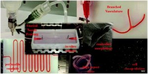
To address the current challenges, Ibrahim Ozbolat’s research group at the University of Iowa designed a novel system for bioprinting vascular conduits. Bioprinting allows for precise 3D fabrication of cell constructs, usually using a sacrificial support biomaterial. In his study, Dr. Ozbolat’s system consists of a coaxial nozzle in which cell-loaded alginate is dispensed through the sheath, and a crosslinking calcium chloride solution through the core, to allow for instantaneous formation of hollow fiber conduits without the need for post-fabrication procedures. The nozzle is under precise robotic control, allowing fabrication of conduits of desired dimension and geometry.

In the current work, the applicability of the bioprinting unit is demonstrated using human umbilical vein smooth muscle cells (HUVSMCs) embedded in alginate as the vessel wall material. By varying alginate and calcium chloride concentrations, conduit properties could be controlled. Relevant properties reported here include vascular lumen and wall dimensions, burst pressures and wall permeability to allow nutrient diffusion. Especially the latter is of paramount importance for long-term functioning of complex engineered tissues. Importantly, this report also shows that despite significant loss of viability during the bioprinting process, alginate-encapsulated cell recovered completely and proliferated well, laying down extracellular matrix throughout the vessel wall as evidenced by histology. Perhaps most intriguing is the demonstrated formation of branched vascular conduits using Dr. Ozbolat’s system.

The straight-forward bioprinting system reported here, allowing for tight control of vascular conduit dimensions, mechanical and perfusion properties, represents a highly promising platform for incorporating effective media exchange and nutrient transport in 3D engineered tissues. Especially the unique lack of post-fabrication requirements and capability for printing branched vessels increase the applicability of this particular design.

In vitro study of directly bioprinted perfusable vasculature conduits
Yahui Zhang, Yin Yu, Adil Akkouch, Amer Dababneh, Farzaneh Dolati and Ibrahim T. Ozbolat

Biomater. Sci., 2015, Advance Article DOI: 10.1039/C4BM00234B


Biomaterials Science web writer Robert van LithRobert van Lith is currently a Post-Doc in the Biomedical Engineering department at Northwestern University, developing intrinsically antioxidant biomaterials. He recently received his Ph.D. from Northwestern University for his work on citrate-based antioxidant polyesters, receiving an American Heart Association Fellowship and Society for Biomaterials award for his work. He was trained in the Netherlands, holding an M.S. degree in Biomedical Engineering from Eindhoven University of Technology. Read more about Robert’s research publications here.

Digg This
Reddit This
Stumble Now!
Share on Facebook
Bookmark this on Delicious
Share on LinkedIn
Bookmark this on Technorati
Post on Twitter
Google Buzz (aka. Google Reader)

Self assembling substrates probe impact of ligands on stem cell fate

By precisely controlling the number of various adhesive ligands in integrin-sized nanodomains, this study provides important insights about the impact of local ligand redundancy on mesenchymal stem cell adhesion and phenotype.

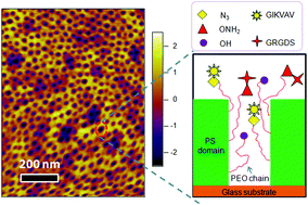
Graphical abstract: Changing ligand number and type within nanocylindrical domains through kinetically constrained self-assembly – impacts of ligand ‘redundancy’ on human mesenchymal stem cell adhesion and morphology

In regenerative medicine, one of the limiting factors has been the number, complex isolation and limited lifespan of patients’ differentiated cell types for seeding scaffolds and subsequent cultivation of a functional tissue. The use of patient-specific stem cell populations, which have a prolonged life-span and can potentially be differentiated into any cell type desired, has emerged as the prevailing tissue engineering paradigm in the past decade. Despite its tremendous potential, steering stem cell differentiation towards a specific phenotypical outcome has been challenging. Polymer surfaces have been used extensively to elucidate the permissive cues required to drive selective differentiation, such as surface functionalities and adhesive molecules. Especially the impact of surface distribution and concentration of adhesive ligands remains a question mark. Yet, it is challenging to tether multiple distinct functionalities in a controllable manner and assess their respective effects on stem cell adhesion and differentiation.

Based at the University of Queensland, Justin Cooper-White and Haiqing Li sought to increase the understanding of surface ligand-dependent stem cell fate by precisely controlling the concentration and spatial organization of various ligands. To this end, they analyzed the effects of two well-known cell adhesion ligands, IKVAV and RGD, on mesenchymal stem cell adhesion and morphology.

Polystyrene-polyethyleneoxide (PS-PEO) block copolymers were shown to self-assemble into polymer films with a uniform distribution of cylindrical nanodomains of PEO, approximately the size of adhesion-controlling cell integrin pairs. By adding varying percentages of azide- or aminooxy-terminated PEO, orthogonal click chemistry was used to sequentially functionalize those cylindrical nanodomains with a controllable local density of grafted adhesion sequences IKVAV and RGD. Then, the researchers assessed the effects of varying local IKVAV and RGD densities in the nanodomains on human mesenchymal stem cell adhesion, spreading morphology and focal adhesion complex formation. They found that with increasing IKVAV or RGD ligand density, leading to ligand ‘redundancy’ for integrin pairs, stem cells showed increased attachment, spreading and stress fiber formation. Moreover, an increase in ratio between RGD and IKVAV densities increased stem cell adhesion.

Together, the researchers found that PS-PEO block co-polymers with functional end-groups, permitting orthogonal chemistry, allowed tight control of ligand decoration within cylindrical nanodomains. Furthermore, using IKVAV and RGD as exemplars, they showed the effect of varying ligand redundancy within nanodomains on stem cell fate. The reported self-assembling substrates offer a highly flexible platform technology to investigate and elucidate the impact of various ligands and their density on integrin binding, which also determines cell phenotype.

Changing ligand number and type within nanocylindrical domains through kinetically constrained self-assembly – impacts of ligand ‘redundancy’ on human mesenchymal stem cell adhesion and morphology
H. Li and J. J. Cooper-White
Biomater. Sci., 2014, Advance Article DOI: 10.1039/C4BM00109E

Robert van Lith is currently a Ph.D. Candidate in the Biomedical Engineering department at Northwestern University, working on novel biomaterials to modulate oxidative stress in tissues. He received an American Heart Association Fellowship and Society for Biomaterials award for his work. He holds B.S. and M.S. degrees in Biomedical Engineering from Eindhoven University of Technology, the Netherlands. Read more about Robert’s research publications here.

Digg This
Reddit This
Stumble Now!
Share on Facebook
Bookmark this on Delicious
Share on LinkedIn
Bookmark this on Technorati
Post on Twitter
Google Buzz (aka. Google Reader)

Nanovehicles for targeted bone metastasis chemotherapy

The cover story for the July 2014 issue of Biomaterials Science highlights the use of nanovehicle particles for the targeted delivery of chemotherapy drugs to tumor cells in bone.

Bone metastasis, or the spreading of tumor cells from the initial tumor site to bone tissue, marks the stage of cancer progression where the diseased is deemed incurable. Currently, the main method to treat bone metastasis is to utilize aggressive chemotherapy drugs and destroy metastatic tumor cells.  Unfortunately, untargeted chemotherapy treatments result in death of both cancerous and healthy tissue. Treatments could be significantly improved with the technology to specifically target the destruction of tumor cells without affecting the surrounding bone tissue.

Researchers at the University of Utah, University of Iowa, and Wuhan University in China are making this desired technology a reality. Wang et al. have developed a nanovehicle capable of targeting tumor cells within bone tissue.  Nanovehicles (NVs) are essentially nano-sized cargo containers capable of delivering drugs to a desired destination. The cleverly designed NVs are designed to target diseased bone tissue to deliver chemotherapy drugs at a specific site.

The NVs developed in this study have three design features:

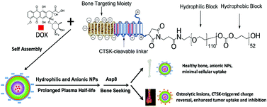
(1) The NVs core is made of hydrophilic and hydrophobic block co-polymers able to form a shell-like structure known as a micelle. The micelle serves as the cargo vessel that contains the chemotherapy drug doxorubicin.

(2) The outermost segments of the NVs are negatively charged bone-targeting peptides. The peptide is responsible for the accumulation of NVs in skeletal tissue, and the negative charge prevents cellular uptake of the NVs into healthy tissue.

(3) For uptake into cancer cells, the particle must be positively charged. Therefore, a positively charged peptide linker was added in between the micelle core and the negatively charged bone-targeting region. The peptide linker is cleavable by cathepsin K enzyme (CTSK), which is overexpressed in bone metastatic lesions. When the NV enters a metastatic lesion, the CTSK cleaves off the negatively charged peptides and exposes the positively charged peptides. This charge reversal allows for tumor cells present in the metastatic lesion to uptake the NV.

After extensively characterizing the nanoparticles with NMR and zeta potential measurements, the NVs were used to observe decreased tumor cell viability under CTSK-rich conditions in vitro. More strikingly, in vivo tests of the NVs in a mouse model of bone metastasis showed significant increases in mouse survival and decreases in overall tumor burden.

Collectively, these ingeniously designed nanovehicles show great promise in providing an effective targeted therapy for bone metastasis.

Peptide decoration of nanovehicles to achieve active targeting and pathology-responsive cellular uptake for bone metastasis chemotherapy
X. Wang, Y. Yang, H. Jia, W. Jia, S. Miller, B. Bowman, J. Feng, and F. Zhan
Biomater. Sci., 2014, Advance Article DOI: 10.1039/C4BM00020J

Brian Aguado is currently a Ph.D. Candidate and NSF Fellow in the Biomedical Engineering department at Northwestern University. He holds a B.S. degree in Biomechanical Engineering from Stanford University and a M.S. degree in Biomedical Engineering from Northwestern University. Read more about Brian’s research publications here.

Follow the latest journal news on Twitter @BioMaterSci or go to our Facebook page.

Digg This
Reddit This
Stumble Now!
Share on Facebook
Bookmark this on Delicious
Share on LinkedIn
Bookmark this on Technorati
Post on Twitter
Google Buzz (aka. Google Reader)

Protein corona key to safe, personalized nanomedicine

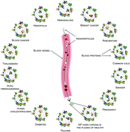
This study introduces the concept of the “personalized protein corona” as a determinant factor in nano-biomedicine, specifically regarding the biological identity and fate of nanoparticles.

Graphical abstract: Personalized protein coronas: a “key” factor at the nanobiointerfaceThe concept of personalized medicine has become increasingly relevant in healthcare over the past decade. Ever since the human genome was unraveled, the idea of tailoring therapies specific to the individual patient has been at the forefront of medicine. Diagnosis, prognosis and most importantly treatment are thought to eventually be adapted to each individual’s genetic and pathophysiological makeup. Nonetheless, biomaterials for invasive use (e.g. implantation or injection) are still largely being designed for general use. This study argues for a much more specific approach of biomaterials design, looking at the nanoparticles (NPs) in particular.

A collaborative effort headed by Dr. Mahmoudi from Stanford University, US, and the University of Medical Sciences in Tehran, Iran, Hajipour et al. evaluated effects of the altered protein composition of human plasma in different disease states on NP corona formation. Two NP materials were chosen for their analysis, hydrophobic polystyrene and hydrophilic silica, which are widely investigated for biomedical material approaches and currently evaluated for safety purposes. 

 NPs were exposed to plasma solutions of different patient populations to form hard protein coronas, after which the composition of NP protein coronas was analyzed using SDS-page, densitometry and zeta-potential measurements. Interestingly, the authors found that changes in plasma protein composition due to underlying conditions alter the protein decoration on NPs, resulting in an unpredictable biologic identity of the NPs and thus their biological fate. The severity of the condition was also found to impact protein deposition. Intriguingly, even among healthy subjects there was considerable variation in corona composition, creating even more of an impetus for personalized NP design.

Collectively, the researchers found that NP protein corona distinctly depends on particular condition, severity of condition and combination of conditions, with relatively low variation between subjects with similar health profiles. Moving forward, identifying the personalized protein corona for individual patients or at least the expected protein corona in specific patient populations may become an integral aspect of nanoparticle-based biomaterial therapies.

Personalized protein coronas: a “key” factor at the nanobiointerface 
M. J. Hajipour, S. Laurent, A. Aghaie, F. Rezaee and M. Mahmoudi
Biomater. Sci., 2014, Advance Article DOI: 10.1039/c4bm00131a

Robert van Lith


Robert van Lith is currently a Ph.D. Candidate in the Biomedical Engineering department at Northwestern University, working on novel biomaterials to modulate oxidative stress in tissues. He received an American Heart Association Fellowship and Society for Biomaterials award for his work. He holds B.S. and M.S. degrees in Biomedical Engineering from Eindhoven University of Technology, the Netherlands. Read more about Robert’s research publications here.

Digg This
Reddit This
Stumble Now!
Share on Facebook
Bookmark this on Delicious
Share on LinkedIn
Bookmark this on Technorati
Post on Twitter
Google Buzz (aka. Google Reader)