Interfacing living cells with inorganic nanowire (NW) array substrates is one of the latest areas of exploration in life sciences with potential applications in electrical stimulation, biosensors, cell injection, axonal guidance and so on. A growing body of evidence has identified the role of substrate nanotopography in regulating various cellular phenotypes including cell morphology, adhesion, proliferation, differentiation and intracellular signaling. However, cellular interactions with high surface area vertical nanowires are relatively unexplored and further studies are necessary to fully reveal the correlations between NW array geometry and stem cell behavior.
Researchers from Germany have recently interfaced human induced pluripotent stem cell (hiPSC)-derived neurons with tailor-made silicon nitride NW array substrates, achieving highly efficient neuronal differentiation and generating electrophysiologically mature neurons within 4 weeks of culture.
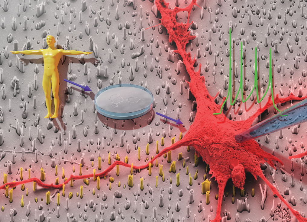
First, they fabricated NW arrays using a top-down dry reactive ion etching (RIE) approach in 3 different arrangements – random, hexagonal and rectangular. The NW lengths were fixed to 1.2 μm with pitches of 1.8 μm and 4 μm, resulting in low density (LD) and high density (HD) arrangements respectively. The cells were transferred onto the NW substrates after 14 days in vitro (DIV) and cultured for another 14-16 DIV before performing functional characterization.
Next, they assessed viability of cells cultured on NW substrates and found that both material used as well as substrate topology had no negative impact on cell viability compared with controls cultured on glass coverslips. Furthermore, on studying cellular outgrowth and morphology, they observed that cells rested on NW tips in the case of HD arrays whereas they encapsulated the NWs in LD arrays, thus indicating the effect of NW density on regulating the settling regime of the cells.
Finally, they tested the electrophysiological integrity of the hiPSC-derived neurons via patch clamping and observed that the neurons cultured on the NW substrates fired characteristic action potentials and demonstrated no significant differences in electrophysiological parameters compared with controls.
Taken together, the results indicate the potential of this platform in stem cell research and regenerative medicine for interfacing human stem cell-derived neurons with tailor-made nanostructured substrates to achieve desired cell behaviors.
To find out more please read:
Jann Harberts, Undine Haferkamp, Stefanie Haugg, Cornelius Fendler, Dennis Lam, Robert Zierold, Ole Pless and Robert H. Blick
Biomater. Sci., 2020, 8, 2434-2446, DOI: 10.1039/D0BM00182A


")