Archive for January, 2018

Bio-mimicking melanin-manganese nanoparticles for tumor targeting MRI agent

Good quality MRI contrast agents require profound tumor-targeting ability, well relaxivity, along with rapid clearance properties. In this context, endogenous biological mimicking biomaterials, with high biodegradability and biocompatibility hold enormous potential for the development of clinically translatable nanotheranostics platforms. Manganese plays a crucial role in mitochondrial and cellular function and recently, manganese (Mn)-based contrast agents have been receiving significant attention, due to improved biosafety and superior contrast abilities. However, the long term toxicity and non-biodegradability of these inorganic nanoplatforms have significantly halted their clinical progress. In contrast to this, melanin, an asymmetrical natural biopolymer, has garnered enormous attention due to good biocompatibility, biodegradability and MRI contrast imaging abilities. Hence, exploring endogenous natural materials with high contrast properties seems promising as clinically translatable in vivo MRI imaging contrast agent.

melanin-manganese nanoparticles for tumor targeting MRI agent

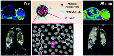
The Wang group developed an ultra small and water soluble Mn2+ chelating pegylated melanin nanoparticles (MNP-PEG-Mn) demonstrating excellent tumor-targeting Magnetic Resonance Imaging (MRI) ability. The MNP-PEG-Mn nanoparticles show a size of 5.6 nm displaying high chelating stability and low cytotoxicity. Interestingly, the MNP-PEG-Mn nanoparticles show improved longitudinal relaxivity compared to clinically approved MRI contrast agent Gadodiamide. In vivo studies further showcased excellent tumor targeting abilities upon intravenous administration of MNP-PEG-Mn nanoparticles in mouse model. The author further showed that the MNP-PEG-Mn nanoparticles could be excreted via hepatobiliary and renal routes. In this process negligible toxicity was generated to body tissues that indicate high biocompatibility. Altogether, these results clinically validate the tumor targeted T1 MRI contrast properties of bio-mimicking melanin conjugated manganese nanoparticles.

Melanin-manganese nanoparticles with ultrahigh efficient clearance in vivo for tumor-targeting T1 magnetic resonance imaging contrast agent . Biomater. Sci., 2018, 6, 207-215.

This article is free to read until 28 February!


About the Web writer:

Dr. Sudip MukherjeeDr. Sudip Mukherjee is a Web Writer for Biomaterials Science. He is currently a Postdoctoral Research Associate working at the Department of Bioengineering at the Rice University. His research is involved in the development of advanced nanomaterials for drug/gene delivery in cancer theranostics, immunotherapy, immunomodulatory applications & angiogenesis. He published a total of ~30 research articles/patents. He serves as International Advisory Board Member for ‘Materials Research Express‘, IOP Sciences. He is an associate member (AMRSC) of The Royal Society of Chemistry, UK. He serves as reviewer for several international journals like Chem Comm, J Mater Chem A, J Mater Chem B, Journal of Biomedical Nanotechnology, RSC Advances, IOP Nanotechnology etc. He can be contacted by email at sudip.mukherjee@rice.edu or on Twitter.

Digg This
Reddit This
Stumble Now!
Share on Facebook
Bookmark this on Delicious
Share on LinkedIn
Bookmark this on Technorati
Post on Twitter
Google Buzz (aka. Google Reader)

Focus on: Controlled micro-/nano-geometry

The geometry of micro- or nano-materials determine their physico-chemical properties and play crucial roles in directing an array of in vivo biological activities. Historically, the significance of micro-/nano-geometry for biomaterials has not been adequately appreciated due to the limitations in manufacturing techniques. Recent years have witnessed the exponential growth of micro-/nano-fabrication technologies which allow closer scrutiny of the relationship between the micro-/nano-geometry and the biological effects of materials. This month the focus is on three articles published in Biomaterials Science reporting the recent advances to achieve controlled micro-/nano-geometry of biomedical devices including microdisks, nanosheets, and microcapsules.

Focus on controlled micro/nano-geometry

1. Advanced manufacturing of microdisk vaccines for uniform control of material properties and immune cell function

Qin Zeng, Peipei Zhang, Xiangbin Zeng, Lisa H. Tostanoski and Christopher M. Jewell
Biomater. Sci., DOI: 10.1039/c7bm00520b

The heterogeneity in the geometry and loading level represents one major drawback of synthetic vaccine constructs. Therefore, taking advantage of the facile and flexible technique of soft lithography, the authors designed and fabricated PLGA microdisks with controllable dimension and payload loading. Compared to the conventional PLGA microparticles made by emulsion, microdisks possessed highly uniform diameter with less variation. Importantly, microdisks could safely co-deliver vaccine antigen and molecular adjuvants to primary dendritic cells. As a result, dendritic cells could be activated according to the composition of antigen and adjuvants. This controllable and programmable microdisk system will serve as platform to probe the relationship between the vaccine design parameters and immune responses in vivo.

2. The biodistribution, excretion and potential toxicity of different-sized Pd nanosheets in mice following oral and intraperitoneal administration

Xiaolan Chen, Jingchao Li, Yizhuan Huang, Jingping Wei, Duo Sun and Nanfeng Zheng
Biomater. Sci., 2017, 5, 2448. DOI: 10.1039/c7bm00769h

To elucidate the in vivo behaviors of Pd nanosheets (NSs), the authors synthesized size-specific Pd NSs (with 5 nm, 30 nm, or 80 nm in diameter) by controlling the chemical reaction conditions. Those NSs were then administrated via oral feeding or intraperitoneal injection to reveal different biodistribution, excretion, and toxicity profiles of those size-specific NSs. Interestingly, when intraperitoneally delivered, larger sized Pd NSs (80 nm) had higher accumulation in liver and spleen than the smaller sized Pd NSs (5 nm), which had higher accumulation in tumor tissue. In addition, the smaller sized NSs (5 nm) exhibited more excretion through urine than larger sized Pd NSs (30 and 80 nm). Overall, this study indicated that the size of nanomaterials could have significant influence on their biodistribution and bioavailability in vivo.

3. Development of drug-loaded polymer microcapsules for treatment of epilepsy

Yu Chen, Qi Gu, Zhilian Yue, Jeremy M. Crook, Simon E. Moulton, Mark J. Cook and Gordon G. Wallace
Biomater. Sci.,
2017, 5, 2159. DOI: 10.1039/c7bm00623c

To achieve the goal of developing controllable local delivery system for treating epilepsy, the authors developed drug-loaded PLGA based microspheroids, microspheres, and microfibers by tuning the solution concentration during electrojetting (electrospinning and/or electrospraying) process. All the resultant microcapsules had excellent shape and size uniformity with high controllability and low variance. Different microcapsule geometry led to various sustained drug release profiles in vitro without compromising their cytocompatibility. This study highlighted the potential of programmable and controllable delivery system and suggested the potential role of geometry in controlling the drug release profile of micron-sized biomedical device.

Read these articles for free until 3 February

About the webwriter

Yingfei XueYingfei Xue is a web writer for Biomaterials Science. Currently, he is a PhD candidate and graduate student researcher in Dr. Shilpa Sant lab at the University of Pittsburgh, USA.  His research focus on nano-/micro-technology in novel heart valve therapy. Find him on Twitter: @Phil_Xue  or connect with him on ResearchGate.

Digg This
Reddit This
Stumble Now!
Share on Facebook
Bookmark this on Delicious
Share on LinkedIn
Bookmark this on Technorati
Post on Twitter
Google Buzz (aka. Google Reader)