Archive for December, 2013

Happy holidays from Biomaterials Science!

All of us in the Biomaterials Science Editorial team would like to wish you all a merry Christmas and a happy new year! The Editorial office will be closed from 24th December 2013 and will reopen on 2nd January 2014.

We’re really looking forward to 2014, which will see more high quality articles from top international biomaterials scientists, some great themed issues and the first Biomaterials Science lectureship.  Thank you for all of your contributions which have helped make 2013 a great first year for Biomaterials Science.

Don’t miss out on all the journal news – follow us on twitter @BioMaterSci and like us on Facebook!

Digg This
Reddit This
Stumble Now!
Share on Facebook
Bookmark this on Delicious
Share on LinkedIn
Bookmark this on Technorati
Post on Twitter
Google Buzz (aka. Google Reader)

Dextran polyampholyte hydrogels for cell cryopreservation

One of the goals of tissue engineers is to manufacture “spare body parts” made of cells and biomaterials to repair damaged or diseased tissues. A current challenge for commercializing cell-biomaterial constructs is maintaining their viability during shipping and/or storage. The storage of cells in liquid suspension is relatively simple with the use of common cryoprotectants (such as dimethyl sulfoxide), but current preservation methods for large cell-biomaterial constructs yield low cell viability in comparison.  More effective preservation methods for cell-biomaterial constructs are needed to produce “off the shelf” tissue engineered products and accelerate the translation of tissue engineered products to the clinic.

In a current study conducted at the Japan Advanced Institute of Science and Technology (JAIST), researchers developed a dextran-based polyampholyte (charged polymer) hydrogel with cryoprotective properties. First, dextran polymers were functionalized with azide groups (azide-Dex) to provide sites for crosslinking. Then, positively charged amine groups were introduced on the dextran using poly-L-lysine to form azide-amino-Dex. To add negatively charged carboxyl groups, succinic anhydride was added at various concentrations to azide-amino-Dex to convert amine groups to carboxyl groups. This newly formed dextran polyampholyte (azide-Dex-PA) polymer was utilized for cryopreservation studies. After encapsulating and freezing cells with various azide-Dex-PA solutions, cell viability post-cryopreservation was shown to increase with higher concentrations of azide-Dex-PA. Viability was also dependent on the ratio of amine and carboxyl groups on the polymer, indicating that the cryoprotective properties of the polyampholyte are likely due to polymer charge.

The azide groups on the azide-Dex-PA were utilized to crosslink the dextran polyampholytes to form hydrogels. After synthesizing alkyne-functionalized dextran with dibenzylcyclooctyne (DBCO-Dex), the two components were mixed with cells to form hydrogels with azide-alkyne click chemistry. Encapsulated cells in the hydrogel were cryopreserved, thawed, and evaluated for viability. Compared to the 0% viability observed with collagen hydrogel and non-polyampholyte dextran hydrogel controls, the dextran polyampholyte hydrogels substantially increased cell viability to 93-94%. The increased viability post-cryopreservation may be due to the polyampholyte polymer adsorbing onto the cell membrane, providing a polymer “shell” of protection during thawing. Images of cells coated with FITC labeled azide-Dex-PA provide evidence to support this hypothesis.

All in all, dextran polyampholyte hydrogels have cryoprotective properties, and are able to preserve cell viability during the cryopreservation process. The dramatic increase in cell viability post-cryopreservation justifies future studies for using these hydrogels for preserving larger cell-biomaterial constructs for long term storage.

Hydrogelation of dextran-based polyampholytes with cryoprotective properties via click chemistry
Minkle Jain, Robin Rajan, Suong-Hyu Hyon, and Kazuaki Matsumura
Biomaterials Science, 2014, Advance Article DOI: 10.1039/C3BM60261C

Brian Aguado is currently a Ph.D. Candidate and NSF Fellow in the Biomedical Engineering department at Northwestern University. He holds a B.S. degree in Biomechanical Engineering from Stanford University and a M.S. degree in Biomedical Engineering from Northwestern University. Read more about Brian’s research publications here.

To keep up-to-date with all the latest research, sign-up to our RSS feed or Table of contents

Digg This
Reddit This
Stumble Now!
Share on Facebook
Bookmark this on Delicious
Share on LinkedIn
Bookmark this on Technorati
Post on Twitter
Google Buzz (aka. Google Reader)

Editor-in-Chief Phillip Messersmith interviewed in Chemistry World

Phillip Messersmith, Biomaterials Science co-Editor-in-Chief, has been interviewed in Chemistry World about his work on biological adhesives to develop new biomaterials for the repair, replacement, or augmentation of human tissue.

Here are some highlights from the interview:

…What are the main applications for your synthetic polymers, are they just biomedical?
Not exclusively, but the funding sources right now are primarily in health related areas. We have a lot of funding from the National Institutes of Health in the US and obviously their main interest at the end of the day is to contribute to basic understanding as well as the applications of new materials, new devices and new therapies. So we work through the government funding as well as some corporate and institutional funding towards applications. The application you mentioned before, fetal surgery, has been a great passion for me over the last few years. I only really became involved in this three–four years ago but it’s become really important to me.
It’s the kind of medical problem that has too small a market to interest big companies and so the surgeons work in this area and do wonderful things without having all the tools they would like. One example of a tool they need is for sealing ruptures in the fetal membrane that occur spontaneously or after an interventional procedure. The ruptures can lead to leakage of amniotic fluid and when that happens you have two major problems. First, is the risk of infection and second is the premature induction of labour. Either way you have a very serious medical problem for the mother and the fetus and there aren’t many ways to treat this apart from bed rest.
There’s a small community of fetal surgeons that have trained for many years to try and avoid these ruptures but if it happens there’s not a lot they can do. So we’re developing materials to try and seal the membranes after rupture. Here obviously the tissue is wet and there’s a large volume of high ionic strength fluid. This is not very different from the conditions encountered by mussels- thus providing a great argument for learning how mussels and other marine organisms can accomplish wet adhesion.

How easy is it to make these materials biocompatible?
That’s a great question and something we spend a lot of time thinking about. Biocompatibility is an all-encompassing word: but it’s all about context. All we can say is we try to develop systems based on biocompatible polymers and DOPA and then formulate them in a way that doesn’t induce a severe inflammatory response. But any synthetic material has some level of that response. There’s an interesting give and take between in vitro results and in vivo results. A positive in vitro result won’t necessarily translate to a positive in vivo result. One of the interesting things is that the opposite is also true. Sometimes in vivo cell toxicity assays give a borderline response but in vivo we see really good results. We choose the polymers and how we go about the functionalisation and purification very carefully and then we do in vitro and in vivo tests.

Going back slightly, what made you get into bioadhesion?
The guy I mentioned earlier, Herbert Waite. When I was a young faculty member I used to block off one full day a month and just go to the library and look at all the new journal issues that had come in. And I used to try and make a point of trying to read out of my comfort zone, in areas I really wasn’t trained in. And one of those times I encountered one of his papers which described these proteins, the mussel adhesive proteins. And I said, wow, this is really interesting. Then I started looking for more of his papers and it just struck me, as a materials scientist, as an interesting translation opportunity, which it’s turned out to be. To this day I often tell my students that story because I don’t think they really appreciate how important it is not just to read the literature, but to read the literature outside of what you happen to be looking for that day, that hour…

Read the full interview with Laura Howes here

Follow the latest journal news on Twitter @BioMaterSci or go to our Facebook page.

To keep up-to-date with all the latest research, sign-up to our RSS feed or Table of contents

Digg This
Reddit This
Stumble Now!
Share on Facebook
Bookmark this on Delicious
Share on LinkedIn
Bookmark this on Technorati
Post on Twitter
Google Buzz (aka. Google Reader)

Decreasing Tumor Growth with Magnetic Nanohydrogels

Cancer is the second leading cause of death worldwide, and continues to be a challenging disease to treat therapeutically. One strategy to treat cancer patients is to surgically remove the primary tumor and attempt to prevent the spread of cancer cells to other organs in the body. Unfortunately, surgical resection of tumors may be difficult in sensitive organs (e.g. the brain), and tumors may need to be treated directly with chemotherapy drugs to inhibit growth.

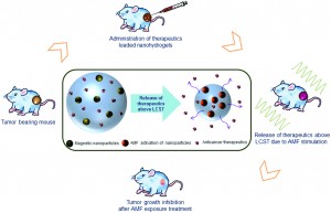
Recently, there has been an increased interest in utilizing biomaterials to deliver chemotherapy drugs directly to the primary tumor. Current research at the Indian Institute of Technology explores the use of nanohydrogels supplemented with iron oxide magnetic nanoparticles as a vehicle to deliver chemotherapy drugs to the primary tumor. Interestingly, these thermo-responsive nanohydrogels could release their anticancer drug cargo when heated with a magnetic field. Although this study did not use chemotherapy drugs, the magnetic hydrogels were used to heat the tumor locally to reduce tumor size – upon stimulation with a magnetic field. This study focused on characterizing the material properties of the magnetic nanohydrogels, in addition to analyzing the effects of the nanohydrogel on inhibiting tumor growth.

The magnetic nanohydrogel is composed of iron oxide nanoparticles, chitosan, and poly-N-isopropylacrylamide (NIPAAm).  The chitosan-poly-(NIPAAm) nanohydrogels are hydrophilic below the lower critical solution temperature (LCST) of 42oC. When stimulated with the appropriate magnetic field, the magnetic nanoparticles heat the hydrogel above the LCST. When the temperature is increased above the LCST, the polymers in the hydrogel change from “expanded coils” to “compact globules,” and cause the release of the aqueous contents of the hydrogel. The hydrophilic to hydrophobic transition is the main mechanism by which the anticancer drug cargo could be released from the hydrogel.

The biocompatibility and efficacy of the hydrogel in reducing tumor growth in vivo were analyzed. Using a mouse model of fibrosarcoma, the magnetic nanohydrogels were delivered via injection to the primary tumor site, and then stimulated with a magnetic field. Various biocompatibility studies were conducted, showing that serum protein concentrations and blood cell counts were not affected. The biodistribution of the magnetic hydrogel was also quantified, with minimal accumulation of the nanohydrogel in various organs (14 days post-delivery). Additionally, the magnetic nanohydrogel decelerated the growth of the primary tumor. After magnetic stimulation, the magnetic particles in the nanohydrogel successfully heated the tumor locally with minimal damage to the surrounding tissues. Despite not containing chemotherapy drugs, the magnetic nanohydrogels were still efficient in inhibiting the primary tumor growth.

This study describes the thermo-responsive properties of magnetic nanohydrogels, and demonstrates the ability to reduce tumor size using nanohydrogels heated with a magnetic field. Future studies are needed to show efficacy in delivering chemotherapy drugs using the magnetic nanohydrogels as a delivery vehicle. Still, the magnetic nanohydrogels are a promising platform for a minimally invasive method of decreasing tumor growth in vivo.

Biocompatibility, biodistribution and efficacy of magnetic nanohydrogels in inhibiting growth of tumors in experimental mice models
Manish K. Jaiswal, Manashjit Gogoi, Haladhar Dev Sarma, Rinti Banerjee and D. Bahadur
Biomater. Sci., 2014, Advance Article DOI: 10.1039/C3BM60225G

Brian Aguado is currently a Ph.D. Candidate and NSF Fellow in the Biomedical Engineering department at Northwestern University. He holds a B.S. degree in Biomechanical Engineering from Stanford University and a M.S. degree in Biomedical Engineering from Northwestern University. When he’s not in the lab, Brian enjoys traveling, cooking, swimming, and spending time with family and friends. Read more about Brian’s research publications here.

To keep up-to-date with all the latest research, sign-up to our RSS feed or Table of contents

Digg This
Reddit This
Stumble Now!
Share on Facebook
Bookmark this on Delicious
Share on LinkedIn
Bookmark this on Technorati
Post on Twitter
Google Buzz (aka. Google Reader)

Achieving improved separation of output signals in ‘sense and treat’ biosensors via an enzymatic filter

The next generation of diagnostic devices is currently being realized in the form of ‘smart’ biosensors.  These sensors use logic gates to digitalize biochemical signals and produce a binary output that either indicates the presence or absence of a disease.  The biochemical signals, which are often the product of enzymatic cascades, are also capable of triggering drug release from stimuli-responsive materials, thus creating a device that is capable of both sensing and treating a particular condition.

However, these integrated sense and treat biosensors have a major obstacle to overcome if they are to be successfully implemented.  Many of the enzymatic cascades used for input will produce low, but non-zero concentrations of the sensing analytes under normal, physiological conditions.  Over time, the accumulation of these background signals can rise to a level that is high enough to trigger drug release, resulting in the dosage of a perfectly healthy patient which often has adverse consequences.  In this work, a team from Clarkson University and Oak Ridge National Laboratory propose a novel solution to this problem which involves the incorporation of an enzymatic filter into these devices.

The group created a system that was designed to detect and treat liver injury using input from two known biomarkers of liver disease: alanine transaminase (ALT) and aspartate transaminase, type I (AST).  When both ALT and AST are present in elevated concentrations, they will produce high levels of molecules which can be used to synthesize citric acid and another enzyme known as CoA-SH.  While CoA-SH can be readily detected and used to generate a positive signal indicating liver injury, citric acid can be used to trigger drug release by dissolving the iron-cross-linked alginate beads which were used in this study to simulate drug release capsules.  The group then introduced an enzymatic filter, malate dehydrogenase (MDH), into the system.  MDH is able to regulate the production of citric acid, thus controlling the degradation of the alginate beads and subsequent drug release.  In the test cases listed, the modified system generated the correct diagnostic result 100% of the time.  However, the filter was not able to completely eliminate bead dissolution and drug release under conditions associated with a normal, healthy environment.

To tackle this problem, the group decided to try improving the stability of the beads themselves.  By alternately depositing layers of poly-L-lysine with additional layers of alginate, PLA bi-layers were formed on the surface of the alginate cores.   The group found that, as more layers were added, the speed of bead dissolution and the amount of drug released were reduced.  The combination of the filter and the PLA coatings showed enhanced suppression of the negative signals while continuing to have a minimal effect on the positive signal.

Although the group was unable to completely eliminate drug release under normal conditions due to long-term noise accumulation, they were able to reduce it significantly.  Their approach marks a promising step forwards in addressing one of the major obstacles faced by smart biosensor systems.

Enzymatic filter for improved separation of output signals in enzyme logic systems towards ‘sense and treat’ medicine
Shay Mailloux, Oleksandr Zavalov, Nataliia Guz, Evgeny Katz and Vera Bocharova  
Biomater. Sci., 2014, Advance Article DOI: 10.1039/C3BM60197H

Ellen Tworkoski is a guest web-writer for Biomaterials Science.  She is currently a second year Ph.D. student in the biomedical engineering department at Northwestern University (Evanston, IL, USA).

Follow the latest journal news on Twitter @BioMaterSci or go to our Facebook page.

To keep up-to-date with all the latest research, sign-up to our RSS feed or Table of contents alert.

Digg This
Reddit This
Stumble Now!
Share on Facebook
Bookmark this on Delicious
Share on LinkedIn
Bookmark this on Technorati
Post on Twitter
Google Buzz (aka. Google Reader)