Enhancing Dopamine Sensitivity by Reducing Surface Fouling

Laura Ruvuna is guest web writer for Analyst. She is currently a PhD student at Northwestern University, Evanston, Illinois, USA.

Researchers have adapted a gold electrode to enhance electrochemical dopamine measurements and overcome the fouling problems that typically occur on the surface when using this technique.
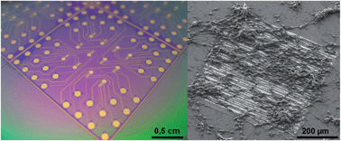
Cells on doped overoxidised PPy modified electrodes

Many diseases such as Parkinson’s and schizophrenia are caused by irregularities in the neurotransmitter dopamine. Each disease affects dopamine production differently, however in all cases studying the process both inside and outside the body has presented many challenges.

Jenny Emnéus at the Technical University of Denmark and collaborators in Italy improved the detection of dopamine by placing a doped overoxidised polypyrrole (PPy) film on the electrode surface. The film was doped with different counter ions to inhibit dopamine polymerisation and the binding of negatively charged species. Although the overoxidation of PPy did affect the conductivity of the film, it also became more sensitive to dopamine, suggesting that doped overoxidised PPy can be used as sensors for dopamine.

To learn more about the techniques the authors used to reduce surface fouling and detect dopamine release from live cells, check out the article below. It will be free to read until May 28th .

Doped overoxidized polypyrrole microelectrodes as sensors for the detection of dopamine released from cell populations
Luigi Sasso, Arto Heiskanen, Francesco Diazzi, Maria Dimaki, Jaime Castillo-León, Marco Vergani, Ettore Landini, Roberto Raiteri, Giorgio Ferrari, Marco Carminati, Marco Sampietro, Winnie E. Svendsen and   Jenny Emnéus  
Analyst, 2013, Advance Article
DOI: 10.1039/C3AN00085K

Digg This
Reddit This
Stumble Now!
Share on Facebook
Bookmark this on Delicious
Share on LinkedIn
Bookmark this on Technorati
Post on Twitter
Google Buzz (aka. Google Reader)

Gordon F. Kirkbright Bursary Award, 2014

The Gordon F. Kirkbright bursary award is a prestigious annual award that enables a promising student/non-tenured young scientist of any nation to attend a recognised scientific meeting or visit a place of learning.
The fund for this bursary was established in 1985 as a memorial to Professor Gordon Kirkbright in recognition of his contributions to analytical spectroscopy and to science in general. Although the fund is administered by the Association of British Spectroscopists (ABS) Trust, the award is not restricted to spectroscopists.

Applications are invited for the 2014 Gordon Kirkbright Bursary.

For further information contact John Chalmers at, email: vibspecconsult@aol.com

The closing date for entries is 31 December 2013.

Digg This
Reddit This
Stumble Now!
Share on Facebook
Bookmark this on Delicious
Share on LinkedIn
Bookmark this on Technorati
Post on Twitter
Google Buzz (aka. Google Reader)

Analyst Issue 11 out now!

Wang and Schultz, Analyst, 2013, 138, 3150-3157

The latest issue of Analyst is now online and ready for you to enjoy. We have three new covers and a couple of HOT papers free for you to read.

On the outside cover is an image from a paper by Zachary Schultz and Hao Wang from the University of Notre Dame, USA, showing functionalised nanoparticles detected with surface-enhanced Raman scattering (SERS) and Tip-enhanced Raman scattering (TERS) using streptavidin and biotin as a model system for protein-ligand binding. The researchers examined the chemical origin of enhanced signals from SERS and TERS detection and observed that different sizes of functionalised nanoparticles show changes in intensities in both types of measurements.

The chemical origin of enhanced signals from tip-enhanced Raman detection of functionalized nanoparticles
Hao Wang and   Zachary D. Schultz  
Analyst, 2013,138, 3150-3157
DOI: 10.1039/C3AN36898J

Yun et al., Analyst, 2013, 138, 3196-3200

Our inside front cover features a study on digital imaging of cultured cells enhanced by augmented reality technologies. Won Gu Lee and colleagues from Seoul National University, South Korea, present a new method to look at specimens under the microscope using augmented indicators. This new technique enables observation of long term live cells and identification of the exact location of individual cells within a biological sample.

Microscopic augmented-reality indicators for long-term live cell time-lapsed imaging
Kyungwon Yun, Jungman Chung, Yong Park, Byungjoo Lee, Won Gu Lee and   Hyunwoo Bang  
Analyst, 2013,138, 3196-3200
DOI: 10.1039/C3AN00124E

Ishimoto et al., Analyst, 2013, 138, 3158-3162

Finally, our outisde back cover showcases research on the simplification of conventional neuraminidase inhibitor assays. Hideaki Hisamoto and his group at Osaka Prefecture University, Japan, successfully integrated the assay into a single step operation using a combinable poly(dimethylsiloxane) (PDMS) capillary sensor array.

Integration of neuraminidase inhibitor assay into a single-step operation using a combinable poly(dimethylsiloxane) capillary sensor
Tadashi Ishimoto, Kaede Jigawa, Terence G. Henares, Tatsuro Endo and   Hideaki Hisamoto  
Analyst, 2013,138, 3158-3162
DOI: 10.1039/C3AN36785A

Now take a look at the selected HOT papers below. They will be free to read until May 30th.

Tagging the rolling circle products with nanocrystal clusters for cascade signal increase in the detection of miRNA
Jingjing Yao, Kenneth Flack, Liangzi Ding and   Wenwan Zhong  
Analyst, 2013,138, 3121-3125
DOI: 10.1039/C3AN00398A

Isotachophoresis with ionic spacer and two-stage separation for high sensitivity DNA hybridization assay
Charbel Eid, Giancarlo Garcia-Schwarz and   Juan G. Santiago 
Analyst, 2013,138, 3117-3120
DOI: 10.1039/C3AN00374D

Digg This
Reddit This
Stumble Now!
Share on Facebook
Bookmark this on Delicious
Share on LinkedIn
Bookmark this on Technorati
Post on Twitter
Google Buzz (aka. Google Reader)

FTIR spectral signature of anticancer drug effects on cancer cells: any influence of the cell cycle?

Jennifer A. Dougan is a guest web-writer for Analyst. She is currently a Post-Doctoral Research Associate at Imperial College London, UK.

Visible image, infrared image, propidium iodide fluorescence image of a cell smear

Erik Goormaghtigh and co-workers from the Centre for Structural Biology and Bioinformatics at the Unversité Libre de Bruxelles have investigated the use of Fourier transformfo infrared (FTIR) spectroscopy r studying the effect of an anti-cancer drug, paclitaxel, on a human prostate cancer cell line.

The researchers combined fluorescence spectroscopy with FTIR spectroscopy using a fluorescent DNA intercalator to identify cell cycle stage.  By doing so, they sought to understand the drug’s effect on cell cycle and/or metabolic perturbation. The authors discuss both the application and limitation of this approach to cell studies.

Read more about this study in this HOT Analyst paper, free for you until May 24th .

FTIR spectral signature of anticancer drug effects on PC-3 cancer cells: is there any influence of the cell cycle?
Allison Derenne, Alix Mignolet and   Erik Goormaghtigh  
Analyst, 2013, Advance Article
DOI: 10.1039/C3AN00225J

Digg This
Reddit This
Stumble Now!
Share on Facebook
Bookmark this on Delicious
Share on LinkedIn
Bookmark this on Technorati
Post on Twitter
Google Buzz (aka. Google Reader)

Top ten most accessed articles in March

This month sees the following articles in Analyst that are in the top ten most accessed:-

Construction of NIR and ratiometric fluorescent probe for Hg2+ based on a rhodamine-inspired dye platform 
Jing Liu, Yuan-Qiang Sun, Pi Wang, Jingyu Zhang and Wei Guo 
Analyst, 2013,138, 2654-2660 
DOI: 10.1039/C3AN00061C  

A turn-on and reversible Schiff base fluorescence sensor for Al3+ ion 
Chang-Hung Chen, De-Jhong Liao, Chin-Feng Wan and An-Tai  
Analyst, 2013,138, 2527-2530 
DOI: 10.1039/C3AN00004D  

Quantum dot enabled detection of Escherichia coli using a cell-phone 
Hongying Zhu, Uzair Sikoraa and Aydogan Ozcan  
Analyst, 2012,137, 2541-2544 
DOI: 10.1039/C2AN35071H  

Upconversion nanoparticles in biological labeling, imaging, and therapy 
Feng Wang, Debapriya Banerjee, Yongsheng Liu, Xueyuan Chen and Xiaogang Liu  
Analyst, 2010,135, 1839-1854 
DOI: 10.1039/C0AN00144A  

A quantum dot-based “off–on” fluorescent probe for biological detection of zinc ions 
Hu Xu, Zhiping Wang, Yan Li, Shijian Ma, Peiyi Hua and Xinhua Zhong 
Analyst, 2013,138, 2181-2191 
DOI: 10.1039/C3AN36742H  

ATR-FTIR spectroscopic imaging: recent advances and applications to biological systems 
Sergei G. Kazarian and K. L. Andrew Chan 
Analyst, 2013,138, 1940-1951 
DOI: 10.1039/C3AN36865C  

Highly sensitive label-free fluorescent detection of Hg2+ ions by DNA molecular machine-based Ag nanoclusters 
Jinjin Yin, Xiaoxiao He, Xuekun Jia, Kemin Wang and Fengzhou Xua   
Analyst, 2013,138, 2350-2356
DOI: 10.1039/C3AN00029J  

Antibody orientation on biosensor surfaces: a minireview 
Anke K. Trilling, Jules Beekwilder and Han Zuilhof  
Analyst, 2013,138, 1619-1627 
DOI: 10.1039/C2AN36787D  

Quantum dots for fluorescent biosensing and bio-imaging applications 
Jingjing Lia and Jun-Jie Zhu   
Analyst, 2013,138, 2506-2515 
DOI: 10.1039/C3AN36705C  

Protein amyloids develop an intrinsic fluorescence signature during aggregation 
Fiona T. S. Chan, Gabriele S. Kaminski Schierle, Janet R. Kumita, Carlos W. Bertoncini, Christopher M. Dobson and Clemens F. Kaminski 
Analyst, 2013,138, 2156-2162 
DOI: 10.1039/C3AN36798C  

Why not take a look at the articles today and blog your thoughts and comments below.

Fancy submitting an article to Analyst? Then why not submit to us today or alternatively email us your suggestions.

Digg This
Reddit This
Stumble Now!
Share on Facebook
Bookmark this on Delicious
Share on LinkedIn
Bookmark this on Technorati
Post on Twitter
Google Buzz (aka. Google Reader)

The 36th Annual Meeting of the Brazilian Chemical Society – Aguas de Lindoia, Brazil 25-28 May 2013

The 36th Annual Meeting of the Brazilian Chemical Society is being held this month in Aguas de Lindoia, Brazil, 25th-28th May. Click here for more information.

Analyst and Analytical Methods are pleased to announce a cross journal web-themed issue on Analytical Sciences in Brazil. Interdisciplinary analytical science is particularly welcome. Guest Editor Ivo M. Raimundo Jr. will be in attendance at the Brazilian Chemical Society annual meeting.

Submission deadline: 31st Jan 2014
Articles published online in a web collection

Guest Editors: Boris Mizaikoff, University of Ulm
Ivo M. Raimundo Jr., UNICAMP

For more information please contact analyst-rsc@rsc.org

Digg This
Reddit This
Stumble Now!
Share on Facebook
Bookmark this on Delicious
Share on LinkedIn
Bookmark this on Technorati
Post on Twitter
Google Buzz (aka. Google Reader)

New Hot Articles in Analyst

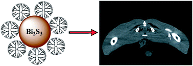
Synthesis, characterisation, tomography imaging of bismuth sulfide nanoparticles

Take a look at our HOT papers now available online . Tomography imaging applications, serum micro-Raman spectroscopy and photoelectrochemical studies are among the topics featured in these articles.

To read the full papers, please access the links below. They will be free for you until  May 20th.

Dendrimer-stabilized bismuth sulfide nanoparticles: synthesis, characterization, and potential computed tomography imaging applications
Yi Fang, Chen Peng, Rui Guo, Linfeng Zheng, Jinbao Qin, Benqing Zhou, Mingwu Shen, Xinwu Lu, Guixiang Zhang and   Xiangyang Shi  
Analyst, 2013, Advance Article
DOI: 10.1039/C3AN00237C

Diagnosis of hepatocellular carcinoma in cirrhotic patients: a proof-of-concept study using serum micro-Raman spectroscopy
I. Taleb, G. Thiéfin, C. Gobinet, V. Untereiner, B. Bernard-Chabert, A. Heurgué, C. Truntzer, P. Hillon, M. Manfait, P. Ducoroyc and   G. D. Sockalingum  
Analyst, 2013, Advance Article
DOI: 10.1039/C3AN00245D

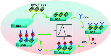
Carbon nanotubes biosensors for malondialdehyde detection

Tagging the rolling circle products with nanocrystal clusters for cascade signal increase in the detection of miRNA
Jingjing Yao, Kenneth Flack, Liangzi Ding and   Wenwan Zhong
Analyst, 2013, Advance Article
DOI: 10.1039/C3AN00398A

Label-free and facile electrochemical biosensing using carbon nanotubes for malondialdehyde detection
Ling Yuan, Yaqian Lan, Min Han, Jianchun Bao, Wenwen Tu and   Zhihui Dai
Analyst, 2013, Advance Article
DOI: 10.1039/C3AN00041A

Tailoring zinc porphyrin to the Ag nanostructure substrate: an effective approach for photoelectrochemical studies in the presence of mononucleotides
Farkhondeh Fathi, Cong Kong, Yueqiang Wang, Yongshu Xie, Yi-Tao Long and   Heinz-Bernhard Kraatz  
Analyst, 2013, Advance Article
DOI: 10.1039/C3AN00156C

Isotachophoresis with ionic spacer and two-stage separation for high sensitivity DNA hybridization assay
Charbel Eid, Giancarlo Garcia-Schwarz and   Juan G. Santiago 
Analyst, 2013, Advance Article
DOI: 10.1039/C3AN00374D

Biological and biomedical 14C-accelerator mass spectrometry and graphitization of carbonaceous samples
Ill-Min Chung and   Seung-Hyun Kim
Analyst, 2013, Advance Article
DOI: 10.1039/C3AN00077J

An AC electrokinetics facilitated biosensor cassette for rapid pathogen identification
Mengxing Ouyang, Ruchika Mohan, Yi Lu, Tingting Liu, Kathleen E. Mach, Mandy L. Y. Sin, Mason McComb, Janhvi Joshi, Vincent Gau, Pak Kin Wong and   Joseph C. Liao 
Analyst, 2013, Advance Article
DOI: 10.1039/C3AN00259D

Digg This
Reddit This
Stumble Now!
Share on Facebook
Bookmark this on Delicious
Share on LinkedIn
Bookmark this on Technorati
Post on Twitter
Google Buzz (aka. Google Reader)

Quicker checks for safer water

Scientists in the Netherlands have developed and tested a new method that uses Raman spectroscopy to quickly identify harmful bacteria in drinking water. This could prevent contaminated drinking water from making people ill by detecting deadly bacteria before it ever reaches the tap.

Many of the standard methods used to test water quality rely on selective plating and enzyme activity tests which take a long time to get results. ‘The bottleneck between collection and results is because microbes need time to grow, or nucleic acids need time to be processed,’ says Jack van de Vossenberg, from the KWR Watercycle Research Institute, who led the research.

To read the full article please visit Chemistry World.

Identification of bacteria in drinking water with Raman spectroscopy
Jack van de Vossenberg, Heli Tervahauta, Kees Maquelin, Carola H. W. Blokker-Koopmans, Marijan Uytewaal-Aarts, Dick van der Kooij, Annemarie van Wezel and Bram van der Gaag  
Anal. Methods, 2013, Accepted Manuscript
DOI: 10.1039/C3AY40289D

Digg This
Reddit This
Stumble Now!
Share on Facebook
Bookmark this on Delicious
Share on LinkedIn
Bookmark this on Technorati
Post on Twitter
Google Buzz (aka. Google Reader)

DNA, Russian opera and blue suede shoes

Duncan Graham is Professor of chemistry at the University of Strathclyde, UK. He has recently been named as the next Chair of the Editorial Board for Analyst, and will start this role in 2014. His research areas include nucleic acid chemistry and synthetic chemistry for bioanalysis. Currently he is working on functionalised metallic nanoparticles that can be used for the diagnosis and treatment of disease.

What motivated you to specialise in SERS (surface enhanced Raman spectroscopy)?

Duncan Graham

 To read the full article please visit Chemistry World.

Digg This
Reddit This
Stumble Now!
Share on Facebook
Bookmark this on Delicious
Share on LinkedIn
Bookmark this on Technorati
Post on Twitter
Google Buzz (aka. Google Reader)

Probing Alzheimer’s disease Plaques using Synchrotron FTIR

Laura Ruvuna is guest web writer for Analyst. She is currently a PhD student at Northwestern University, Evanston, Illinois, USA.

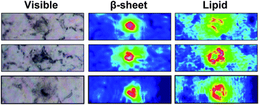
Visible and FTIR images showing plaques and lipids in mice tissues

Alzheimer’s disease (AD) affects millions of patients worldwide, and with much still unknown, few treatments options are available for the long duration of this disease. Although opinions differ on the cause of AD, histological staining shows the presence of neurotoxic plaques and tangles in deceased patients and serves as the primary diagnostic method.

Researchers at the University of Manitoba, Canada, used high resolution Fourier Transform Infrared Spectroscopy (FTIR) to image brain tissue in mice and human samples in order to study plaque formation at the sub cellular level. Different components in the cell such as DNA, proteins or sugars, produce unique chemical signatures which correspond to vibrational bands. The researchers discovered infiltration of lipid membrane components surrounding the plaques, which increase the signal as the plaques become larger. The spatial resolution of the system used here enables detection of lipids that a staining would miss, but it is important to understand disease progression. Although the relationship between plaque formation and lipid concentration remains unclear, the authors have developed a new method towards understanding plaque formation in AD.

To read the full article, please access the link below. This paper will be free to read until May 14th .

Synchrotron FTIR reveals lipid around and within amyloid plaques in transgenic mice and Alzheimer’s disease brain
Catherine R. Liao, Margaret Rak, Jillian Lund, Miriam Unger, Eric Platt, Benedict C. Albensi, Carol J. Hirschmugl and   Kathleen M. Gough
Analyst, 2013, Advance Article
DOI: 10.1039/C3AN00295K

Digg This
Reddit This
Stumble Now!
Share on Facebook
Bookmark this on Delicious
Share on LinkedIn
Bookmark this on Technorati
Post on Twitter
Google Buzz (aka. Google Reader)