HOT articles in Analyst

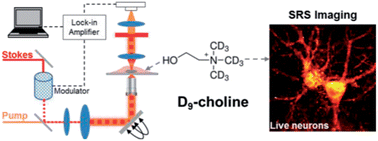
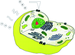
Take a look at our new HOT articles just published in Analyst and free for you for the next couple of weeks: Graphical abstract: Live-cell vibrational imaging of choline metabolites by stimulated Raman scattering coupled with isotope-based metabolic labeling

Live-cell vibrational imaging of choline metabolites by stimulated Raman scattering coupled with isotope-based metabolic labeling
Fanghao Hu, Lu Wei, Chaogu Zheng, Yihui Shen and Wei Min
Analyst, 2014, Advance Article
DOI: 10.1039/C3AN02281A, Communication

Chip based single cell analysis for nanotoxicity assessment
Pratikkumar Shah, Ajeet Kaushik, Xuena Zhu, Chengxiao Zhang and Chen-Zhong Li
Analyst, 2014, Advance Article
DOI: 10.1039/C3AN02280C, Minireview

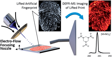
Chemical imaging of artificial fingerprints by desorption electro-flow focusing ionization mass spectrometry
Thomas P. Forbes and Edward Sisco
Analyst, 2014, Advance Article
DOI: 10.1039/C4AN00172A, Communication

Binding assay for low molecular weight analytes based on reflectometry of absorbing molecules in porous substrates
Milena Stephan, Corinna Kramer, Claudia Steinem and Andreas Janshoff
Analyst, 2014, Advance Article
DOI: 10.1039/C4AN00009A, Paper

Quantized double layer charging of Au130(SR)50 nanomoleculesGraphical abstract: Chemical imaging of artificial fingerprints by desorption electro-flow focusing ionization mass spectrometry
Vijay Reddy Jupally, Jacob G. Thrasher and Amala Dass
Analyst, 2014, Advance Article
DOI: 10.1039/C3AN02204H, Communication

Robotic plasma probe ionization mass spectrometry (RoPPI-MS) of non-planar surfaces
Rachel V. Bennett, Ezequiel M. Morzan, Jacob O. Huckaby, María Eugenia Monge, Henrick I. Christensen and Facundo M. Fernández
Analyst, 2014, Advance Article
DOI: 10.1039/C4AN00277F, Communication

Digg This
Reddit This
Stumble Now!
Share on Facebook
Bookmark this on Delicious
Share on LinkedIn
Bookmark this on Technorati
Post on Twitter
Google Buzz (aka. Google Reader)

Kick Off and the Power of Twitter

Matthew J. Baker is a web-writer for Analyst. He is currently Senior Lecturer in Toxicology and Analytical Chemistry at the University of Central Lancashire (UCLan), UK

I was waiting for a train on a cold and rainy Friday morning in February at Preston train station (this will be a shock for those that know the Preston climate as it is actually subtropical) and tweeting from @ChemistryBaker to my colleagues @RoyGoodacre and @AlexHenderson00 about the upcoming kick off meeting of the Clinical Infrared and Raman Spectroscopy network. Analyst (@analystrsc) picked this tweet up and through a few tweets back and forth agreed to host a blog on the subject and here it is; to show a different side of research and the way a fledgling network starts and, hopefully, becomes successful. Success for us is closely linked to a clinical impact that would have a massive positive effect on many many people’s lives.

The UK is a world leader in the field of vibrational biomedical spectroscopy with excellent research being performed by many groups based in our academic and governmental research systems. Diagnostic and prognostic tools based on these technologies have the potential to revolutionise our clinical systems leading to improved patient outcome, a more efficient healthcare service and significant economic savings. However, despite these strong reasons and the research that has occurred to date, this technology has not truly made the jump into the clinic. There was a need to bring many researchers, clinicians and industrialists together in order to provide a critical mass that will overcome these clinical implementation barriers.

It was for these reasons, that in 2012, several of us got together to speak about the possibility of developing a bid for an EPSRC network grant. Importantly network grants provide no money for research projects rather funding to enable collaborative multidisciplinary meetings targeted to tackle problems and drive a field forwards. Luckily EPSRC agreed (thank you EPSRC) that our bid, plan and field deserved funding and the EPSRC Clinical Infrared and Raman SPECtroscopy (CLIRSPEC) network was set up.

Even though the meeting that February day was long and actually quite tiring it was also fun and welcoming. Our field is known to be a supportive field. Essentially the aims of the meeting was to get people together to discuss and finalise our plans to overcome the challenges we identified, agree how we assess bids to the training fund for PhDs and PDRAs, conference locations and summer school locations.  The main aim was to ask already overworked people to do more work for free, which thankfully they agreed to.

The work of the network will be achieved through six working parties that aim to overcome the barriers that challenge our field:

1)   To develop our understanding of the interaction of light with clinical samples

2)   To develop internationally recognized protocols for the preparation of cells, tissue and biofluids for clinical spectroscopy

3)   To provide evidence of the power of spectroscopy for use in the clinical arena

4)   To determine the requirements of instrumentation suitable for use in the clinic

5)   To develop strategies for pre-processing and statistical analysis in clinical spectroscopy

6)   To develop inter-group data sharing protocols and portal

I would like to use this first blog to ask if there are any clinicians, scientists or industrialists out there who feel they can contribute to the cause. We currently have international and national partners and would welcome more. If you feel you can contribute then please visit our website. We do not want to be a closed shop but rather harness the power of many to achieve our aims.

Until the next time…

Please find links to recent papers below that describe the research currently on-going in the field to demonstrate the application of spectroscopy to the clinical environment as well as tackling the current research challenges.

The inherent problem of transflection-mode infrared spectroscopic microscopy and the ramifications for biomedical single point and imaging applications

Paul Bassan, Joe Lee, Ashwin Sachdeva, Juliana Pissardini, Konrad M. Dorling, John
S. Fletcher, Alex Henderson and Peter Gardner
Analyst, 2013,138, 144-157

A comparison of Raman, FTIR and ATR-FTIR micro spectroscopy for imaging human skin tissue sections
S. M. Ali, F. Bonnier, H. Lambkin, K. Flynn, V. McDonagh, C. Healy, T. C. Lee, F. M. Lyng and H. J. Byrne
Anal. Methods, 2013,5, 2281-2291

Effect of substrate choice and tissue type on tissue preparation for spectral histopathology by Raman microspectroscopy
Leanne M. Fullwood, Dave Griffiths, Katherine Ashton, Timothy Dawson, Robert W. Lea, Charles Davis, Franck Bonnier, Hugh J. Byrne and Matthew J. Baker
Analyst, 2014,139, 446-454

Identification of different subsets of lung cells using Raman microspectroscopy and whole cell nucleus isolation
Jacek K. Pijanka, Nicholas Stone, Abigail V. Rutter, Nicholas Forsyth, Ganesh D. Sockalingum, Ying Yang and Josep Sulé-Suso
Analyst, 2013,138, 5052-5058

ATR-FTIR spectroscopic imaging: recent advances and applications to biological systems
Sergei G. Kazarian and K. L. Andrew Chan
Analyst, 2013,138, 1940-1951

Illuminating disease and enlightening biomedicine: Raman spectroscopy as a diagnostic tool

David I. Ellis, David P. Cowcher, Lorna Ashton, Steve O’Hagan and Royston Goodacre
Analyst, 2013,138, 3871-3884

Simultaneous detection and quantification of three bacterial meningitis pathogens by SERS
Kirsten Gracie, Elon Correa, Samuel Mabbott, Jennifer A. Dougan, Duncan Graham, Royston Goodacre and Karen Faulds
Chem. Sci., 2014,5, 1030-1040

Resonance Raman microscopy in combination with partial dark-field microscopy lights up a new path in malaria diagnostics
Bayden R. Wood, Antje Hermelink, Peter Lasch, Keith R. Bambery, Grant T. Webster, Mehdi Asghari Khiavi, Brian M. Cooke, Samantha Deed, Dieter Naumann and Don McNaughton
Analyst, 2009,134, 1119-1125

Extracting biological information with computational analysis of Fourier-transform infrared (FTIR) biospectroscopy datasets: current practices to future perspectives
Júlio Trevisan, Plamen P. Angelov, Paul L. Carmichael, Andrew D. Scott and Francis L. Martin
Analyst, 2012,137, 3202-3215

Emerging concepts in deep Raman spectroscopy of biological tissue
Pavel Matousek and Nicholas Stone
Analyst, 2009,134, 1058-1066

Vibrational spectroscopy: a clinical tool for cancer diagnostics
Catherine Kendall, Martin Isabelle, Florian Bazant-Hegemark, Joanne Hutchings, Linda Orr, Jaspreet Babrah, Rebecca Baker and Nicholas Stone
Analyst, 2009,134, 1029-1045

Also please see the latest themed issue on Optical Diagnostics

Digg This
Reddit This
Stumble Now!
Share on Facebook
Bookmark this on Delicious
Share on LinkedIn
Bookmark this on Technorati
Post on Twitter
Google Buzz (aka. Google Reader)

SPEC 2014 -Shedding New Light on Disease- submissions open!

logo

Submissions now open! Abstracts must be submitted online via the abstract registration form.

Deadline for submission is 15th May 2014.

The aim of this international conference series is to bring together clinicians and scientists who have joined forces in the quest for novel biomedical applications of Infrared and Raman spectroscopy to improve patient care. Recent advances in the biological sciences and medicine have led to an increasing demand for real time and minimally invasive chemical and structural information on biological materials. Due to its unique fingerprinting capability, vibrational spectroscopy plays a significant role in histopathology, cytology, biopsy targeting, surgical targets, treatment monitoring and drug studies. This, the 8th in the series, aims to highlight further advances in state of the art and emerging biomedical applications of Vibrational Spectroscopy, while critically reviewing the challenges in the context of other emerging technologies.

Date: 17-22 August 2014
Location: Krakow, Poland
Contact: Katarzyna Marzec
Email: marzec@chemia.uj.edu.pl
Website

Digg This
Reddit This
Stumble Now!
Share on Facebook
Bookmark this on Delicious
Share on LinkedIn
Bookmark this on Technorati
Post on Twitter
Google Buzz (aka. Google Reader)

Femtoliter-Scale Separation and Sensitive Detection Using Nanofluidics

Matthew J. Baker is a web-writer for Analyst. He is currently Senior Lecturer in Toxicology and Analytical Chemistry at the University of Central Lancashire (UCLan), UK

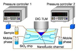
An image of liquid chromatography using nanofluidic chip and DIC-TLM

Liquid chromatography using nanofluidic chip and DIC-TLM

The field of separation science has been continually evolving to enable the separation of smaller volumes. This interdisciplinary field has required developments in capillary electrophoresis, microfluidics and new detection technologies such as plasmonic sensing and thermal lensing. 

Researchers from the Department of Applied Chemistry, University of Tokyo lead by Prof. Kitamori have shown the ability to perform femtolitre (1 x 10-15) separation and detection of nonfluorescent samples in a nanofluidic device. Using an extended nanofluidic device the researchers were able to separate and detect nonfluorescent molecules for the first time. Previous detection methodologies relied mainly on laser-induced fluorescence. 

The researchers showed the ability of nanofluidic separation science combined with differential interference contrast thermal lens microscopy (DIC-TLM) to enable detection of 370 molecules in a 2.3 μm wide by 350 nm deep nanochannel with a separation efficiency of 150,000 plates/m. DIC-TLM is thermal lens microscopy with an increased detection limit by realising background-free photodetection using differential interference contrast. 

Amazingly the injection volume and number of molecules were estimate to be 21 fL (1 x 10-15) and 250 zmol at the limit of detection. This limit of detection provides separation of a sample 8 orders of magnitude smaller that that possible with high-performance liquid chromatography. This injection volume is much smaller than a single cell and detection limit is similar to the number of typical protein molecules in a single cells. Providing a promising tool for single cell research. 

To read more about this research download the HOT article which is free to access until April 5th 

Femtoliter-Scale Separation and Sensitive Detection of Nonfluorescent Samples in an Extended-Nano Fluidic Device
Hisashi Shimizu, Kazuma Mawatari and Takehiko Kitamori 
Analyst, 2014, Accepted Manuscript

Digg This
Reddit This
Stumble Now!
Share on Facebook
Bookmark this on Delicious
Share on LinkedIn
Bookmark this on Technorati
Post on Twitter
Google Buzz (aka. Google Reader)

Shedding New Light on Old Treasures

Analytical techniques are shedding new light on old treasures.

Madame Leon Clapisson, 1883

Analytical chemists are making breakthroughs in the science of heritage and conservation. Using new techniques, previously lost nuances in the works of cultural artifacts are being rediscovered, enriching our understanding of our shared culture, and recasting our relationship with the past.

Dr Richard P. Van Duyne recently used Raman spectroscopy to reveal the stunning original colours of Renoir’s “Madame Valentine Clapisson” painted over a century ago. Using Surface Enhanced Raman Spectroscopy (SERS), a technique widely used in biomedical research that Van Duyne has spent the past few years adapting for the study of organic paints, the lush reds of the origiGraphical abstract: Mapping of egg yolk and animal skin glue paint binders in Early Renaissance paintings using near infrared reflectance imaging spectroscopynal could be acurately recreated. SERS involves adsorbing tiny paint samples to metal nanoparticles, which enhances the Raman signal so trace amounts of the pigment molecules can be detected. Routine art analysis techniques like x-ray fluorescence scanning are unsuitable for this kind of paint, as they rely on elemental signatures from metals, without Dr Van Duyne’s developments the original colours envisioned by Renoir would remain unknown.
In a recent themed collection in Analyst and Analytical Methods cutting edge developments in analytical science used in heritage and conservation were drawn together. Guest edited by Dr Francesca Casadio and Dr Richard P. Van Duyne, the collection of articles highlights work on historic buildings, illuminated manuscripts and paintings by artists such as Dali and Matisse, as well as developments on the underpinning analytical techniques that are revealing ground-breaking insights into our rich cultural heritage.

In their editorial, Dr Francesca Casadio and Dr Richard P. Van Duyne set out the case for the cooperation between the chemical sciences and heritage work, and highlight the need for future research into this area to guarantee the conservation and understanding of our shared history.

The collection is open for submissions. Submit your work to the collection, or read it online today.

Digg This
Reddit This
Stumble Now!
Share on Facebook
Bookmark this on Delicious
Share on LinkedIn
Bookmark this on Technorati
Post on Twitter
Google Buzz (aka. Google Reader)

SPEC 2014

logo

The aim of this international conference series is to bring together clinicians and scientists who have joined forces in the quest for novel biomedical applications of Infrared and Raman spectroscopy to improve patient care. Recent advances in the biological sciences and medicine have led to an increasing demand for real time and minimally invasive chemical and structural information on biological materials. Due to its unique fingerprinting capability, vibrational spectroscopy plays a significant role in histopathology, cytology, biopsy targeting, surgical targets, treatment monitoring and drug studies. This, the 8th in the series, aims to highlight further advances in state of the art and emerging biomedical applications of Vibrational Spectroscopy, while critically reviewing the challenges in the context of other emerging technologies.

Date: 17-22 August 2014
Location: Krakow, Poland
Contact: Katarzyna Marzec
Email: marzec@chemia.uj.edu.pl
Website

Digg This
Reddit This
Stumble Now!
Share on Facebook
Bookmark this on Delicious
Share on LinkedIn
Bookmark this on Technorati
Post on Twitter
Google Buzz (aka. Google Reader)

Novel analytical technique for profiling enteric coated drugs

An image of enteric coated drug delivery spheres dissolving while being profiled by Broadband Acoustic Resonance Dissolution Spectroscopy (BARDS)

The authors work features on the front cover of Issue 5

This blog has been written by Adam Davis, a Guest Web-Writer for Analyst and Analytical Methods.

Enteric coatings are used to protect acid labile active pharmaceutical ingredients upon entry into the stomach, allowing for controlled and sustained release of active ingredients to the small intestine later. Enteric coated formulations typically consist of a sugar core, a layer of active ingredient followed by the enteric coating. Using current analysis methods determining the thickness of these layers can take up to 24hrs.

In this article, which appears in Issue 5 of Analyst, researchers from University College Cork, Ireland and Kinetox, Netherlands, have demonstrated how Broadband Acoustic Resonance Dissolution Spectroscopy (BARDS) can be used for rapid profiling of enteric coated spheres. The BARDS technique is a novel approach which utilises the acoustic signature of cores spheres and enteric coating as they dissolve, which arise as result of the changes in compressibility of the solvent as a compound dissolves in it. This alters the speed of sound through the solvent, which in turn changes the frequency in solution. The use of BARDS has important implications for manufacturing, quality assurance and stability testing for pharmaceuticals.

Rapid profiling of enteric coated drug delivery spheres via Broadband Acoustic Resonance Dissolution Spectroscopy (BARDS)
D. Fitzpatrick, R. Evans-Hurson, Y. Fu, T. Burke, J. Krüse, B. Vos, S. G. McSweeney, P. Casaubieilh and J. J. Keating
Analyst, 2014, 139, 1000-1006
DOI: 10.1039/C3AN01809A

Digg This
Reddit This
Stumble Now!
Share on Facebook
Bookmark this on Delicious
Share on LinkedIn
Bookmark this on Technorati
Post on Twitter
Google Buzz (aka. Google Reader)

HOT articles in Analyst

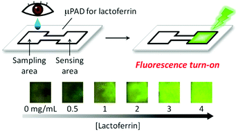
Take a look at our new HOT articles just published in Analyst and free for you for the next couple of weeks: Graphical abstract: An antibody-free microfluidic paper-based analytical device for the determination of tear fluid lactoferrin by fluorescence sensitization of Tb3+

An antibody-free microfluidic paper-based analytical device for the determination of tear fluid lactoferrin by fluorescence sensitization of Tb3+
Kentaro Yamada, Shunsuke Takaki, Nobutoshi Komuro, Koji Suzuki and Daniel Citterio
Analyst, 2014, Advance Article
DOI: 10.1039/C3AN01926H, Paper

Protein binding for detection of small changes on a nanoparticle surface
Shang Zeng, Yu-ming M. Huang, Chia-en A. Chang and Wenwan Zhong
Analyst, 2014,139, 1364-1371
DOI: 10.1039/C3AN02155F, Paper

Proteomic evaluation of citrate-coated silver nanoparticles toxicity in Daphnia magna
Louis-Charles Rainville, Darragh Carolan, Ana Coelho Varela, Hugh Doyle and David Sheehan
Analyst, 2014, Advance Article
DOI: 10.1039/C3AN02160B, Paper

Oxidized polyethylene films for orienting polar molecules for linear dichroism spectroscopy
Kasra Razmkhah, Nikola Paul Chmel, Matthew I. Gibson and Alison Rodger
Analyst, 2014,139, 1372-1382
DOI: 10.1039/C3AN02322B, Paper Graphical abstract: Making a big thing of a small cell – recent advances in single cell analysis

Solid-phase extraction and purification of membrane proteins using a UV-modified PMMA microfluidic bioaffinity μSPE device
Katrina N. Battle, Joshua M. Jackson, Małgorzata A. Witek, Mateusz L. Hupert, Sally A. Hunsucker, Paul M. Armistead and Steven A. Soper
Analyst, 2014,139, 1355-1363
DOI: 10.1039/C3AN02400H, Paper

Non-invasive mouthguard biosensor for continuous salivary monitoring of metabolites
Jayoung Kim, Gabriela Valdés-Ramírez, Amay J. Bandodkar, Wenzhao Jia, Alexandra G. Martinez, Julian Ramírez, Patrick Mercier and Joseph Wang
Analyst, 2014, Advance Article
DOI: 10.1039/C3AN02359A, Paper

Making a big thing of a small cell – recent advances in single cell analysis
Kerstin Galler, Katharina Bräutigam, Christina Große, Jürgen Popp and Ute Neugebauer
Analyst, 2014,139, 1237-1273
DOI: 10.1039/C3AN01939J, Critical Review

Advances in nanowire transistors for biological analysis and cellular investigation
Bor-Ran Li, Chiao-Chen Chen, U. Rajesh Kumar and Yit-Tsong Chen
Analyst, 2014, Advance Article
DOI: 10.1039/C3AN01861J, Critical Review

High resolution magic angle spinning NMR as a tool for unveiling the molecular enantiorecognition of omeprazole by amylose-based chiral phase
Juliana Cristina Barreiro, Tiago de Campos Lourenço, Lorena Mara A. Silva, Tiago Venâncio and Quezia Bezerra Cass
Analyst, 2014,139, 1350-1354
DOI: 10.1039/C3AN02029K, Paper Graphical abstract: Intra-molecular reactions as a new approach to investigate bio-radical reactivity: a case study of cysteine sulfinyl radicals

Intra-molecular reactions as a new approach to investigate bio-radical reactivity: a case study of cysteine sulfinyl radicals
Kirt L. Durand, Xiaoxiao Ma and Yu Xia
Analyst, 2014,139, 1327-1330
DOI: 10.1039/C3AN02307A, Communication

Digg This
Reddit This
Stumble Now!
Share on Facebook
Bookmark this on Delicious
Share on LinkedIn
Bookmark this on Technorati
Post on Twitter
Google Buzz (aka. Google Reader)

Top ten most accessed Analyst articles from October to December 2013

Here are the top ten most accessed Analyst articles for October, November and December 2013.

Imaging live cells grown on a three dimensional collagen matrix using Raman microspectroscopy
F. Bonnier, P. Knief, B. Lim, A. D. Meade, J. Dorney, K. Bhattacharya, F. M. Lyng and H. J. Byrne
Analyst, 2010,135, 3169-3177
DOI: 10.1039/C0AN00539H, Paper

Detection of calcium phosphate crystals in the joint fluid of patients with osteoarthritis – analytical approaches and challenges
Alexander Yavorskyy, Aaron Hernandez-Santana, Geraldine McCarthy and Gillian McMahon
Analyst, 2008,133, 302-318
DOI: 10.1039/B716791A, Critical Review

Gold-coated polycarbonate membrane filter for pathogen concentration and SERS-based detection
Krista Rule Wigginton and Peter John Vikesland
Analyst, 2010,135, 1320-1326
DOI: 10.1039/B919270K, Paper

Capillary zone electrophoresis-multiple reaction monitoring from 100 pg of RAW 264.7 cell lysate digest
Liangliang Sun, Yihan Li, Matthew M. Champion, Guijie Zhu, Roza Wojcik and Norman J. Dovichi
Analyst, 2013,138, 3181-3188
DOI: 10.1039/C3AN00287J, Paper

Upconversion nanoparticles in biological labeling, imaging, and therapy
Feng Wang, Debapriya Banerjee, Yongsheng Liu, Xueyuan Chen and Xiaogang Liu
Analyst, 2010,135, 1839-1854
DOI: 10.1039/C0AN00144A, Critical Review

Single-channel electrophysiology of cell-free expressed ion channels by direct incorporation in lipid bilayers
Mark S. Friddin, Natalie P. Smithers, Maïwenn Beaugrand, Isabelle Marcotte, Philip T. F. Williamson, Hywel Morgan and Maurits R. R. de Planque
Analyst, 2013,138, 7294-7298
DOI: 10.1039/C3AN01540H, Communication

A non enzymatic glucose biosensor based on an ultrasensitive calix[4]arene functionalized boronic acid gold nanoprobe for sensing in human blood serum
Alok Pandya, Pinkesh G. Sutariya and Shobhana K. Menon
Analyst, 2013,138, 2483-2490
DOI: 10.1039/C3AN36833E, Paper

Mid-infrared spectroscopy for gases and liquids based on quantum cascade technologies
Pierre Jouy, Markus Mangold, Béla Tuzson, Lukas Emmenegger, Yu-Chi Chang, Lubos Hvozdara, Hans Peter Herzig, Philip Wägli, Alexandra Homsy, Nico F. de Rooij, Alexander Wirthmueller, Daniel Hofstetter, Herbert Looser and Jérôme Faist
Analyst, 2014, Advance Article
DOI: 10.1039/C3AN01462B, Paper

A pyrene-based simple but highly selective fluorescence sensor for Cu2+ ions via a static excimer mechanism
Soma Sarkar, Swapnadip Roy, Anindita Sikdar, R. N. Saha and Sujit S. Panja
Analyst, 2013,138, 7119-7126
DOI: 10.1039/C3AN00928A, Paper

A novel 2,6-diformyl-4-methylphenol based chemosensor for Zn(II) ions by ratiometric displacement of Cd(II) ions and its application for cell imaging on human melanoma cancer cells
Atanu Jana, Pradip K. Sukul, Sushil K. Mandal, Saugata Konar, Sangita Ray, Kinsuk Das, James A. Golen, Arnold L. Rheingold, Sudipa Mondal, Tapan K. Mondal, Anisur R. Khuda-Bukhsh and Susanta K. Kar
Analyst, 2014,139, 495-504
DOI: 10.1039/C3AN01750H, Paper

We hope you enjoyed reading these articles, and if you have any thoughts or comments, please leave these in the comment box below.

Fancy submitting an article to Analyst? Why not submit to us here today or alternatively email us your suggestions!

Digg This
Reddit This
Stumble Now!
Share on Facebook
Bookmark this on Delicious
Share on LinkedIn
Bookmark this on Technorati
Post on Twitter
Google Buzz (aka. Google Reader)

The Ongoing Hunt for Deadly Microbes – A New Lateral Flow Assay for Pathogen Detection

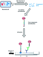
Early and accurate monitoring of pathogens such as Escherichia coli is critical for food and water safety, clinical diagnosis, and biosecurity. However there are a number of limitations to current detection methods such as cell culturing, and immunoassays. These drawbacks include non-specificity, insensitivity, as well as the requirement of long analysis time, intensive labor, and extensive sample purification. To address some of these limitations, Christopher Pöhlmann and his colleagues from the University of Bayreuth in Germany have developed a lateral flow device based on a sandwich assay design using gold nanoparticle-oligonucleotides to detect ribosomal RNA of E. coli. The new device is able to provide specific detection of E. coli within 25 minutes and requires no signal amplification step.

A Lateral Flow Assay for Detection of E. coli Ribosomal RNA

A Lateral Flow Assay for Detection of E. coli Ribosomal RNA

Find out more about this discovery by reading the full paper below, which is free to download until March 7th:

A lateral flow assay for identification of Escherichia coli by ribosomal RNA hybridization
Christopher Pöhlmann, Irina Dieser and Mathias Sprinzl
Analyst, 2014, 139, 1063-1071
DOI: 10.1039/C3AN02059B

Let us know what your thoughts are by leaving a comment!

Digg This
Reddit This
Stumble Now!
Share on Facebook
Bookmark this on Delicious
Share on LinkedIn
Bookmark this on Technorati
Post on Twitter
Google Buzz (aka. Google Reader)