Author Archive

Nanoparticles for platelet adhesion and aggregation

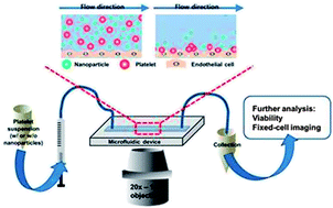
On-chip evaluation of platelet adhesion and aggregation

The use of nanoparticles in diagnostic and therapeutic roles requires that we understand their interactions and behaviour within complex biological systems. Where clinical applications would involve intravenous injection of nanoparticle-based therapeutics, it will be necessary to understand the interaction between nanoparticles and native blood components.

To address this issue, Christy Haynes and coworkers from the University of Minnesota, USA, have created a microfluidic device coated with endothelial cells to mimic the walls of the vascular system. This initial in vitro approach was carried out with both activated and unactivated platelets in the presence of various (therapeutically-relevant) concentrations of fluorescently-labelled, mesophorous silica nanoparticles. The impact of nanoparticles on the critical platelet functions of adhesion and aggregation was assessed.

Microfluidic platforms are readily customised and future work would be expected to expand upon these initial conditions to advance our understanding of nanotoxicology and interaction studies.

To read more about this study and the impact of nanoparticles on platelet interactions you can access this Analyst HOT Article free until 6 January 2014:

On-chip evaluation of platelet adhesion and aggregation upon exposure to mesoporous silica nanoparticles
Donghyuk Kim, Solaire Finkenstaedt-Quinn, Katie R. Hurley, Joseph T. Buchman and Christy L. Haynes 
Analyst, 2014, Advance Article
DOI: 10.1039/C3AN01679J

Digg This
Reddit This
Stumble Now!
Share on Facebook
Bookmark this on Delicious
Share on LinkedIn
Bookmark this on Technorati
Post on Twitter
Google Buzz (aka. Google Reader)

Analysis of thyroid tissues by FT-IR microscopectroscopy


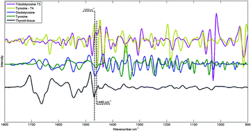
FTIR spectra of hormones triiodothyronine, thyroxine, diiodotyrosine, tyrosine and thyroid tissue

The combination of vibrational spectroscopy with mapping and imaging techniques to address important biochemical questions is an area of active and expanding research.  By subjecting adjacent tissue sections to standard histopathological screening and spectroscopic imaging, a greater understanding of the biochemical processes underlying tissue form and function can be achieved.

Researchers from Northeastern University, USA, and the Institute of Energy and Nuclear Research (IPEN), Sao Paolo, have used Fourier transform infrared (FTIR) microspectroscopy to examine healthy thyroid tissues. FTIR allows spatially resolved images, or maps, to be obtained by use of focal plane array detectors and spectral processing of the data then reveals chemical images.  In this HOT Analyst paper, Denise Zezell and coworkers present an example from their investigation of 80 different patient samples, which were analysed by transflection-mode FTIR mapping.  The approach was  applied to the study of healthy thyroid tissue and, with reference to thyroid specific hormones, iodination state. 

To know more about this research, click on the links below. This paper will be free to read for the next three weeks:

The characterization of normal thyroid tissue by micro-FTIR spectroscopy
Thiago M. Pereira, Denise M. Zezell, Benjamin Bird, Milos Miljković and Max Diem
Analyst, 2013,138, 7094-7100
DOI: 10.1039/C3AN00296A

Digg This
Reddit This
Stumble Now!
Share on Facebook
Bookmark this on Delicious
Share on LinkedIn
Bookmark this on Technorati
Post on Twitter
Google Buzz (aka. Google Reader)

Intracellular enzymatic activity by SERRS

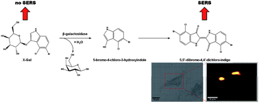
Analysis of intracellular enzyme activity by SERS

Enzymatic activity in cells has been revealed by surface enhanced Raman scattering (SERRS) spectroscopic studies, according to new research led by Duncan Graham at the University of Strathclyde.

X-Gal, a SERRS inactive precursor and a common marker for galactosidase enzymes, was introduced to macrophage cells along with metal nanoparticles. On incubation, enzymatic activity of galactosidase on X-Gal produces a SERRS active trans-alkene dimerised product. This product adsorbs onto the delivered citrate capped gold nanoparticles producing a distinctive SERRS signal. Using SERRS mapping to evaluate the formation of this product, enzyme activity was assessed, not only across a cell population, but also at sub-cellular levels. The authors note that the SERRS activity in this study may not reflect the only sites of galactosidase activity, but rather the sites where both galactosidase and the nanoparticles are co-located. Nevertheless, approaches towards sub-cellular analysis of enzyme activity are important.

Advances in targeted nanoparticle uptake combined with the multiplexing capability of SERRS make this an interesting approach to sub-cellular studies of biochemical activity.

To read the full article, please click on the link below. This paper will be free to read until October 18th.

Analysis of intracellular enzyme activity by surface enhanced Raman scattering
Ross Stevenson, Sarah McAughtrie, Laura Senior, Robert J. Stokes, Helen McGachy, Laurence Tetley, Paola Nativo, James M. Brewer, James Alexander, Karen Faulds and   Duncan Graham
Analyst, 2013, Advance Article
DOI: 10.1039/C3AN00729D

Digg This
Reddit This
Stumble Now!
Share on Facebook
Bookmark this on Delicious
Share on LinkedIn
Bookmark this on Technorati
Post on Twitter
Google Buzz (aka. Google Reader)

An new aptasensing platform for detection of multiple analytes

Detecting multiple target species simultaneously via a single detection mechanism remains an attractive goal for biologically relevant studies. 

Ruo Yuan and Jingdong Peng from the College of Chemistry and Chemical Engineering, Southwest University, China, have successfully developed a multiplexed electrochemical sensor for two biological targets.

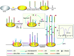
Multiplexed electrochemical sensor for two biological targets

In the study, gold nanoparticles were electrochemically deposted on a highly polished glass carbon electrode and functionalised by thrombin- and ochratoxin A-binding apatmers. In the presence of the targets, an exonuclease digestion occurred allowing the target molecules to be recycled. The single stranded product of the exonuclease digestions were then used as initiation sequences for concatamer reaction designed to introduce two distinct electrochemically active species in a sequence specific manner. Only by the initial presence of either target would the related concatamer and, by extention, the electroactive species be present.

By this elaborate system of aptamer binding, exonuclease digestion, concatamer reaction and electrochemical detection the researchers were able to simultaneously detect thrombin, a blood-clotting protein, over the range 0.1 pM – 40 nM and ochratoxin A, a nephrotic toxin, over the range 0.4 pM – 35 nM. 

To read the full article, please click on the link below:

An aptasensing platform for simultaneous detection of multiple analytes based on the amplification of exonuclease-catalyzed target recycling and DNA concatemers
Liping Jiang, Jingdong Peng, Ruo Yuan, Yaqin Chai, Yali Yuan, Lijuan Bai and   Yan Wang
Analyst, 2013, Advance Article
DOI: 10.1039/C3AN00757J

Digg This
Reddit This
Stumble Now!
Share on Facebook
Bookmark this on Delicious
Share on LinkedIn
Bookmark this on Technorati
Post on Twitter
Google Buzz (aka. Google Reader)

FTIR spectral signature of anticancer drug effects on cancer cells: any influence of the cell cycle?

Visible image, infrared image, propidium iodide fluorescence image of a cell smear

Erik Goormaghtigh and co-workers from the Centre for Structural Biology and Bioinformatics at the Unversité Libre de Bruxelles have investigated the use of Fourier transformfo infrared (FTIR) spectroscopy r studying the effect of an anti-cancer drug, paclitaxel, on a human prostate cancer cell line.

The researchers combined fluorescence spectroscopy with FTIR spectroscopy using a fluorescent DNA intercalator to identify cell cycle stage.  By doing so, they sought to understand the drug’s effect on cell cycle and/or metabolic perturbation. The authors discuss both the application and limitation of this approach to cell studies.

Read more about this study in this HOT Analyst paper, free for you until May 24th .

FTIR spectral signature of anticancer drug effects on PC-3 cancer cells: is there any influence of the cell cycle?
Allison Derenne, Alix Mignolet and   Erik Goormaghtigh  
Analyst, 2013, Advance Article
DOI: 10.1039/C3AN00225J

Digg This
Reddit This
Stumble Now!
Share on Facebook
Bookmark this on Delicious
Share on LinkedIn
Bookmark this on Technorati
Post on Twitter
Google Buzz (aka. Google Reader)

A point-of-care testing system for cardiac troponin

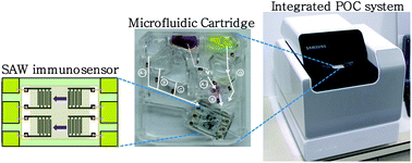
Detection of cardiac troponin with a surface acustive wave immunosensor

Youn-Suk Choi and Soo Suk Lee from the Samsung Advanced Institute of Technology, Republic of Korea, have developed a point of care diagnostic test for cardiac troponin I (cTnI).

The researchers fabricated a bubble-free microfluidics device based on a gold-nanoparticle immunoassay and a surface acoustic wave immunosensor. The authors highlight a limit of detection of  6.7 pg mL-1 as a clinically relevant result and a first for centrifugally-based microfluidic devices. In this work, the researchers developed the device for the detection of cTnI, a biomarker for acute myocardial infarction for which rapid analysis would be beneficial for patient prognosis. The group is now extending studies to alternative systems.

To read more about the current results, access this Analyst HOT article clicking on the link below. This paper will be free to read for the next 10 days.

A centrifugally actuated point-of-care testing system for the surface acoustic wave immunosensing of cardiac troponin I
Woochang Lee ,  Jaeyeon Jung ,  Young Ki Hahn ,  Sang Kyu Kim ,  Yeolho Lee ,  Joonhyung Lee ,  Tae-Han Lee ,  Jin-Young Park ,  Hyejung Seo ,  Jung Nam Lee ,  Jin Ho Oh ,  Youn-Suk Choi and Soo Suk Lee
Analyst, 2013,138, 2558-2566
DOI: 10.1039/C3AN00182B

Digg This
Reddit This
Stumble Now!
Share on Facebook
Bookmark this on Delicious
Share on LinkedIn
Bookmark this on Technorati
Post on Twitter
Google Buzz (aka. Google Reader)

Analysis of single particle photodegradation using photothermal infrared microspectroscopy

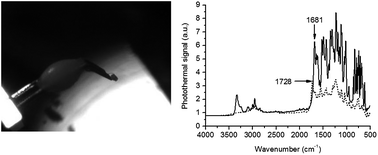
Photomicrograph of a nifedipine particle and photermal spectra

Duncan Craig from the University College London and collaborators from the Universities of Cambridge and East Anglia have applied Photothermal Infrared Microscopy (PTMS) to study drug degradation.

A combination of Fourier Transform Infrared Spectroscopy (FT-IR) and a modification of atomic force microscopy, PTMS, was used to pick up microparticles of a drug known for its photo-instability and analyse its degradation by changes in the FT-IR spectrum after exposure to light.  In this HOT new Analyst paper the researchers not only show the application of this new technique but discuss some limitations and areas for future development.

To know more about the study, please access the link below. This paper will be free to read until April 4th.

Analysis of single particle photodegradation using photothermal infrared microspectroscopy
Jonathan G. Moffat ,  Mark D. Eddleston ,  Peter S. Belton ,  William Jones and Duncan Q. M. Craig
Analyst, 2013, Advance Article
DOI: 10.1039/C3AN36686C

Digg This
Reddit This
Stumble Now!
Share on Facebook
Bookmark this on Delicious
Share on LinkedIn
Bookmark this on Technorati
Post on Twitter
Google Buzz (aka. Google Reader)

A new switchable sensor for phosphate in water

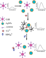
Schematic interaction between Ce3+ and phosphate

Guang-Li Wang and co-workers, from the Jiangnan University, China, have developed an assay to detect the level of phosphate in water. 

Ce3+ ions are used to aggregate citrate-capped silver nanoparticles with cysteine-capped quantum dots, thereby causing efficient quenching of the fluorescence signal.  However, when phosphate is introduced to the system it interacts with the Ce3+ ions causing the quenched aggregates to redisperse and switching the fluorescent signal from “off” to “on”.  This work has potential applications for the environmental analysis of groundwater.

To read more about this work, please access this Analyst HOT article below. It will be free to read until March 8th.

Novel switchable sensor for phosphate based on the distance-dependant fluorescence coupling of cysteine-capped cadmium sulfide quantum dots and silver nanoparticles
Guang-Li Wang ,  Huan-Jun Jiao ,  Xiao-Ying Zhu ,  Yu-Ming Dong and Zai-Jun Li
Analyst, 2013, Advance Article
DOI: 10.1039/C3AN36878E

Digg This
Reddit This
Stumble Now!
Share on Facebook
Bookmark this on Delicious
Share on LinkedIn
Bookmark this on Technorati
Post on Twitter
Google Buzz (aka. Google Reader)

Efficient detection of glutathione and cysteine in human serum

Ruqin Yu and colleagues from the University of Hunan, China, have developed a label-free fluorescent detection system for glutathione (GSH) and cysteine (Cys). 

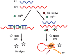
Hg2+ mediated fluorescent sensing strategy for detection of GSH and Cys

Three key interactions are necessary for the development of this assay: two single stranded DNA with thymine-thymine (T•T) mismatches stabilised in the presence of Hg2+, GSH and Cys interaction with Hg2+ with much higher binding affinity than T•T mismatches, and interaction of N-methyl porphyrin IX (MMP) with G-quadruplex structures, which leads to increased fluorescence.
The researchers  judiciously designed two sequences of DNA such that inter-molecular T•T mismatches were stabilised in the presence of Hg2+, inhibiting one of the stands from forming a G-quadruplex.  When the Hg2+ interacts with GSH or Cys, an intramolecular G-quadruplex is formed.  The G-quadruplex interacts with the NMM producing a marked increase in fluorescence.  Contrarily, in the absence of GSH and Cys, the Hg2+ is available to stabilise the mismatched duplex, the G-quadruplex is not formed, no NMM binding occurs and no fluorescent increase is observed.

This system was successfully used to detect GSH or Cys from protein extracted from human serum samples. To read more about this work, please access the full article below. It will be free to read until February 28th.

A Hg2+-mediated label-free fluorescent sensing strategy based on G-quadruplex formation for selective detection of glutathione and cysteine
Jingjin Zhao ,  Chunfei Chen ,  Liangliang Zhang ,  Jianhui Jiang ,  Guoli Shen and Ruqin Yu
Analyst, 2013, Advance Article
DOI: 10.1039/C3AN36657J

Digg This
Reddit This
Stumble Now!
Share on Facebook
Bookmark this on Delicious
Share on LinkedIn
Bookmark this on Technorati
Post on Twitter
Google Buzz (aka. Google Reader)

AFM and Raman to understand cancer cell behaviour

In 2004 the World Health Organisation estimated that over half a million women died from breast cancer.  A great deal of research is now conducted to improve the diagnostics and prognosis of cancers.  One avenue of research is to improve our understanding of cancers at a cellular level. Anhong Zhou and coworkers from Utah State University have recently used atomic force microscopy (AFM) and Raman spectroscopy to attempt just that.

McEwen et al., Analyst 2013

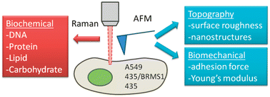
Combined Raman spectroscopy and AFM to detect differences in cancer cells

Their preliminary study, in this new Analyst HOT paper, examines the use of atomic force microscopy (AFM) and Raman spectroscopy to study a breast cancer cell line and the effect of the presence or absence of a metastasis suppressor gene on cell behaviour.  They have also compared various cancer cell lines to examine the differences in behaviour at the cellular level between cancer types. The authors provide a comprehensive analysis of both the practical and data processing techniques required to differentiate the cell types.  The use of both AFM and Raman reveals information about the biochemical and biomechanical attributes of the cell lines and is an approach that could increase our understanding of cancer cell behaviour and tumour development.

Subcellular spectroscopic markers, topography and nanomechanics of human lung cancer and breast cancer cells examined by combined confocal Raman microspectroscopy and atomic force microscopy
Gerald D. McEwen, Yangzhe Wu, Mingjie Tang, Xiaojun Qi, Zhongmiao Xiao, Sherry M. Baker, Tian Yu, Timothy A. Gilbertson, Daryll B. DeWald and Anhong Zhou
Analyst, 2013, Advance Article
DOI: 10.1039/C2AN36359C

This article will be free to read for the next two weeks.

Digg This
Reddit This
Stumble Now!
Share on Facebook
Bookmark this on Delicious
Share on LinkedIn
Bookmark this on Technorati
Post on Twitter
Google Buzz (aka. Google Reader)