"Bringing optical histopathology into the clinic would have a huge impact for surgeons, pathologists and patients," says Dr Bevin Lin
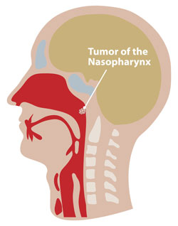
Detecting early-stage nasopharyngeal carcinomas via a non-invasive technique which could improve the survival rates of patients has been reported by scientists from Singapore. Diagnosing nasopharyngeal carcinomas before they have become life-threatening is very difficult, with five-year survival rates only around 34 per cent.
A team led by Bevin Lin, from the National University of Singapore, has developed a technique that uses a bifurcated fibre optic probe to collect nasopharyngeal tissue data. By combining ultraviolet auto fluorescence excitation-emission matrix (EEM) spectroscopy and parallel factor analysis calculations, they can then diagnose early-stage carcinomas.
To find out more, including comments from Dr Lin, read Jennifer Newton‘s article in Chemistry World, and access the paper for free using the link below.
Diagnosis of early stage nasopharyngeal carcinoma using ultraviolet autofluorescence excitation–emission matrix spectroscopy and parallel factor analysis
Bevin Lin, Mads Sylvest Bergholt, David P. Lau and Zhiwei Huang
Analyst, 2011
DOI: 10.1039/c1an15525c


