Author Archive

Electrochemical approach to distinguish bovine DNA

Can this technique answer the need for a method to acurately identify adultered food products?

This paper presents an electrochemical way of discriminating between the DNA of different bovine species. It targets the mitochondrial Cox-1 gene which is considered to be a standard ‘barcode sequence’, having low variation within species but high degrees of variation between taxa.  The benefit of targeting mitochondrial DNA is that each cell will have hundreds to thousands of copies, facilitating the use of very small samples.

Electrochemical impedance spectroscopy is central to the method and was used to probe for charge resistance transfer differences due to mismatched DNA. Zinc ions were added when DNA hybrids (matched or mismatched) formed on the gold electrode surface, expediting charge transfer to the solution phase redox probe. The zinc ions effectively adjust the charge transfer in situations where complementary sequences exist.  It is therefore possible to use charge resistance transfer differences to tell if the correct DNA had been captured. The authors also carried out a dehybridization study to show if the sensor could be reused.

Find out more by reading the article for free until 4th October.

Electrochemical identification of artificial oligonucleotides related to bovine species. Potential for identification of species based on mismatches in the mitochondrial cytochrome C1 oxidase gene
Mohtashim Hassan Shamsi and Heinz-Bernhard Kraatz
Analyst
DOI: 10.1039/C1AN15414A

Recap on some related papers below…

The effects of oligonucleotide overhangs on the surface hybridization in DNA films: an impedance study
Mohtashim Hassan Shamsi and Heinz-Bernhard Kraatz
Analyst, 2011, 136, 3107-3112
DOI: 10.1039/C1AN15253J

Probing nucleobase mismatch variations by electrochemical techniques: exploring the effects of position and nature of the single-nucleotide mismatch
Mohtashim H. Shamsi and Heinz-Bernhard Kraatz
Analyst, 2010, 135, 2280-2285
DOI: 10.1039/C0AN00184H

Enzymatically modified peptide surfaces: towards general electrochemical sensor platform for protein kinase catalyzed phosphorylations

Sanela Martic, Mahmoud Labib and Heinz-Bernhard Kraatz
Analyst, 2011, 136, 107-112
DOI: 10.1039/C0AN00438C

Digg This
Reddit This
Stumble Now!
Share on Facebook
Bookmark this on Delicious
Share on LinkedIn
Bookmark this on Technorati
Post on Twitter
Google Buzz (aka. Google Reader)

Thermochemiluminescence patterns act as fingerprints to accurately classify proteins

Albumin solutions were added directly onto each sensing element. When heated, the albumins are thermally oxidised, generating distinct thermochemiluminescence patterns.

We’ve just published the latest paper from Analyst‘s Associate Editor for Asia, Xinrong Zhang, and colleagues from Tsinghua Universtiy, China.  It details an innovative nanomaterial array which uses chacteristic thermochemiluminescence signals to recognise protein subtypes and denatured shapes.

Read the paper for free until 8th September.

A thermochemiluminescence array for recognition of protein subtypes and their denatured shapes
Hao Kong, He Wang, Sichun Zhang and Xinrong Zhang
Analyst
DOI: 10.1039/C1AN15382J

Recap on Xinrong Zhang’s previous paper on an aerosol chemiluminescence-based sensor which functions as a mammalian tongue….

Recognition of organic compounds in aqueous solutions by chemiluminescence on an array of catalytic nanoparticles

Hao Kong, Sichun Zhang, Na Na, Da Liu and Xinrong Zhang
Analyst, 2009, 134, 2441-2446
DOI: 10.1039/B917538E

Digg This
Reddit This
Stumble Now!
Share on Facebook
Bookmark this on Delicious
Share on LinkedIn
Bookmark this on Technorati
Post on Twitter
Google Buzz (aka. Google Reader)

Grand challenges: Surface enhanced optical spectroscopies for bioanalysis

Surface enhancement can provide improved detection sensitivity in a range of optical spectroscopies.

Read this comprehensive review on surface enhanced optical spectroscopies and their application to bioanalysis by Analyst Editorial Board member Duncan Graham and his colleague Iain Larmour.  They consider the “grand challenges” that need to be overcome before widespread clinical use of surface enhanced techniques can be achieved.

The review will be free until the 6th September.

Surface enhanced optical spectroscopies for bioanalysis

Iain A. Larmour and Duncan Graham
Analyst
DOI: 10.1039/C1AN15452D

This paper is part of an ongoing collection of articles looking at Grand Challenges in analytical science.  In celebration of the International Year of Chemistry, leading scientists (including our own Board members) have put together papers outlining the current challenges faced in analytical science, and how these might be tackled, some of which were published in issue 15.

Below are a selection of recent Analyst papers mentioned in the review:

Evaluation of tip-enhanced Raman spectroscopy for characterizing different virus strains
Peter Hermann, Antje Hermelink, Veronika Lausch, Gudrun Holland, Lars Möller, Norbert Bannert and Dieter Naumann
Analyst, 2011, 136, 1148-1152
DOI: 10.1039/C0AN00531B

Surface enhanced Raman evidence for Ag+ complexes of adenine, deoxyadenosine and 5′-dAMP formed in silver colloids
Evanthia Papadopoulou and Steven E. J. Bell
Analyst, 2010, 135, 3034-3037
DOI: 10.1039/C0AN00612B

Competitive surface-enhanced Raman scattering assay for the 1,25-dihydroxy metabolite of vitamin D3
Eric J. Dufek, Brian Ehlert, Michael C. Granger, Tanya M. Sandrock, Samuel L. Legge, Mark G. Herrmann, A. Wayne Meikle and Marc D. Porter
Analyst, 2010, 135, 2811-2817
DOI: 10.1039/C0AN00354A

Short-wave infrared excited SERS
Kirstin A. Lynn, Graeme McNay, David A. Eustace, Neil C. Shand and W. Ewen Smith
Analyst, 2010, 135, 1904-1905
DOI: 10.1039/C0AN00096E

Digg This
Reddit This
Stumble Now!
Share on Facebook
Bookmark this on Delicious
Share on LinkedIn
Bookmark this on Technorati
Post on Twitter
Google Buzz (aka. Google Reader)

Ricin biosensor outperforms current methods to detect food contamination

Ricin, a toxin derived from the castor bean plany may be easily amendable as a deliberate food biocontaminate

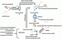
This study describes the characterisation and selection of an aptamer against the toxin, ricin B.  The aptamer functioned well in liquid food matrices and was tested against a commercially available ELISA kit.

Read the paper for free until 30th August to learn more…

A single DNA aptamer functions as a biosensor for ricin
Elise A. Lamont, Lili He, Keith Warriner, Theodore P. Labuza and Srinand Sreevatsan
Analyst
DOI: 10.1039/C1AN15352H

You might also be interested in these papers:

Aptamer-based surface-enhanced Raman scattering detection of ricin in liquid foods
Lili He, Elise Lamont, Belamaranahally Veeregowda, Srinand Sreevatsan, Christy L. Haynes, Francisco Diez-Gonzalez and Theodore P. Labuza
Chem. Sci., 2011, 2, 1579-1582
DOI: 10.1039/C1SC00201E

Simultaneous quantification of five bacterial and plant toxins from complex matrices using a multiplexed fluorescent magnetic suspension assay
Diana Pauly, Sebastian Kirchner, Britta Stoermann, Tanja Schreiber, Stefan Kaulfuss, Rüdiger Schade, Reto Zbinden, Marc-André Avondet, Martin B. Dorner and Brigitte G. Dorner
Analyst, 2009, 134, 2028-2039
DOI: 10.1039/B911525K

Digg This
Reddit This
Stumble Now!
Share on Facebook
Bookmark this on Delicious
Share on LinkedIn
Bookmark this on Technorati
Post on Twitter
Google Buzz (aka. Google Reader)

DESI and PADI ambient mass spectrometry analysis of anti-aging creams on model skin surfaces

DESI and PADI are useful when determining the siloxane contents of anti-aging creams.

Tara Salter and colleagues present a preliminary study for a non-invasive in vivo investigation of personal care products on fixed fibroblast cells. They carried our mass spectrometry analyses of 13 molecules commonly used in personal care products, including organosiloxanes, with both PADI and DESI to determine the different sensitivities of the techniques.

Read the paper for free until 16th August.

Analysis of personal care products on model skin surfaces using DESI and PADI ambient mass spectrometry
Tara L. Salter, Felicia M. Green, Nilofar Faruqui and Ian S. Gilmore
Analyst
DOI: 10.1039/C1AN15138J

On a similar note, why not check out the themed issue on ambient mass spectrometry that we published last year, in particular this paper from Graham Cooks and colleagues

Desorption electrospray ionization and other ambient ionization methods: current progress and preview

Demian R. Ifa, Chunping Wu, Zheng Ouyang and R. Graham Cooks
Analyst, 2010, 135, 669-681
DOI: 10.1039/B925257F

Digg This
Reddit This
Stumble Now!
Share on Facebook
Bookmark this on Delicious
Share on LinkedIn
Bookmark this on Technorati
Post on Twitter
Google Buzz (aka. Google Reader)

High miscibility of water in the hydrophobic PEDOT:PSS layer prevents separation of undesirable water layer

On exposure to water, the "soaking" action of the polymer is analogous to the behaviour of a sponge.

On exposure to water, the "soaking" action of the polymer is analogous to the behaviour of a sponge.

This paper explains why the conducting polymer poly(3,4-ethylenedioxythiophene):poly(styrene sulfonate), regularly used for the preparation of solid-contact ion-selective electrodes, does not show evidence for a water layer.  One of the referees commented that ‘this manuscript clarifies an issue that has been puzzling me for a few years now.’  These findings will aid those working in the chemical sensors community by helping to understand the physical state of the polymer layers in solid-contact ion-selective electrodes when they are exposed to solutions.

Read the paper for free until 17th August.

Water uptake in the hydrophilic poly(3,4-ethylenedioxythiophene):poly(styrene sulfonate) solid-contact of all-solid-state polymeric ion-selective electrodes
Jean-Pierre Veder, Roland De Marco, Graeme Clarke, San Ping Jiang, Kathryn Prince, Ernö Pretsch and Eric Bakker
Analyst
DOI: 10.1039/C1AN15267J

You might also find this Analyst paper interesting….

Tuning the gas sensing performance of single PEDOT nanowire devices

Carlos M. Hangarter, Sandra C. Hernandez, Xueing He, Nicha Chartuprayoon, Yong Ho Choa and Nosang V. Myung
Analyst, 2011, 136, 2350-2358
DOI: 10.1039/C0AN01000f

Digg This
Reddit This
Stumble Now!
Share on Facebook
Bookmark this on Delicious
Share on LinkedIn
Bookmark this on Technorati
Post on Twitter
Google Buzz (aka. Google Reader)

Proteotyping appraoch for monitoring evolution of bird flu

This study could help predict future bird flu outbreaks.

This study could help predict future bird flu outbreaks.

In this paper, a team from Sydney, combine bioinformatics and high resolution mass spectrometry to identify the strain, time period and region of avian flu hemagglutinin.

Read the paper for free until 15th August.

Evolution of H5N1 influenza virus through proteotyping of hemagglutinin with high resolution mass spectrometry
Ji-Won Ha and Kevin M. Downard
Analyst
DOI: 10.1039/C1AN15354D

Digg This
Reddit This
Stumble Now!
Share on Facebook
Bookmark this on Delicious
Share on LinkedIn
Bookmark this on Technorati
Post on Twitter
Google Buzz (aka. Google Reader)

Detecting chemical and physical changes in thick tissue with X-ray excited luminescent sensors

Scanning XEOL provides new capabilities for chemical analysis in tissue

Scanning XEOL provides new capabilities for chemical analysis in tissue

The benefits of non-invasive biomedical imaging techniques in studying disease are obvious but developing sensors which can measure chemical concentrations around and on implants is challenging.  This is because optical imaging through tissue results in light scattering and in turn, images with poor resolution. Jeffrey Anker and colleagues use a narrow scanning X-ray beam to excite luminescence from X-ray scintillators in this hybrid technique for high-resolution imaging of local optial absorption through tissue.  The scintillators serve as light sources with spectra and intensity that depend on local absorption.

Download the paper, which is free to access until 22nd July, to learn more…

Optical imaging in tissue with X-ray excited luminescent sensors
Hongyu Chen, David E. Longfield, Venkata S. Varahagiri, KhanhVan T. Nguyen, Amanda L. Patrick, Haijun Qian, Donald G. VanDerveer and Jeffrey N. Anker
Analyst
DOI: 10.1039/C0AN00931H

This paper will be included in a themed issue on Emerging Investigators, Guest Edited by Perdita Barran and Christy Haynes, to be published later this year.

Digg This
Reddit This
Stumble Now!
Share on Facebook
Bookmark this on Delicious
Share on LinkedIn
Bookmark this on Technorati
Post on Twitter
Google Buzz (aka. Google Reader)

Swimming with sensors

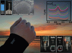
The sensor is worn on the wetsuit's sleeve and the potentiostat and battery are kept in a watertight compartment integrated into the wetsuit

Sensors printed onto the sleeves of wetsuits could alert the wearer to contaminated water. Navy divers could also use the sensors to locate underwater explosives, such as mines.

Joseph Wang at the University of California in San Diego, US, and colleagues have demonstrated that their sensors can be printed directly onto neoprene, a synthetic rubber commonly used in wet- and dry-suits.

Read Tamsin Phillips’ news piece in Chemistry World or access the full article for free using the link below.

Wearable electrochemical sensors for in situ analysis in marine environments
Kerstin Malzahn, Joshua Ray Windmiller, Gabriela Valdés-Ramírez, Michael J. Schöning and Joseph Wang
Analyst
DOI: 10.1039/C1AN15193B

Digg This
Reddit This
Stumble Now!
Share on Facebook
Bookmark this on Delicious
Share on LinkedIn
Bookmark this on Technorati
Post on Twitter
Google Buzz (aka. Google Reader)

“Canary in a coal mine chip”

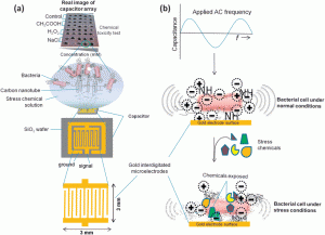
(a) Capacitor array biochip immobilized with viable E. coli and (b) response of E. coli and surface charge distribution under the applied AC electrical frequency.

Javed H. Niazi and colleagues from Sabanci University, Turkey, have developed a new capactitive biochip capable of detecting cellular stress caused by chemicals thanks to immobilised bacteria.

The biosensor works by using non-Faradaic electrochemical impedance spectroscopy to monitor changes in surface capacitance which occur when chemicals, such as acetic acid and hydrogen peroxide, disrupt the membrane function of the immobilised E. coli cells.

One referee described it as a “canary in a coal mine chip”.

The team still have some challenges to overcome but once properly developed, it could potentially be used as a cytotoxicity indicator.

It’s a must-read for anyone involved in drug, food and water safety research and will be free to access until 1st July.

Probing chemical induced cellular stress by non-Faradaic electrochemical impedance spectroscopy using an Escherichia coli capacitive biochip
Anjum Qureshi, Yasar Gurbuz and Javed H. Niazi
Analyst
DOI: 10.1039/C1AN15202E

Digg This
Reddit This
Stumble Now!
Share on Facebook
Bookmark this on Delicious
Share on LinkedIn
Bookmark this on Technorati
Post on Twitter
Google Buzz (aka. Google Reader)