Archive for November, 2013

Sulfated hyaluronic acid hydrogels for protein delivery

Hyaluronic acid (HA) is a biological polymer found ubiquitously in the extracellular matrix (ECM) of several tissues. Given the biocompatibility of HA, there is great interest in utilizing HA polymers to generate hydrogels for protein/drug delivery and tissue engineering applications.

The ability for a hydrogel to bind proteins (such as growth factors or cytokines) is an important design parameter to consider for protein-mediated tissue remodeling. For instance, incorporating sulfate groups in hydrogels increases the binding of proteins to a hydrogel through electrostatic interactions between sulfates and amino acids. Unfortunately, HA does not readily bind protein because the polymer does not have sulfate groups.

To address this limitation of HA, recent research in the Burdick Lab at the University of Pennsylvania focuses on designing HA polymers with sulfate groups to create robust hydrogels capable of protein binding and release. HA has an abundance of carboxylic acid groups along the polymer chain, making HA amenable to a variety of chemical modifications – including the addition of sulfate groups.

Researchers took advantage of carboxylic acid residues on HA to generate modified HA polymers with hydroxyethyl methacrylate (HEMA-HA). The methacrylate groups on HEMA-HA allow for free radical cross-linking of the polymers. The ester groups present in the cross-links also make the gel susceptible to hydrolytic degradation, which is an important feature for temporal release of bound protein from the hydrogel. Additionally, HEMA-HA polymers were allowed to react with SO3 to form sulfated HEMA-HA (HEMA-SHA). The resulting HEMA-HA and HEMA-SHA polymer products were characterized extensively with NMR and molecular weight/polydispersity measurements. 

The binding affinities of HEMA-HA and HEMA-SHA were characterized using protein-binding assays of stromal cell-derived factor-1α (SDF-1α). Interestingly, SDF-1α binding to sulfated HA was comparable to heparin controls, and virtually no SDF-1α was bound to non-sulfated HA.  The enhanced binding to HEMA-SHA is likely due to the increased negative charge from the sulfate groups on the HEMA-SHA (confirmed with zeta potential measurements).

After characterizing the binding efficiency of the polymers, HEMA-SHA and HEMA-HA were used to fabricate hydrogels. Using a 90%-10% blend of HEMA-HA and HEMA-SHA and a 100% solution HEMA-HA as a control, the polymer solutions were cross-linked into hydrogels using a redox initiator system for free radical cross-linking. Rheometry data show that the presence of sulfate groups in the hydrogel does not alter gel cross-linking kinetics and gel stiffness. Additionally, the sulfated HA hydrogel showed prolonged release of SDF-1α over time compared to the non-sulfated hydrogel control. These results show the ability to generate a robust sulfated HA hydrogel capable of controlled release of encapsulated protein.

This work describes a novel method to easily incorporate sulfate groups into HA hydrogels for enhanced binding and prolonged release of protein. The described technique is poised for use in future HA hydrogel designs for a wide array of drug delivery and tissue engineering applications. 

Incorporation of sulfated hyaluronic acid macromers into degradable hydrogel scaffolds for sustained molecule delivery
Brendan P. Purcell, Iris L. Kim, Vanessa Chuo, Theodore Guenin, Shauna M. Dorsey and Jason A. Burdick  
Biomater. Sci., 2014, Advance Article DOI: 10.1039/C3BM60227C

Brian Aguado is currently a Ph.D. Candidate and NSF Fellow in the Biomedical Engineering department at Northwestern University. He holds a B.S. degree in Biomechanical Engineering from Stanford University and a M.S. degree in Biomedical Engineering from Northwestern University. When he’s not in the lab, Brian enjoys traveling, cooking, swimming, and spending time with family and friends. Read more about Brian’s research publications here.

To keep up-to-date with all the latest research, sign-up to our RSS feed or Table of contents alert.

Digg This
Reddit This
Stumble Now!
Share on Facebook
Bookmark this on Delicious
Share on LinkedIn
Bookmark this on Technorati
Post on Twitter
Google Buzz (aka. Google Reader)

Patterned 3D cornea replacements

Hasirci and colleagues have developed a new type of engineered corneal construct using a combination of cells derived from the cornea as well as organized elastin-based biomaterials.  

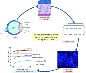
The stroma is a layer in our cornea that consists of highly organized layers of collagen fibers and keratocytes, a special type of corneal cell.  The cells and the collagen layers are aligned with one another and follow a specific pattern of organization.   This organized structure allows the cornea to be transparent and to have the mechanical strength necessary to function correctly.

This study used specific biomaterials that draw inspiration from nature.  Specifically, the researchers used elastin, a commonly found biomaterial, as a template, and created a biomaterial with a similar protein composition. They modified a traditional elastin-based sequence to include the amino acid chain YIGSR, which is known to help cells attach to biomaterial structures.  They blended their elastin-based biomaterial with collagen, and used a silicon template to mold these biomaterials into sheets with surfaces that contain micron-scale grooves or micropatterns that cells can sense and align to. 

The biomaterial sheets were dehydrated completely under vacuum, causing the films to form internal bonds or crosslinks.  The crosslinking confers mechanical stability to the sheets, ensuring that the sheets do not degrade too quickly in response to enzymes secreted by keratocytes.  To test this idea, the biomaterial sheets were then subjected to a collagen-degrading enzyme, collagenase II.  The crosslinked sheets were better able to resist degradation for 24 hours as compared to their non-crosslinked counterparts.  This suggested that the keratocytes could form organized layers on the crosslinked biomaterials before degrading away the original structures. To further test these materials, the biomaterial sheets were then immersed in a saline solution for 4 weeks to study non-enzyme based degradation, and the crosslinked sheets again resisted degradation better than the non-crosslinked sheets. The crosslinked sheets were also able to transmit light, which is crucial for their application as corneal replacements.

Finally, the researchers tested these biomaterial sheets with human corneal keratocytes. The cells were able to survive on the biomaterial and grew well on the micropatterned biomaterial sheets. The cells also followed the alignment of the micropatterns on the surface of the biomaterials. The researchers then placed multiple layers of the micropatterned scaffolds together such that each successive patterned layer was perpendicular to the previous layer, and ‘glued’ the scaffolds together using droplets of collagen.  Cells were then seeded onto each layer using a syringe. They found that the keratocytes remained aligned for a period of 21 days in culture. The material was transparent even with the presence of cells for up to 4 weeks. In fact, the presence of keratocytes contributed to the construct’s overall transparency.

This work is an elegant demonstration of how biomaterials and cells can be combined to produce a viable tissue-engineered construct that may one day be used as corneal replacement therapy.

A collagen-based corneal stroma substitute with micro-designed architecture
Cemile Kilic, Alessandra Girotti, J. Carlos Rodriguez-Cabello and Vasif Hasirci  
Biomater. Sci., 2014, Advance Article DOI: 10.1039/C3BM60194C

Debanti Sengupta recently completed her PhD in Chemistry from Stanford University.  She is currently a Siebel postdoctoral scholar at the University of California, Berkeley.  

Follow the latest journal news on Twitter @BioMaterSci or go to our Facebook page.

To keep up-to-date with all the latest research, sign-up to our RSS feed or Table of contents alert.

Digg This
Reddit This
Stumble Now!
Share on Facebook
Bookmark this on Delicious
Share on LinkedIn
Bookmark this on Technorati
Post on Twitter
Google Buzz (aka. Google Reader)

Honeycomb surfaces for stem cell differentiation

The ability for a stem cell to differentiate into a specific cell type depends on the cell’s surrounding microenvironment. Variables such as substrate stiffness, presence of certain growth factors, fluid flow, and extracellular matrix composition in the cell culture environment all play a role in the cell’s ability to differentiate. Additionally, there is great interest in studying the effects of surface topology on stem cell differentiation in vitro to more accurately mimic the in vivo microenvironment. Using various micro-fabrication technologies, 2D and 3D substrates with varying topographic features including grooves, pits, pores, and posts can be fabricated and tested with cells to evaluate cell response.

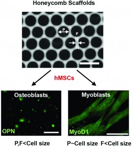
Researchers at Tohoku University in Japan have fabricated “honeycomb” topological substrates to regulate the differentiation of mesenchymal stem cells. The substrates are manufactured using a chloroform solution of polystyrene and an amphiphilic co-polymer, poly(N-dodecyl acrylamide-co-6-acrylamidehexanoic acid), or dioleoylphosphatidylethanolamine (DOPE). The solutions were cast on a glass slide and exposed to a flow of humid air to generate polystyrene films that resemble honeycomb from a beehive. The honeycomb substrates had varying pore size and strut width depending on the chloroform evaporation time.

After obtaining four different honeycomb films with varying pore size and strut width, human mesenchymal stem cells (hMSCs) were seeded on the surfaces to evaluate cell differentiation into osteo-specific (bone) and myo-specific (muscle) cells. To determine the cell differentiation state, cells were stained with osteoblast and myoblast markers, osteopontin and MyoD1, respectively.

Interestingly, when both the pore size and strut width were smaller than hMSC size, cells showed globular morphologies and increased expression of osteopotin (consistent with osteo-specific differentiation). On the other hand, when the pore size was the same size as hMSCs, the cells expressed MyoD1 and had elongated morphologies (consistent with myo-specific differentiation). Given the promising morphological and staining results, further studies are required to fully characterize the molecular mechanisms of the differentiation process.

This study demonstrates that geometries of honeycomb topographical substrates may have a direct influence on stem cell differentiation. Surface topology of biomaterial surfaces must be taken into account for future designs of cell culture environments for stem cell differentiation studies.

Honeycomb-shaped surface topography induces differentiation of human mesenchymal stem cells (hMSCs): uniform porous polymer scaffolds prepared by the breath figure technique
Takahito Kawano, Madoka Sato, Hiroshi Yabu and Masatsugu Shimomura 
Biomater. Sci., 2014, Advance Article DOI: 10.1039/C3BM60195A

Brian Aguado is currently a Ph.D. Candidate and NSF Fellow in the Biomedical Engineering department at Northwestern University. He holds a B.S. degree in Biomechanical Engineering from Stanford University and a M.S. degree in Biomedical Engineering from Northwestern University. When he’s not in the lab, Brian enjoys traveling, cooking, swimming, and spending time with family and friends. Read more about Brian’s research publications here.

To keep up-to-date with all the latest research, sign-up to our RSS feed or Table of contents alert.

Digg This
Reddit This
Stumble Now!
Share on Facebook
Bookmark this on Delicious
Share on LinkedIn
Bookmark this on Technorati
Post on Twitter
Google Buzz (aka. Google Reader)