Carbon Nanotube Vehicles for Cellular Delivery

New Nanoscale Communication

Polyethylenimine–carbon nanotube nanohybrids for siRNA-mediated gene silencing at cellular level

Stéphanie Foillarda, Guy Zuber and Eric Doris

Nanoscale DOI:10.1039/C0NR01005G

Carbon nanotube (CNT) based structures which can act as ‘nanohybrid vehicles’ for the delivery of functional molecules into cells have been developed by scientists in France.

Synthetic interfering RNA (siRNA) is able to inhibit the expression of a targeted gene by triggering enzymatic and sequence-selective degradation of the corresponding mRNAs, which holds great promise for the highly selective treatment of medical disorders at a genetic level. However, nucleic acids require a ‘delivery vehicle’ to take carry them through the cellular membrane and to the sites where they are required. The CNT hybrid nanostructure developed by this group is intended to do exactly this.

Eric Doris and co-workers at CEA, Service de Chimie Bioorganique et de Marquage, covalently modified short CNTs (~200 nm) with the cationic polymer polyethylenimine (PEI), which were then able to bind siRNA. The intention was to build a nanostructure which could enter cells by endocytosis but escape endosomal capture to increase the biological activity of the payload. They appear to be successful in this as they show that their system performs better than a reference lipid carrier.

To read this article, click here.

Digg This
Reddit This
Stumble Now!
Share on Facebook
Bookmark this on Delicious
Share on LinkedIn
Bookmark this on Technorati
Post on Twitter
Google Buzz (aka. Google Reader)

Anisotropic Gold Nanoparticles: Controlling growth with surfactants

New Nanoscale Feature Article

Surfactant-assisted, shape-controlled synthesis of gold nanocrystals

Junyan Xiao and Limin Qi

Nanoscale, DOI: 10.1039/c0nr00814a

This week at Nanoscale we have a published a Feature Article on the shape control of gold nanoparticles using surfactant systems. In this work, the authors Junyan Xiao and Limin Qi provide a comprehensive review of the techniques used to grow anisotropic gold nanoparticles, starting with an overview of the general strategies, before delving deeper into the role of surfactants in the production of some truly remarkable nanostructures. The descriptions of the various growth mechanisms are accompanied by excellent schematic diagrams which provide a valuable insight into the complexities of crystal formation and growth.

It is well known that gold nanoparticles have many unique chemical and physical properties, and there is much interest in applying them in a wide range of exciting applications. For example, they have been studied for use in nanoelectronics, drug delivery, catalysis, sensing, and photothermal therapy, to name but a few. Importantly, they exhibit particularly strong absorption and scattering of light due to localized surface plasmon resonance, a property which will be harnessed in the development of some revolutionary bioimaging devices. However, if these potential applications are to be realized, the growth of gold nanoparticles needs to be highly controlled in order exploit different properties which arise as we change their size, shape and surface chemistry.

Surfactants are vital in nanoparticle synthesis. Not only do they provide a protective capping layer and a means of conjugation, but they play an active role in particle nucleation and growth. Therefore, the choice of surfactant, or the design of a surfactant system, is crucial. In this review, the authors focus on gold nanocrystal synthesis assisted by single surfactants, mixed surfactants, supramolecular surfactants, as well as metal–surfactant complex templates.

To read this article, click here.

Digg This
Reddit This
Stumble Now!
Share on Facebook
Bookmark this on Delicious
Share on LinkedIn
Bookmark this on Technorati
Post on Twitter
Google Buzz (aka. Google Reader)

Transplant tracking

Transplant tracking

Harriet Brewerton

Magnetic nanoparticles could be used to track neural stem cells after a transplant in order to monitor how the cells heal spinal injuries, say UK scientists.

Neural stem cells are a promising treatment for repairing spinal cord injuries as they have the ability to generate tissue, but there is no effective way of monitoring the cells for long periods of time after transplantation.

Transplant tracking

Nguyen TK Thanh at the Davy Faraday Research Laboratory, University College London and the Royal Institution, and colleagues, believe they have the answer. They have developed hollow biocompatible cobalt-platinum nanoparticles and attached them to the stem cells. The nanoparticles are stable for months and have a high magnetic moment – tendency to align with a magnetic field – so that low concentrations can be detected using magnetic resonance imaging (MRI).

‘Magnetic nanoparticles are emerging as novel contrast and tracking agents in medical imaging,’ says Samir Pal at the California Institute of Technology, US, an expert in biological-nanoparticle interactions. ‘When used as a contrast agent for MRI, the nanoparticles allow researchers and clinicians to enhance the tissue contrast of an area of interest by increasing the relaxation rate of water.’

Stem cells attached to biocompatible nanoparticles can be visualised by MRI after transplantation into spinal cord slices

The team labelled stem cells with their nanoparticles, injected them into spinal cord slices and took images of their progress over time. They found that low numbers of the nanoparticle-loaded stem cells could still be detected two weeks after transplantation. ‘The new method demonstrates the feasibility of reliable, noninvasive MRI imaging of nanoparticle-labelled cells,’ says Thanh.

Thanh hopes that her stem cell tracking method will be used during stem cell replacement therapy for many central nervous system diseases. Her team is working towards developing nanoparticles that can be used to diagnose and treat these diseases.

Read the article in Nanoscale:

Magnetic CoPt nanoparticles as MRI contrast agent for transplanted neural stem cells detection

Xiaoting Meng, Hugh C. Seton, Le T. Lu, Ian A. Prior, Nguyen T. K. Thanh and Bing Song
Nanoscale, 2011, DOI: 10.1039/C0NR00846J

Read more Chemistry World News here


Digg This
Reddit This
Stumble Now!
Share on Facebook
Bookmark this on Delicious
Share on LinkedIn
Bookmark this on Technorati
Post on Twitter
Google Buzz (aka. Google Reader)

Happy Chinese New Year!

Nanoscale would like to wish all our Chinese friends a happy Chinese New Year!

新年快乐!兔年吉祥!

We hope you enjoy celebrating and we wish you every success in the year of the Rabbit!

  • Chunli BaiNanoscale is a collaboration between RSC Publishing and National Center for Nanoscience and Technology (NCNST), Beijing
  • The Editor-in-Chief, who handles submissions, is Professor Chunli Bai, Executive Vice President of CAS
  • Launched in October 2009; quickly becoming a very high-impact journal for all nanoscience and nanotechnology communities
  • Nanoscale is fully indexed in ISI, JCR, MEDLINE and other leading databases. It reaches the whole nano-research community
  • The first official Impact Factor will be released in June 2011, and is expected to be very high

All submissions handled by leading Editors-in-Chief, committed to rigorous, fair peer-review. The best international balance of any general nano-journal, research from Chinese authors is very important

We invite you to submit your research to Nanoscale.

PCCP ICCAS special collection

Take a look at the great quality work from China published in Nanoscale‘s sister journal PCCP in the special collection of papers from the Institute of Chemistry, Chinese Academy of Sciences (ICCAS), Guest Edited by Professor Li-jun Wan.

Digg This
Reddit This
Stumble Now!
Share on Facebook
Bookmark this on Delicious
Share on LinkedIn
Bookmark this on Technorati
Post on Twitter
Google Buzz (aka. Google Reader)

Interlocking Gold and Silver Nanoparticles with Supramolecular Linkers

New Nanoscale communication

Mechanically interlocked gold and silver nanoparticles using metallosupramolecular catenane chemistry

Carl A. Otter, Philipus J. Patty, Martin A. K. Williams, Mark R. Waterland and Shane G. Telfer

Nanoscale, DOI: 10.1039/c0nr00801j

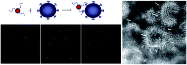
A specially designed metallosupramolecular linker has been used to ‘interlock’ gold and silver nanoparticles into aggregate assemblies. Waterland, Tefler and coworkers at Massey University in New Zealand used a PEGthiol-functionalised bis(phenanthroline)copper(I) complex which acted to ‘catenate’ the nanoparticles into mechanically interlocked structures, which formed a stable yet chemically modifiable linking mechanism with well defined particle separation.

The mechanism behind this particle interlocking is simple and extremely effective (see figure). The copper(I) centre in the complex arranges two phenanthroline ligands in an orthogonal arrangement., while two phenyl substituents direct the polyethylene glycol (PEG) chains away from each side. The PEG chains, which bestow water solubility on the complex, are terminated with thiol groups which have a strong affinity for the nanoparticle surfaces. Once these thiol groups are attached to the nanoparticles, the system locks two particles together into a physically entwined aggregate. The group employed DLS, SERS and TEM in their analyses, three complementary experimental techniques which allowed them to develop a comprehensive picture of their interlocking nanoparticle systems.

The controlled assembly of nanoparticles into complex structures is extremely important in the quest to design and synthesise complex, efficient and multifunctional nanostructures. Strategies such as those employed in this work, which combines the exciting fields of supramolecular chemistry and nanoparticle design, will be extremely important in the future development of novel nanotechnologies.

To read this communication, click here.

Digg This
Reddit This
Stumble Now!
Share on Facebook
Bookmark this on Delicious
Share on LinkedIn
Bookmark this on Technorati
Post on Twitter
Google Buzz (aka. Google Reader)

Hot Article: Biomimetic superhydrophobic surfaces

Hot Article out now

Fabrication of biomimetic superhydrophobic surfaces inspired by lotus leaf and silver ragwort leaf

Jinyou Lin, Yu Cai, Xianfeng Wang, Bin Ding, Jianyong Yu and Moran Wang

Nanoscale, 2011, DOI: 10.1039/C0NR00812E, Paper

Inspired by the self-cleaning lotus leaf and silver ragwort leaf, the authors demonstrate the fabrication of biomimetic superhydrophobic fibrous mats via electrospinning polystyrene (PS) solution in the presence of silica nanoparticles. The  fibers they obtain present a fascinating structure with the combination of nano-protrusions and numerous grooves due to the rapid phase separation in electrospinning. The content of silica nanoparticles incorporated into the fibers proves to be the key factor affecting the fiber surface morphology and hydrophobicity. Read more

Digg This
Reddit This
Stumble Now!
Share on Facebook
Bookmark this on Delicious
Share on LinkedIn
Bookmark this on Technorati
Post on Twitter
Google Buzz (aka. Google Reader)

Hot Article: Gold nanostars with magnetic cores

Hot Article

A facile synthetic route for the preparation of gold nanostars with magnetic cores and their reusable nanohybrid catalytic properties

Xiumin Miao, Tingting Wang, Fang Chai, Xiuli Zhang, Chungang Wang and Wendong Sun

Nanoscale, 2011, Advance Article, DOI: 10.1039/C0NR00704H

Chungang Wang and colleagues develop a new synthetic route to prepare gold nanostars (GNSs) with Fe3O4 cores under mild conditions. The result is an easy way to obtain magnetic GNSs with tunable optical properties from the visible to near-infrared. Even more, the particles described also show catalytic activity and can be easily recycled using an external magnet.

Read the article now

Digg This
Reddit This
Stumble Now!
Share on Facebook
Bookmark this on Delicious
Share on LinkedIn
Bookmark this on Technorati
Post on Twitter
Google Buzz (aka. Google Reader)

Top Ten most-read Nanoscale articles in December

The latest top ten most accessed Nanoscale articles

See the most-read papers of December 2010 here:

Wolfgang Schärtl, Nanoscale, 2010, 2, 829-843
DOI: 10.1039/C0NR00028K
 
Benjamin Weintraub, Zhengzhi Zhou, Yinhua Li and Yulin Deng, Nanoscale, 2010, 2, 1573-1587
DOI: 10.1039/C0NR00047G
 
Wufeng Chen and Lifeng Yan, Nanoscale, 2010, 2, 559-563
DOI: 10.1039/B9NR00191C
 
Idalia Bilecka and Markus Niederberger, Nanoscale, 2010, 2, 1358-1374
DOI: 10.1039/B9NR00377K
 
 
Poulomi Roy, Doohun Kim, Kiyoung Lee, Erdmann Spiecker and Patrik Schmuki, Nanoscale, 2010, 2, 45-59
DOI: 10.1039/B9NR00131J
 
Hualan Wang, Qingli Hao, Xujie Yang, Lude Lu and Xin Wang, Nanoscale, 2010, Advance Article
DOI: 10.1039/C0NR00224K
 
Jong Bum Lee, Michael John Campolongo, Jason Samuel Kahn, Young Hoon Roh, Mark Richard Hartman and Dan Luo, Nanoscale, 2010, 2, 188-197
DOI: 10.1039/B9NR00142E
 
Yen Hsun Su, Sheng-Lung Tu, Shih-Wen Tseng, Yun-Chorng Chang, Shih-Hui Chang and Wei-Min Zhang, Nanoscale, 2010, 2, 2639-2646
DOI: 10.1039/C0NR00330A
 
Frederik C. Krebs, Thomas Tromholt and Mikkel Jørgensen, Nanoscale, 2010, 2, 873-886
DOI: 10.1039/B9NR00430K

Digg This
Reddit This
Stumble Now!
Share on Facebook
Bookmark this on Delicious
Share on LinkedIn
Bookmark this on Technorati
Post on Twitter
Google Buzz (aka. Google Reader)

Quantum Dot Quality Control

New Nanoscale communication

New integrated elemental and molecular strategies as a diagnostic tool for the quality of water soluble quantum dots and their bioconjugates

Laura Trapiella-Alfonso, Antonio R. Montoro Bustos, Jorge Ruiz Encinar, Jose M. Costa-Fernandez, Rosario Pereiro and Alfredo Sanz-Medel

Nanoscale, DOI: 10.1039/c0nr00822b

Alfredo Sanz-Medel and co-workers at the University of Oviedo, Spain have developed a new strategy for analysing the quality of water soluble and bioconjugated quantum dots.

Their work is a novel concept based on size exclusion chromatography (SEC) coupled with elemental and fluorescence detection. Traditionally, it has been difficult to analyse the quality of quantum dot constructs in a quantitative fashion with methods such as transmission electron microscopy and photoluminescence spectroscopy. The current work aims to provide a general platform which allows a much greater understanding of the quality of quantum dots after water solubilisation and bioconjugation.

Although quantum dots exhibit many excellent properties for use in biomedical imaging, such as bright fluorescence and narrow emission lines, it is vital that these innately hydrophobic particles can be dispersed in water using a reliable capping procedure. Furthermore, for them to be used as functional probes in biological environments they need to be conjugated to other molecules, such as proteins or antibodies. As these steps are vital for development of quantum dot-based biomedical probes, there is an urgent need for advanced methods of purification and characterization of quantum dot bioconjugates, such as the one developed by this group.

Read more about this exciting work here.

Digg This
Reddit This
Stumble Now!
Share on Facebook
Bookmark this on Delicious
Share on LinkedIn
Bookmark this on Technorati
Post on Twitter
Google Buzz (aka. Google Reader)

Chemical Nanoscience Symposium

Chemical Nanoscience Symposium

30th March 2011
Newcastle University, Newcastle-Upon-Tyne, UK

Organised by: RSC Chemical Nanoscience & Nanotechnology Interest Group

Speakers:

Thomas Wandlowski, Univeristy of Bern, Switzerland

Paula Mendes, University of Birmingham, UK

Andrew Pike, Newcastle University, UK

Felix Zamora, Universidad Autonoma de Madrid, Spain

Richard Nichols, University of Liverpool, UK

Karl Coleman, Durham University, UK


Free registration: closes the 6th March

More information on how to register for this one day symposium can be found here

Digg This
Reddit This
Stumble Now!
Share on Facebook
Bookmark this on Delicious
Share on LinkedIn
Bookmark this on Technorati
Post on Twitter
Google Buzz (aka. Google Reader)