Archive for the ‘Web writers’ Category

HOT article: Photo-fluorescent and magnetic properties of iron oxide nanoparticles for biomedical applications

As cancer treatments evolve, focus has shifted to techniques and tools, which provide tumor-targeting capabilities and are uncoupled from common detrimental side effects.  The efficient delivery of new therapeutics has been a common area of interest, with studies focused on nanoparticle delivery systems being an eminent research area.

(a) TEM image of PS/Fe3O4 microspheres showing the amorphous coating entrapping multiple Fe3O4 nuclei. (b) TEM image of PAA/Fe3O4 and (c) cumulant size distribution of PAA/Fe3O4.

In this review article, Shi and co-workers draw attention to the properties of magnetite nanoparticles (Fe3O4), which can be utilised as customized therapeutics.  This review provides an overview of the synthesis of magnetite nanoparticles with an in-depth discussion related to their synthesis, functionalization and applications in a biological environment.

A highlight of this review includes an exploration into the recently discovered photo-luminescence properties of magnetite nanoparticles through studies of the electronic band structures to explain the emission mechanisms occurring.  The implications of thermal and magnetic induced hypothermia treatments utilizing these nanoparticles are also discussed relative to the patients being treated by such techniques.

From this review it can be determined that a new theragnostic platform has emerged with multi-functional capabilities both in terms of imaging and drug delivery.

Photo-fluorescent and magnetic properties of iron oxide nanoparticles for biomedical applications
Donglu Shi, M. E. Sadat, Andrew W. Dunn and David B. Mast
Nanoscale, 2015, 7, 8209-8232. DOI: 10.1039/C5NR01538C

Dr Derek Craig is a guest web writer for the Nanoscale blog. He is a Post Doctoral Research Fellow at the University of St. Andrews based in the fields of Biophotonics and Materials Science. With a background in chemistry, his work mainly focuses on the synthesis of nano to meso materials and the use of imaging techniques to study biological samples.

Digg This
Reddit This
Stumble Now!
Share on Facebook
Bookmark this on Delicious
Share on LinkedIn
Bookmark this on Technorati
Post on Twitter
Google Buzz (aka. Google Reader)

Single-walled carbon nanotubes: Catching some rays

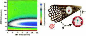
Precision printing and optical modeling of ultrathin SWCNT/C60 heterojunction solar cells

Inexpensive photovoltaics (PV) are an attractive avenue of research in the field of solar cells. In particular, semi-conducting single-walled carbon nanotubes  (s-SWCNTs) are a promising photo-absorbing material due to their strong near-infrared (near-IR) absorption and high carrier mobility. However, most current production methods for SWCNT PVs suffer from high surface roughness and lack nanometer-scale deposition precision, thereby hampering the reproducibility of ultrathin PV devices.

To this end, the authors have utilized ultrasonic spraying in order to tune the thickness of s-SWCNT layers with nanometer-scale precision. The researchers have used a combination of atomic force microscopy (AFM) and optical profilometry to show that their ultrasonic spraying method produces smooth, uniform films with an average roughness of about 5 nm.  The advantage of this low roughness enables fabrication of s-SWCT/C60 bilayer devices with significantly thinner C60 layers than previously reported.

The results reported by the authors help to advance the production of low-cost PV devices by improving the performance and scalability of ultra-thin SWCNT-based solar cells. Ultra-thin SWCNTs reported here could find potential use in other emerging technologies such as vertical field effect transistors and light-emitting diodes incorporating s-SWCNT injection layers.

Precision printing and optical modeling of ultrathin SWCNT/C60 heterojunction solar cells
Sarah L. Guillot, Kevin S. Mistry, Azure D. Avery, Jonah Richard, Anne-Marie Dowgiallo, Paul F. Ndione, Jao van de Lagemaat, Matthew O. Reese and Jeffrey L. Blackburn
Nanoscale, 2015, 7, 6556-6566. DOI: 10.1039/C5NR00205B

Dr Lee Barrett is a guest web writer for the Nanoscale blog. Lee is currently a postdoctoral researcher in the Centre for Molecular Nanometrology at the University of Strathclyde. His research is currently focused on the development of nanoparticle-based sensors and surface enhanced Raman scattering (SERS). Follow him on Twitter: @L_Bargie.

Digg This
Reddit This
Stumble Now!
Share on Facebook
Bookmark this on Delicious
Share on LinkedIn
Bookmark this on Technorati
Post on Twitter
Google Buzz (aka. Google Reader)

HOT article: The support effect on the size and catalytic activity of thiolated Au25 nanoclusters as precatalysts

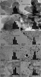
TEM images and size distribution (inset) of various Au25 (1 wt%)/support samples before and after 300 °C calcination.

The use of nanogold (Au NP) catalysts has been widely established for various applications including pharmaceuticals and perfumes.  However, their usage has been limited due to the conventional protocols employed for Au NP synthesis producing polydisperse size ranges, which have proven problematic for identifying catalytic active sites. Recently, this issue has been circumvented through instead using Au nanoclusters (Au NCs), which produce defined cluster sizes with well-defined physiochemical properties. As a direct consequence of being able to exert such control, there have been a number of applications established using such catalysts for processes including CO oxidation, and the oxidation of styrene and cyclohexane. However, whilst the role of the solid support is well established for Au NP catalysis, there currently exists a lack of knowledge on what effect/role the support plays in Au NCs catalysis.

In this study, Fang and co-workers report a systematic assessment of thiolated Au NCs on five different solid supports through examination of the size and electronic structure evolution of the Au NCs during heat treatment, and their catalytic performance when applied to the hydrogenation of nitrobenzene and oxidation of styrene.  The solid supports investigated include hydroxy-apatite (HAP), TiO2 (P25), activated carbon (AC), pyrolyzed grapheme oxide (PGO) and fumed SiO2.

The key findings of these studies establish that AuNCs on HAP and P25 supports presented enhanced activities over the other considered supports. This was found to be due to a number of factors, including reduced NC growth during calcination (heat treatment) following the removal of the thiolated ligands from the nanocluster surface. Further to this, the catalytic activity established during the hydrogenation of nitrobenzene and oxidation of styrene overwhelmingly showed that AuNC-HAP had a greater activity than any of the other supported catalysts. This was found to be due to both the reduced NC growth following heat treatment, and a change in the electronic structure of bound Au, resulting in strengthened metal support interactions.

Although this study has shown HAP to be a superior support in comparison to the others investigated, it should be noted that during heat treatment, an increase in the size of the nanoclusters was still observed.  As a consequence of this, the authors have objectively established improvements which should be made for future studies, including pursuing different support methods and improving the conditions under which the thiolated ligands can be removed from the Au NCs.

The support effect on the size and catalytic activity of thiolated Au25 nanoclusters as precatalysts
Jun Fang, Jingguo Li, Bin Zhang, Xun Yuan, Hiroyuki Asakura, Tsunehiro Tanaka, Kentaro Teramura, Jianping Xie and Ning Yan
Nanoscale, 2015, 7, 6325-6333. DOI: 10.1039/C5NR00549C

Dr Derek Craig is a guest web writer for the Nanoscale blog. He is a Post Doctoral Research Fellow at the University of St. Andrews based in the fields of Biophotonics and Materials Science. With a background in chemistry, his work mainly focuses on the synthesis of nano to meso materials and the use of imaging techniques to study biological samples.

Digg This
Reddit This
Stumble Now!
Share on Facebook
Bookmark this on Delicious
Share on LinkedIn
Bookmark this on Technorati
Post on Twitter
Google Buzz (aka. Google Reader)

HOT article: Radiolabelled magnetic particles for imaging

Radiolabelling of core–shell and dumbbell-like nanoparticles

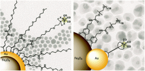
Bi-modal imaging agents are becoming more popular as they can be used to overcome limitations of single imaging modalities. In this work, radiolabelling of gold containing magnetic nanoparticles (Fe3O4-Au) with a nuclear isomer of technetium (99mTc), a commonly used radionuclide for clinical photon emission computed tomography (SPECT), was assessed using two different methods.  In the first approach, Fe3O4-Au core-shell nanoparticles were coated with ligands containing thiol groups to bind to gold and chelator groups for [99mTc(CO)3]+. In the second approach, 99mTc containing ligands were first synthesised, then attached to gold on Fe3O4-Au dumbbell-like nanoparticles. Both synthetic routes were successful in providing a sufficient radiochemical yield on the surface of magnetic nanoparticles, and are ideal candidates as dual magnetic resonance imaging MRI/SPECT imaging agents. In future studies, the authors plan to use the radiolabelled magnetic particles for bimodal imaging of tumours, through attachment of cancer-specific targeting vectors.

99mTc radiolabelling of Fe3O4–Au core–shell and Au–Fe3O4 dumbbell-like nanoparticles
M. Felber and R. Alberto
Nanoscale, 2015, 7, 6653-6660. DOI: 10.1039/C5NR00269A

Dr Mike Barrow is a guest web writer for the Nanoscale blog. He currently works as a Postdoctoral Researcher at the University of Liverpool.

Digg This
Reddit This
Stumble Now!
Share on Facebook
Bookmark this on Delicious
Share on LinkedIn
Bookmark this on Technorati
Post on Twitter
Google Buzz (aka. Google Reader)

HOT article: Scalable and cost effective patterning of graphene layers

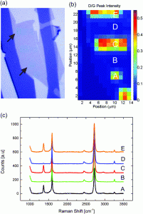
Visual image after EBL treatment, corresponding Raman spectroscopy map of a graphene flake and Raman spectra recorded at different spots on the sample.

Research articles on graphene have been numerously been presented throughout the last decade, indicating the promising future of this material. However, bridging the gap between laboratory research and industrial application remains difficult due to missing specialized large-scale production equipment.

A new article recently published by A. Caglinani et al. introduces an electron beam-based patterning technique using solely widely available clean-room equipment. Based on the findings of the paper, graphene structuring could become a more widespread processing technique.

The researchers developed a patterning process utilizing an industry-standard electron beam lithography system and a standard oven to achieve a resolution of 40 nm.

In the first step, a graphene layer is irradiated by the electron beam which locally generates defects within the crystal lattice. The damage was found by the researchers to be spatially confined to the exposed areas, thus allowing for arbitrary patterns.

The second step comprised etching the irradiated areas by means of hot air at atmospheric pressure. The high defect density (e.g. dangling bonds) induced by the electron beam lead to a large difference in the etch rate compared to the unmodified areas. By exposing the samples for 16 min at 435°C to air, the previously irradiated areas were selectively etched.

In conclusion, the presented process can be used to easily structure graphene layers for future application without the need for specialized equipment. According to the authors, future improvements could reduce the minimum feature size further.

Alberto Cagliani, Niclas Lindvall, Martin Benjamin Barbour Spanget Larsen, David M. A. Mackenzie, Bjarke Sørensen Jessen, Timothy J. Booth and Peter Bøggild
Nanoscale, 2015, 7, 6271-6277. DOI: 10.1039/C4NR07585D

Sebastian Axmann is a guest web-writer for the Nanoscale blog. His interests comprise manufacturing and metrology of nanostructures as well as their usage in current semiconductor devices. He also posts links to interesting research articles on Twitter: @SebastianAxmann.

Digg This
Reddit This
Stumble Now!
Share on Facebook
Bookmark this on Delicious
Share on LinkedIn
Bookmark this on Technorati
Post on Twitter
Google Buzz (aka. Google Reader)

Viruses: Feeling the strain

Viruses are highly complex biological nanostructures.  This complexity has renewed an interest in viruses from the perspective of fundamental physics at the nanoscale. An understanding of the mechanical properties of virus particles at the molecular level can reveal information regarding stiffness, intrinsic elasticity, structural strength and resistance to mechanical fatigue.  This information can provide a basis for researchers to engineer virus-based nanoparticles as nanodevices/ nanocontainers for different biotechnological applications.

The MVM virion.

Castellanos and co-workers have endeavoured to understand the conformational stability and dynamics of the minute virus of mice (MVM), a small ~ 25 nm virus particle, which serves as a model system to understand some of the mechanical properties of viruses. To this end, the researchers have investigated the linkage between the DNA-mediated increase in mechanical stiffness and heat-induced structural changes, and a quantitative relationship between mechanical elasticity and conformational dynamics in MVM nanoparticles.

The researchers used a combination of transmission electron microscopy (TEM) and atomic force microscopy (AFM) for determining the thermal inactivation behaviour and the mechanical stiffness of the viruses, respectively.  By utilizing these techniques, the researchers have deduced that infectious MVM particles may have evolved architectural functions that increase their survival in thermally stressed environments.

This proof-of-principle study has demonstrated that nanoscale features of virus nanoparticles can be probed analytically using AFM and the elucidation of these features have future impact in the field of protein engineering.

Milagros Castellanos, Pablo J. P. Carrillo and Mauricio G. Mateu
Nanoscale, 2015, 7, 5654-5664. DOI: 10.1039/C4NR07046A

Dr Lee Barrett is a guest web writer for the Nanoscale blog. Lee is currently a postdoctoral researcher in the Centre for Molecular Nanometrology at the University of Strathclyde. His research is currently focused on the development of nanoparticle-based sensors and surface enhanced Raman scattering (SERS). Follow him on twitter @L_Bargie.

Digg This
Reddit This
Stumble Now!
Share on Facebook
Bookmark this on Delicious
Share on LinkedIn
Bookmark this on Technorati
Post on Twitter
Google Buzz (aka. Google Reader)

In situ synthesis of luminescent carbon nanoparticles toward target bioimaging

In situ synthesis of luminescent carbon nanoparticles toward target bioimaging

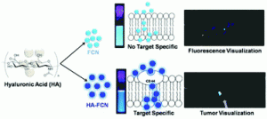
An in-situ synthesis of biocompatible fluorescent carbon nanoparticles (FCNs) is reported for targeted bioimaging. The nanoparticles were formed via dehydration of hyalurinic acid (HA), and through careful alteration of the carbonisation times, the total content of HA and fluorescence in the carbon nanoparticles could be controlled. Sharker et al. then compared two colloidally stable FCN samples; one partially carbonised sample that still contained some HA (HA-FCN), against a “non-specific” fully carbonised sample containing no HA (FCN).

Before in vivo testing, both sets of particles were tested on different cell lines at dosages up to 1.0 mg/ml and were found to not affect cell viability. Interestingly, HA-FCNs showed more uptake than the non-specific FCNs, and were internalised more various cell lines; including cancer cells. This is speculated to be due to the over expression of the CD-44 receptor which can facilitate uptake of particles containing targeting molecules such as HA-FCNs. In vivo bio-distribution studies showed more accumulation of HA-FCNs in tumours pre-implanted into mice compared to FCNs, when particles were injected into the tail vein. This is expected to be of enormous potential in not only bioimaging, but also drug delivery and diagnostics.

In situ synthesis of luminescent carbon nanoparticles toward target bioimaging
Shazid Md. Sharker, Sung Min Kim, Jung Eun Lee, Ji Hoon Jeong, Insik In, Kang Dea Lee, Haeshin Lee and Sung Young Park
Nanoscale, 2015, 7, 5468-5475. DOI: 10.1039/C4NR07422J

Dr Mike Barrow is a guest web writer for the Nanoscale blog. He currently works as a Postdoctoral Researcher at the University of Liverpool.

Digg This
Reddit This
Stumble Now!
Share on Facebook
Bookmark this on Delicious
Share on LinkedIn
Bookmark this on Technorati
Post on Twitter
Google Buzz (aka. Google Reader)

Hybridized doxorubicin-Au nanospheres exhibit enhanced Near-infrared surface plasmon absorption for photothermal therapy applications

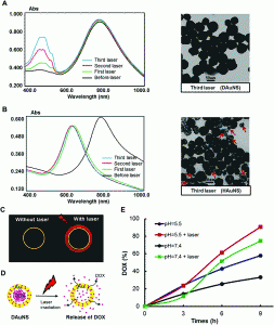
Plasmon absorption stability of DAuNS and the triggered release of DOX

As cancer therapy evolves there is a desire to explore minimally invasive treatments which are applicable to all patients, regardless of concerns with drug resistance and tumour morphology. One such option being investigated is photothermal therapy (PTT), which seeks to achieve these aims through the selective uptake of photosensitizing agents by cancerous cells prior to their abalation using a near infa-red (NIR) light source.  However, currently the efficacy of PTT is reduced due to heterogeneous heat distribution, resulting in the accumulation of sub-lethal doses of the sensitizing agent within areas of the tumor.

Zhou and co-workers have sought to overcome this issue by creating a “double punch” strategy for tumour targeting using PTT. Through the implantation of hollow gold nanoshells with the chemical agent doxorubicin (DAuNS), a novel combination of a photosensitizing agent and a chemical targeting agent has been created. Furthermore, this unique, yet simple synthesis strategy is thought to be interconvertible with other drug and nanomaterial combinations, thus, widening the scope for potential PTT treatments.

Through comparisons with ‘single-strategy’ treatments of bare hollow gold nanoshells (HAuNS) or doxorubicin, the improved efficacy of the DAuNS is well established through both in-vitro and in vivo studies. This significant improvement can be attributed to the enhanced plasmon absorption in the NIR region of DAuNS in comparison to HAuNS (1.5 fold increase), with a more efficient photothermal conversion and a greater efficacy in tumor killing also established. These properties are only enhanced by the combined chemotherapeutic effect achieved through the deployment of the doxorubicin payload.

As this strategy obviates the concerns of genetic drug resistance and is a minimally invasive treatment, it could carry significant potential. This potential is only further enhanced by the ability to exchange different chemotherapeutic reagents, and as such this could be a significant breakthrough which aids future cancer therapies.

Hybridized Doxorubicin-Au Nanospheres Exhibit Enhanced Near-infrared Surface Plasmon Absorption for Photothermal Therapy Applications
Jialin Zhou, Zuhua Wang, Qingpo Li, Fei Liu, Yongzhong Du, Hong Yuan, Fu-Qiang Hu, Yinghui Wei and Jian You
Nanoscale, 2015, Advance Article. DOI: 10.1039/C4NR07279K.

Dr Derek Craig is a guest web writer for the Nanoscale blog. He is a Post Doctoral Research Fellow at the University of St. Andrews based in the fields of Biophotonics and Materials Science. With a background in chemistry, his work mainly focuses on the synthesis of nano to meso materials and the use of imaging techniques to study biological samples.
Digg This
Reddit This
Stumble Now!
Share on Facebook
Bookmark this on Delicious
Share on LinkedIn
Bookmark this on Technorati
Post on Twitter
Google Buzz (aka. Google Reader)

HOT article: Unfolding the secrets of DNA origami nanostructures

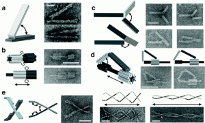
DNA origami machine components

DNA origami is an emerging field of nanotechnology that exploits the unique base-pairing capabilities of the DNA bases to produce complex and mechanically stiff self-assembled nanoscale geometries. Recently, DNA origami has found uses in applications such as single molecule sensing, drug delivery and templating molecular components.

This review, by researchers from Ohio State University, sets out to explore the mechanical nature of DNA nanostructures in order to understand how these structures will respond to physical interactions. The review covers three major areas of progress, including measuring and designing mechanical properties of DNA nanostructures, designing complex nanostructures based on imposed mechanical stresses and, finally, designing and controlling structurally dynamic nanostructures.

For researchers interested in the future possibilities of DNA origami, this review should provide an insightful first stop for discussing the mechanical design of DNA nanostructures.

Mechanical design of DNA nanostructures
Carlos E. Castro, Hai-Jun Su, Alexander E. Marras, Lifeng Zhou and Joshua Johnson
Nanoscale, 2015, Advance Article. DOI: 10.1039/C4NR07153K

Dr Lee Barrett is a guest web writer for the Nanoscale blog. Lee is currently a postdoctoral researcher in the Centre for Molecular Nanometrology at the University of Strathclyde. His research is currently focused on the development of nanoparticle-based sensors and surface enhanced Raman scattering (SERS). Follow him on twitter: @L_Bargie

Digg This
Reddit This
Stumble Now!
Share on Facebook
Bookmark this on Delicious
Share on LinkedIn
Bookmark this on Technorati
Post on Twitter
Google Buzz (aka. Google Reader)

HOT article: Effects of morphology and chemical doping on electrochemical properties of metal hydroxides in pseudocapacitors

With a limited amount of fossil fuels in the ground, developing alterative strategies for energy production is of the utmost importance.  However, without a way to store that energy, these endeavors could be futile.

From the team of Gyeonghee Lee, Chakrapani V. Varansi, and Jie Liu (an Associate Editor here at Nanoscale), comes one of this month’s HOT papers, “Effects of morphology and chemical doping on electrochemical properties of metal hydroxides in pseudocapacitors.” In this paper, the team’s specific metal hydroxide of interest is Ni(OH)2.
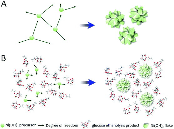
image file: c4nr06997h-s1.tif

Schematic illustration of the proposed mechanism for the morphology modification of Ni(OH)2 by glucose in the solvothermal medium.

Lee et al. state “…that both morphology control and chemical doping positively affect the electrochemical performance of Ni(OH)2 when applied individually.”  In this paper, they set out to determine what happens to the electrochemical performance when morphology and doping are combined instead of applied individually.  In order to investigate the effects of both, they chose a solvothermal synthesis route for metal hydroxide flakes, creating both cobalt-doped (CoxNi1-x(OH)2) and undoped particles (Ni(OH)2) that have varying amounts of D-glucose added.  For both the doped and undoped particles, four sets of particles are created, where 0, 10, 20, and 50% of the urea normally used in the solvothermal process is replaced by the D-glucose.  All of the particles were characterized with x-ray diffraction (XRD) and scanning electron microscopy (SEM), with the SEM showing that increasing amounts of glucose decreased flake size.  XRD and thermogravimetric analysis also revealed that increasing D-glucose amounts created an increase in the amount of interlayer water, with XRD also indicating degree of crystallinity decreased with increasing D-glucose.

In order to understand how the glucose affects morphology, the authors ran the solvothermal synthesis without the metal precursor. XRD and nuclear magnetic resonanace (NMR) data indicates the presence of ethyl-substituted glucose (formed during the synthesis process) instead of D-glucose.   The authors conclude that “…ethyl glucoside molecules can hinder the precursor diffusion and thus inhibit the metal hydroxide growth during synthesis.”

The electrochemical performance of all of the types of particle is evaluated using cyclic voltammetry (CV), galvanostatic charge–discharge, and electrochemical impedance spectroscopy (EIS).  Increasing glucose levels on undoped particles increased specific capacitance and cobalt doping is also shown to improve specific capacitance.  Combining the two showed that specific capacitance increased up to 50% of Co doping, but was less effective above 50%.

Lee et al. find that “…the addition of glucose in the ethanol-mediated solvothermal synthesis effectively reduces the particle size of metal hydroxide flakes. The specific capacitance is improved as a result of the increased surface area and reduced particle size.”  In terms of specific capacitance, the higher interlayer water levels found for increasing glucose levels may make ion mobility easier in the materials, enhancing the specific capacitance of the doped-Ni(OH)2.  In the doped materials, the specific capacitance was increased with low doping and decreased with high doping.

In designing future iterations of Ni(OH)2 batteries, careful attention will need to be placed on both morphology and doping levels in order to find the best balance for electrochemical performance.

Effects of morphology and chemical doping on electrochemical properties of metal hydroxides in pseudocapacitors
Gyeonghee Lee, Chakrapani V. Varanasi and Jie Liu
Nanoscale, 2015, 7, 3181-3188. DOI: 10.1039/C4NR06997H

Stephanie E. Vasko is currently a Senior Research Assistant at The Rock Ethics Institute at the Pennsylvania State University in State College. Her research focuses on science communication, STEM education, and the intersections between art, craft, and science.  You can follow her on Twitter at @stephanievasko.

The opinions and views expressed in this piece are those of the author and do not represent or reflect the opinion, views, or policy of the Pennsylvania State University, the Rock Ethics Institute, or the National Science Foundation.

Digg This
Reddit This
Stumble Now!
Share on Facebook
Bookmark this on Delicious
Share on LinkedIn
Bookmark this on Technorati
Post on Twitter
Google Buzz (aka. Google Reader)