Author Archive

Direct real-time detection of single proteins using silicon nanowire-based electrical circuits

The development of analytical devices that convert biological response into an electrical signal is a very important goal with great potential benefits for clinical diagnostics, environmental science, and defense.

In a recent communication published in Nanoscale, researchers discuss findings from a newly developed single silicon nanowire (SiNW) based biosensor, which is able to directly detect protein adsorption/desorption at single-molecule resolutions.
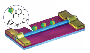
Fig. 1 Schematic demonstration of SiNW FET-based electrical biosensors, where Au electrodes are passivated by using a thermally deposited 50 nm-thick SiO2 layer. The inset shows how His-tag F1-ATPase is immobilized on the surface of SiNWs through Ni2+ chelation.

SiNW’s were synthesised following an Au-catalysed vapor deposition method and then high-density SiNW array devices were fabricated on silicon substrates using photolithography. Subsequently the devices were functionalized in a stepwise manner to impart the biomolecule recognizing Nickel functionality, and characterized with XPS and FTIR spectroscopy.

By combining theses devices with microfluidic systems, the authors were able to achieve real-time, direct detection of the chelation between Nickel and the imidazole of His-tags in the target biomolecules (F1 ATPases) at the single-event level. This nondestructive and label-free sensor shows great promise for number verification and real-time monitoring of proteins in complex biological systems.

Direct real-time detection of single proteins using silicon nanowire-based electrical circuits
Jie Li, Gen He, Hiroshi Ueno, Chuancheng Jia, Hiroyuki Noji, Chuanmin Qi and Xuefeng Guo
Nanoscale, 2016, DOI: 10.1039/C6NR04103E

Alexander Cook is a guest web writer for the RSC journal blogs. He is a PhD researcher in the Perrier group at the University of Warwick, focusing on polymer materials and their use in various applications. Follow him on twitter @alexcook222

Digg This
Reddit This
Stumble Now!
Share on Facebook
Bookmark this on Delicious
Share on LinkedIn
Bookmark this on Technorati
Post on Twitter
Google Buzz (aka. Google Reader)

pH-responsive polyrotaxane self-assembly leads to exciting stimuli-responsive nanoparticles

Polyrotaxanes are an exciting class of materials that in recent years have found applications in self-healing materials, coatings, nanomedicine, and biomaterials. This interesting supramolecular polymer architecture conveys unusual and dynamic properties not found in conventional covalently bonded polymer materials.

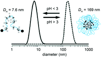
Fig. 1 DLS (number) analysis of PRX 5 (0.1 mg mL−1 ) at pH 7.2 (continuous line) and the self-assembly of PRX 5 into SA-PRX 5 at pH 2 (dashed line).

Commercially available PEG of 10 and 20 kDa were threaded into CDs with two different threading degrees of 28% and 65%. The CDs were modified with succinic anhydride to give approximately 4 carboxylic acid groups per CD ring.

At neutral pH, the 28% threaded polyrotaxanes formed self-assembled nanoparticles due to movement of the CD units along polymer chains to form segregated regions of more hydrophobic CD in the core and PEG chains at the periphery. When the pH of the polymer solution was less than 3 the subsequently charged CDs electrostatically repel each other and the nanoparticles fall apart to give their corresponding unimolecular polyrotaxanes in an extended conformation. By cycling the pH of the polyrotaxane system, nanoparticles can be formed and disassembled up to three times.

These nanoparticles are proposed to be of interest and significance for oral drug delivery systems that could be responsive to the lumen of the stomach (pH 1 – 3.8), and thus release therapeutics when the nanoparticles dissociate at low pH values.

Nanoparticles assembled via pH-responsive reversible segregation of cyclodextrins in polyrotaxanes
Blaise L. Tardy, Shereen Tan, Henk H. Dam, Hirotaka Ejima, Anton Blencowe, Greg G. Qiao and Frank Caruso
Nanoscale, 2016, Advance Article, DOI: 10.1039/C6NR04841B

Alexander Cook is a guest web writer for the RSC journal blogs. He is a PhD researcher in the Perrier group at the University of Warwick, focusing on polymer materials and their use in various applications. Follow him on twitter @alexcook222

Digg This
Reddit This
Stumble Now!
Share on Facebook
Bookmark this on Delicious
Share on LinkedIn
Bookmark this on Technorati
Post on Twitter
Google Buzz (aka. Google Reader)

iPAINT: brushing-up on super resolution microscopy

In recent years, super-resolution microscopy has enabled researchers to explore biological interfaces at the nanoscale. Single-molecule localization methods, such as point accumulation for imaging in nanoscale topography (PAINT), are fundamental techniques for studying the morphology and architecture of living matter. While super-resolution microscopy techniques like PAINT have acquired the interest of researchers in biology, it remains elusive to applications in soft matter and materials science.

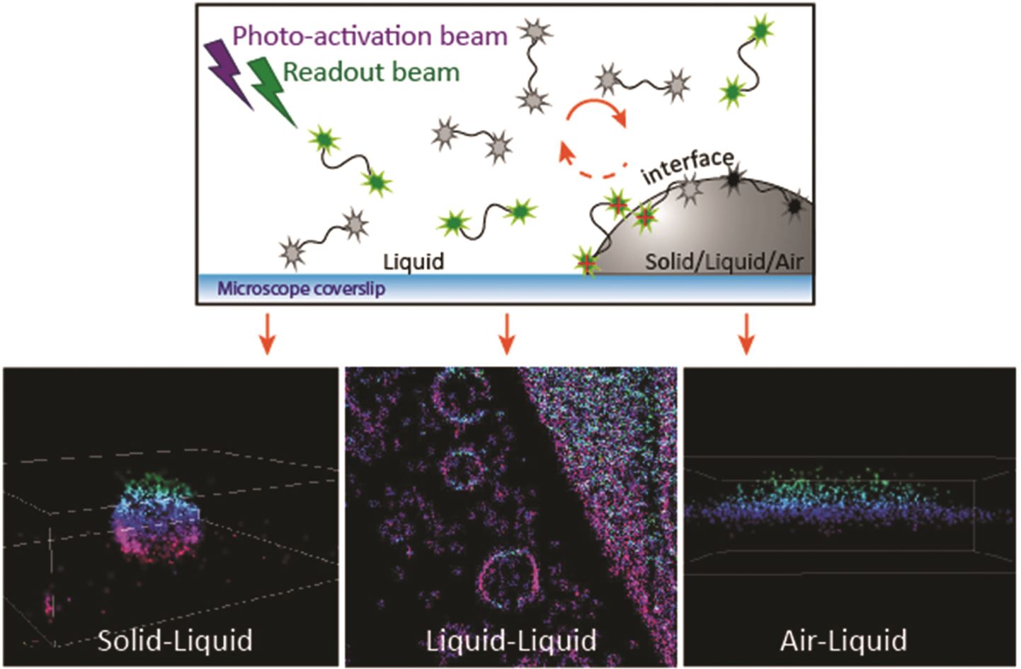
In issue 16 of Nanoscale, researchers from the Netherlands have endeavoured to overcome the limitations of PAINT, such as a pre-requisite for hydrophobic domains or specific ligand/receptor pairs, by introducing interface point accumulation for imaging in nanoscale topography (iPAINT). In short, this new technique enables nanometre resolution imaging of interfaces by non-covalent, continuous labelling during imaging. This was achieved by labelling silica nanoparticles with polyethylene glycol (PEG) end-functionalized with a photoactivatable rhodamine analogue (PEG552) that is able to continuously adsorb and desorb from the interface. This method of labelling is essential for interfaces such as emulsions, foams and crystals like ice.

By employing iPAINT as a generic imaging method, the authors are able to obtain super-solution images at different interfaces in 3D. This innovation allows users to develop PAINT in other fields, such as colloid and interface science, food science, soft matter physics and nanotechnology.

iPAINT: a general approach tailored to image the topology of interfaces with nanometer resolution
A. Aloi, N. Vilanova, L. Albertazzi and I. K. Voets
Nanoscale, 2016, DOI: 10.1039/C6NR00445H

Dr Lee Barrett is a guest web writer for the Nanoscale blog. Lee is currently a postdoctoral researcher in the Centre for Molecular Nanometrology at the University of Strathclyde. His research is currently focused on the development of nanoparticle-based sensors and surface enhanced Raman scattering (SERS). Follow him on twitter @L_Bargie

Digg This
Reddit This
Stumble Now!
Share on Facebook
Bookmark this on Delicious
Share on LinkedIn
Bookmark this on Technorati
Post on Twitter
Google Buzz (aka. Google Reader)

Hybrid nanoparticles: The fight against breast cancer heats up

Gold nanorods with cisplatin-polypeptide wrapping were developed for combinational photothermal therapy and chemotherapy of triple negative breast cancer.

Researchers from China have advanced the fight against breast cancer (BC) by developing a method that targets triple negative breast cancer (TNBC) – a highly aggressive subtype of BC and a form that is challenging to completely eradicate.

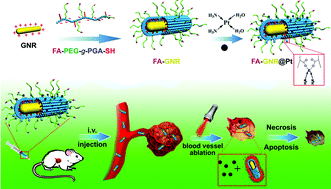
Their method consisted of the formation of gold nanorods (GNRs) with a cisplatin-polypeptide wrapping and folic acid (FA) functionalization (FA-GNR@Pt) for the simultaneous targeted photothermal therapy and chemotherapy. These hybrid nanoparticles combine the photothermal conversion properties of GNRs, superior biocompatibility of polypeptide poly(L-glutamic acid) (PGA), chemotoxicity of cisplatin and the tumour targeting ability of FA.  FA-GNR@Pt nanoparticles exhibited temperature increases both in vitro and in vivo using 655 nm NIR laser irradiation and, in combination with systemic administration in mice, were able to inhibit the proliferation and lung metastisis of the 4T1 breast tumour.

The research presented here takes significant steps in furthering the understanding of breast cancer, particularly TNBC, which have increased risk of metastisis.

Near infrared light-actuated gold nanorods with cisplatin–polypeptide wrapping for targeted therapy of triple negative breast cancer
Bing Feng, Zhiai Xu, Fangyuan Zhou, Haijun Yu, Qianqian Sun, Dangge Wang, Zhaohui Tang, Haiyang Yu, Qi Yin, Zhiwen Zhang and Yaping Li
Nanoscale, 2015, 7, 14854-14864.  DOI: 10.1039/C5NR03693C

Dr Lee Barrett is a guest web writer for the Nanoscale blog. Lee is currently a postdoctoral researcher in the Centre for Molecular Nanometrology at the University of Strathclyde. His research is currently focused on the development of nanoparticle-based sensors and surface enhanced Raman scattering (SERS). Follow him on twitter @L_Bargie.

Digg This
Reddit This
Stumble Now!
Share on Facebook
Bookmark this on Delicious
Share on LinkedIn
Bookmark this on Technorati
Post on Twitter
Google Buzz (aka. Google Reader)