Archive for 2011

Nanoscale junctions for next generation memory

From stochastic single atomic switch to nanoscale resistive memory device

Attila Geresdi, András Halbritter, András Gyenis, Péter Makk and György Mihály

Nanoscale, 2011, DOI:10.1039/C0NR00951B

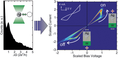
Scientists from Hungary have published some important work in Nanoscale regarding the use of solid state ionic conductors as atomic-sized junctions in non-volatile computer memory devices. By varying the size of the junctions from single-atom-sized to 10 nm, the group concluded that there is a lower size limit of 3 nm for reliable ionic nano-switches, a size which is well below the resolution of recent lithographic techniques.

In this work, Garesdi et al. created the junctions by gently touching a silver thin film with an electrochemically sharpened tungsten tip. Exposure of the silver film to air established the ionic conductor surface layer, and the nanoscale ‘point-contact’ geometry was sufficient to form a reliable switching device above the 3 nm threshold. Below this value, the switching process was much less reliable. The storage density here, even with the 3 nm limit, would be higher than current NAND flash devices and similar to the proposed bit size threshold of magnetic media which arises due to the superparamagnetic limit.

The authors provide a detailed analysis of the physical properties of the nano-junctions, as well as an explanation of the underlying mechanisms. They conclude that their ionic conductor-based devices are good candidates for non-volatile memory cells.

To read this article, click here.

<!–[if !mso]> <! st1\:*{behavior:url(#ieooui) } –>

From stochastic single atomic switch to nanoscale resistive memory device

Attila Geresdi, András Halbritter, András Gyenis, Péter Makk and György Mihály

Nanoscale

DOI:10.1039/C0NR00951B

Scientists from Hungary have published some important results in Nanoscale regarding the use of solid state ionic conductors as atomic-sized junctions in non-volatile computer memory devices. By varying the size of the junctions from single-atom-sized to 10 nm, the group concluded that there is a lower size limit of 3 nm for reliable ionic nano-switches, a size which is well below the resolution of recent lithographic techniques.

In this work, Garesdi et al. created these junctions by gently touching a silver thin film with an electrochemically sharpened tungsten tip. Exposure of the silver film to air established the ionic conductor surface layer, and the nanoscale ‘point-contact’ geometry was sufficient to form a reliable switching device above the 3 nm threshold. Below this value, the switching process was much less reliable. The storage density here, even with the 3 nm limit, would be higher than current NAND flash devices and similar to the proposed bit size limit of magnetic media which arises due to the superparamagnetic limit.

The authors provide a detailed analysis of the physical properties of the nano-junctions, as well as an explanation of the underlying mechanisms. They conclude that their ionic conductor-based devices are good candidates for non-volatile memory cells.

To read this article, click here.

Digg This
Reddit This
Stumble Now!
Share on Facebook
Bookmark this on Delicious
Share on LinkedIn
Bookmark this on Technorati
Post on Twitter
Google Buzz (aka. Google Reader)

New nanocomposites as strong as bone

Scientists in China have made a synthetic bone material using a new technique for creating polymer nanocomposites incorporated with inorganic nanoparticles.

Yuandong Dou, Kaili Lin and Jiang Chang, Nanoscale, 2011, DOI: 10.1039/C1NR10028A

Fluorescent images of the composite films

Fluorescent images of the composite films

Jiang Chang and colleagues created this material after coming up with a new approach to making the  nanocomposites,  allowing them to control both the spatial distribution and orientational organisation of the nanocomponents, a known limitation of current methods of fabrication.

Their method involves using electrospinning and hot pressing techniques. They firstly homogeneously dispersed the nanoparticles within a polymer matrix solution, which was then electrospun into a patterned “nanofibrous mat” using a specifically designed “collector”. This mat was then placed between two sheets of non-woven polymer nanofibre and hot pressed to create the nanocomposite.

Because bone tissue is, generally speaking, structurally similar to these composites (they involve mineral particles preferentially oriented in a collagen matrix), the researchers tested their new fabrication method by creating an synthetic bone material by incorporating calcium silicate hydrate nanowires into a polyvinyl butyral matrix.  Their artificial material showed remarkable mechanical properties, particularly when compared with the pure polymer (for instance, the bending strength of the researchers’ material reached 188 MPa, as compared to the 86 MPa of the polymer), which also matched those of real cortical bone tissue.

You can find out more about this work by reading the article here.

Digg This
Reddit This
Stumble Now!
Share on Facebook
Bookmark this on Delicious
Share on LinkedIn
Bookmark this on Technorati
Post on Twitter
Google Buzz (aka. Google Reader)

Interactions with stem cells

stem cellsHOT Nanoscale paperread it now!

Surface carboxyl groups on polystyrene nanoparticles are able to trigger clathrin-mediated endocytosis and strongly facilitate internalization of negatively charged nanoparticles by mesenchymal stem cells (MSCs).

Specific effects of surface carboxyl groups on anionic polystyrene particles in their interactions with mesenchymal stem cells
Xiue Jiang, Anna Musyanovych, Carlheinz Röcker, Katharina Landfester, Volker Mailänder and G. Ulrich Nienhaus
Nanoscale, 2011, DOI: 10.1039/C0NR00944J

Digg This
Reddit This
Stumble Now!
Share on Facebook
Bookmark this on Delicious
Share on LinkedIn
Bookmark this on Technorati
Post on Twitter
Google Buzz (aka. Google Reader)

Polarisation stabilisation of surface emitting lasers

lasersNanoscale ‘HOT’ paper – read it today!

The electron beam of a scanning electron microscope was used to write a polarisation grating onto vertical surface emitting lasers in a maskless, dry, single step process.

Polarisation stabilisation of vertical cavity surface emitting lasers by minimally invasive focused electron beam triggered chemistry
Ivo Utke, Martin G. Jenke, Christian Röling, Peter H. Thiesen, Vladimir Iakovlev, Alexei Sirbu, Alexandru Mereuta, Andrei Caliman and Eli Kapon

Nanoscale
, 2011, DOI: 10.1039/C1NR10047E

Digg This
Reddit This
Stumble Now!
Share on Facebook
Bookmark this on Delicious
Share on LinkedIn
Bookmark this on Technorati
Post on Twitter
Google Buzz (aka. Google Reader)

Nanoscale Board Member talks at Biophysical Society Meeting

Nanoscale Editorial Board member Molly Stevens (Imperial College London) gave a talk at the Annual Biophysical Society meeting yesterday.

This was the first time Professor Stevens had been invited to talk at the Biophysical Society Meeting, and her lecture on her research into tissue engineering was very well received. Her research focusses on the regeneration of tissue, with a focus on engineering new bone tissue.

She also discussed her collaborative research on sub-nanoscale patterning which she did with Francesco Stellacci’s group (Nanoscale Editor-in-Chief) – find out more about Nanoscale and our Editorial Board at www.rsc.org/nanoscale

Digg This
Reddit This
Stumble Now!
Share on Facebook
Bookmark this on Delicious
Share on LinkedIn
Bookmark this on Technorati
Post on Twitter
Google Buzz (aka. Google Reader)

New Nanoprobes: Silica capped green fluorescent protein

Encapsulated enhanced green fluorescence protein in silica nanoparticle for cellular imaging

Zhengwei Cai, Zhangmei Ye, Xiaowei Yang, Yanli Chang, Haifang Wang, Yuanfang Liu and Aoneng Cao
Nanoscale DOI:10.1039/C0NR00956C

Scientists in China have developed a simple method of capping green fluorescent protein (GFP) in silica, which is a vital step in improving the versatility of fluorescent proteins for use as imaging probes.

Cai et al. at Shanghai University developed a covalent attachment route, which means the capping precursors are chemically bonded to the protein, rather than just providing passive encapsulation. The silica shell is then grown from the precursor layer to provide a solid and stable shell. A simple reverse emulsion method was used, and the group achieved a very high encapsulation efficiency and high protein loading. Their characterisation results suggest that encapsulating GFP in silica significantly increases its fluorescence and stability as the capping provides an effective barrier from external interference, such as protease attack, denaturants, and excessive heating.

Fluorescent probes are widely used to image biological structures and processes, both in vivo and in vitro. The main concerns in the design of these probes are their optical properties and the way they interact with their environment. For example, you may have a probe which exhibits excellent optical properties, but is toxic and therefore adversely affects the things you are trying to image. Conversely, you could have a probe which is non-toxic, but is unstable and loses its fluorescence too quickly under excitation. Traditional organic dyes suffered from various problems, including a lack of stability, broad emission profiles and toxicity issues. These have gradually been replaced with modern ‘nanoprobes’ which consist of either fluorescent nanoparticles or nanoparticulate coatings for fluorescent molecules. Of all the nanoprobes developed, fluorescent quantum dots exhibit the best optical properties, however, as they generally contain heavy metals such as cadmium, there are toxicity concerns in many applications.

The silica encapsulated fluorescent protein nanoparticles developed in this work may prove to be an exciting new probe which exhibits excellent optical and stability properties whilst avoiding problems such as toxicity and instability,

To read this article, click here.

Nanoscale DOI:10.1039/C0NR00956C

Digg This
Reddit This
Stumble Now!
Share on Facebook
Bookmark this on Delicious
Share on LinkedIn
Bookmark this on Technorati
Post on Twitter
Google Buzz (aka. Google Reader)

Nanoscale Editor-in-Chief confirmed as CAS President

Chunli BaiThe Chinese State Council announced yesterday that they have appointed Chunli Bai as the next president of the Chinese Academy of Sciences (CAS).

Professor Bai has been the executive vice president of CAS since 2004, and will take over from former CAS President Lu Yongxiang.

The Nanoscale team would like to congratulate Professor Bai and we wish him all the best in his new role!

Digg This
Reddit This
Stumble Now!
Share on Facebook
Bookmark this on Delicious
Share on LinkedIn
Bookmark this on Technorati
Post on Twitter
Google Buzz (aka. Google Reader)

Simple and scalable graphene patterning method for Schottky solar cells

graphene patterningHOT’ Nanoscale Communication – hot off the press!

A simple, scalable, graphene patterning method has been developed and applied to fabricate CdSe nanobelt/graphene Schottky junction solar cells. A typical as-fabricated solar cell shows an energy conversion efficiency of ~1.25%.

A simple and scalable graphene patterning method and its application in CdSe nanobelt/graphene Schottky junction solar cells
Yu Ye, Lin Gan, Lun Dai, Yu Dai, Xuefeng Guo, Hu Meng, Bin Yu, Zujin Shi, Kuanping Shang and Guogang Qin
Nanoscale, 2011, DOI: 10.1039/C0NR00999G

Digg This
Reddit This
Stumble Now!
Share on Facebook
Bookmark this on Delicious
Share on LinkedIn
Bookmark this on Technorati
Post on Twitter
Google Buzz (aka. Google Reader)

Chickpeas grow taller with carbon nanotubes

Carbon nanotubes can enhance plant growth without damaging plant cells, say scientists from India

Chickpeas grow taller with carbon nanotubes

Sabyasachi Sarkar and colleagues from the Indian Institute of Technology in Kanpur treated chickpea plants with up to 6ug/ml of water soluble carbon nanotubes. They found that the nanotubes increased the growth rate in every part of the plant – in the roots, shoots and branches.

Sarkar thought that the channels could be replicated by carbon nanotubes. ‘We followed Thomas Edison’s recipe to make carbonised filament from bamboo or wood wool in his electric bulb to get the carbon nanotubes,‘ he adds. ‘Of course, we had to derivatise them to make them water soluble.’ The team achieved this by attaching carboxylic acid groups to the surface of the tubes.

The work seems to support the positive effect of carbon nanotubes,’ says Xiaohong Fang, an expert in the use of carbon nanotubes as molecular transporters in plants at the Chinese Academy of Sciences in China. However, she points out that the biological effects on plants may differ depending on the materials’ chemical and physical properties, plant type and cultivation conditions.

Read the rest of the Chemistry World story by Elinor Richards

View the Nanoscale article in full:

Growth stimulation of gram (Cicer arietinum) plant by water soluble carbon nanotubes
Shweta Tripathi, Sumit Kumar Sonkar and Sabyasachi Sarkar
Nanoscale,
2011, DOI: 10.1039/c0nr00722f

Digg This
Reddit This
Stumble Now!
Share on Facebook
Bookmark this on Delicious
Share on LinkedIn
Bookmark this on Technorati
Post on Twitter
Google Buzz (aka. Google Reader)

Surface nanotechnology for biological applications – themed issue

Issue 2 of Nanoscale out now!

This month’s issue includes a collection of articles on the theme surface nanotechnology for biological applications

This themed issue is Guest Edited by Professor Marcus Textor, Professor Darrell Irvine and Professor Xingyu Jiang. It includes a Review by Antonio Nanci et al. on Nanoscale surface modifications of medically relevant metals: state-of-the art and perspectives and a Communication by Molly Stevens, Kinetic investigation of bioresponsive nanoparticle assembly as a function of ligand design, as well as much, much more!

issue 2 coverCover image

The cover image highlights the paper by Nicholas Melosh and colleagues and shows that the stability of nanoscale hydrophobic bands inside the hydrophobic core of lipid membranes depends on their relative size.

Nanoscale patterning controls inorganic–membrane interface structure
Benjamin D. Almquist, Piyush Verma, Wei Cai and Nicholas A. Melosh
Nanoscale, 2011, 3, 391-400

Want to read more about our themed issues? Please visit the ‘ Themed Issues’ page on our website.

Digg This
Reddit This
Stumble Now!
Share on Facebook
Bookmark this on Delicious
Share on LinkedIn
Bookmark this on Technorati
Post on Twitter
Google Buzz (aka. Google Reader)