Once more Lab on a Chip plays a pivotal role in supporting the Lab-on-a-Chip community by providing FREE Access (thanks to CBMS) to microTAS abstracts from 2003 to 2009 (2010 available soon!).
Top ten most accessed articles in September
This month sees the following articles in Lab on a Chip that are in the top ten most accessed:-
Cell lysis and DNA extraction of gram-positive and gram-negative bacteria from whole blood in a disposable microfluidic chip
Madhumita Mahalanabis, Hussam Al-Muayad, M. Dominika Kulinski, Dave Altman and Catherine M. Klapperich
Lab Chip, 2009, 9, 2811-2817, DOI: 10.1039/B905065P , Paper
Programmable diagnostic devices made from paper and tape
Andres W. Martinez, Scott T. Phillips, Zhihong Nie, Chao-Min Cheng, Emanuel Carrilho, Benjamin J. Wiley and George M. Whitesides
Lab Chip, 2010, 10, 2499-2504, DOI: 10.1039/C0LC00021C , Paper
Sickling of red blood cells through rapid oxygen exchange in microfluidic drops
Paul Abbyad, Pierre-Louis Tharaux, Jean-Louis Martin, Charles N. Baroud and Antigoni Alexandrou
Lab Chip, 2010, 10, 2505-2512, DOI: 10.1039/C004390G , Paper
Microstructuring of polymer films for sensitive genotyping by real-time PCR on a centrifugal microfluidic platform
Maximilian Focke, Fabian Stumpf, Bernd Faltin, Patrick Reith, Dylan Bamarni, Simon Wadle, Claas Müller, Holger Reinecke, Jacques Schrenzel, Patrice Francois, Daniel Mark, Günter Roth, Roland Zengerle and Felix von Stetten
Lab Chip, 2010, 10, 2519-2526, DOI: 10.1039/C004954A , Paper
Precompetitive preclinical ADME/Tox data: set it free on the web to facilitate computational model building and assist drug development
Sean Ekins and Antony J. Williams
Lab Chip, 2010, 10, 13-22, DOI: 10.1039/B917760B , Perspective
A microfluidic platform for probing small artery structure and function
Axel Günther, Sanjesh Yasotharan, Andrei Vagaon, Conrad Lochovsky, Sascha Pinto, Jingli Yang, Calvin Lau, Julia Voigtlaender-Bolz and Steffen-Sebastian Bolz
Lab Chip, 2010, 10, 2341-2349, DOI: 10.1039/C004675B , Paper
Predictive model for the size of bubbles and droplets created in microfluidic T-junctions
Volkert van Steijn, Chris R. Kleijn and Michiel T. Kreutzer
Lab Chip, 2010, 10, 2513-2518, DOI: 10.1039/C002625E , Paper
Agarose droplet microfluidics for highly parallel and efficient single molecule emulsion PCR
Xuefei Leng, Wenhua Zhang, Chunming Wang, Liang Cui and Chaoyong James Yang
Lab Chip, 2010, 10, 2841-2843, DOI: 10.1039/C0LC00145G , Communication
Research Highlights
Petra S. Dittrich
Lab Chip, 2010, 10, 2495-2496, DOI: 10.1039/C0LC90045A , Highlight
Electrochemical sensing in paper-based microfluidic devices
Zhihong Nie, Christian A. Nijhuis, Jinlong Gong, Xin Chen, Alexander Kumachev, Andres W. Martinez, Max Narovlyansky and George M. Whitesides
Lab Chip, 2010, 10, 477-483, DOI: 10.1039/B917150A , Paper
Why not take a look at the articles today and blog your thoughts and comments below.
Fancy submitting an article to Lab on a Chip? Then why not submit to us today or alternatively email us your suggestions.
New YouTube Videos
View the new videos on the Lab on a Chip YouTube site using the links below:
Enhancement by optical force of separation in pinched flow fractionation
New Innovator Awardees
Congratulations to the recipients of the 2010 NIH New Innovator Award. Particularly heartening for the Lab on a Chip community is that over 10% of the 50 awardees are working in the nano- and microfluidics arena.
The NIH Director’s New Innovator Award aims to stimulate ‘highly innovative research’ and also provides valuable support to upcoming new investigators. This year’s awardees include: Dino Di Carlo, Amy Elizabeth Herr, Tony Jun Huang, Michelle Khine, Pak Kin Wong, Changhuei Yang to name just a few.
A full list of all this year’s recipients can be viewed on the NIH website and while on the subject why not take a second look at the Lab on a Chip Emerging Investigator issue (Issue 18, 2010).
New YouTube Videos
View the new videos on the Lab on a Chip YouTube site using the links below:
Sub-pixel resolving optofluidic microscope for on-chip cell imaging
A microdroplet-based shift register
Hybrid electronics get twisted
A stretchable radio frequency (RF) radiation sensor that combines a microfluidic antenna and rigid electronic circuits has been developed by scientists in Sweden. This could open the way to reliable and durable second skin sensors for monitoring health.
 Flexible electronics are used in applications such as cameras, computer keyboards and photovoltaic cells. Some success has been found with stretchable antennas but the connection between the stretchable material and the rigid circuits still results in strain and loss of device sensitivity. To make wearable devices, electronics not only need to be flexible but they also need to be stretchable to truly conform to skin. Unfortunately, development from a flexible to a stretchable device has remained an elusive goal.
Now, Shi Cheng and Zhigang Wu from Uppsala University have developed a hybrid technology that combines conventional rigid circuitry with a substrate making a device that can bend, twist and stretch
IMAGE: Flexible microfluidic sensor responds to radio frequency signals
Click here to read the full story
Link to journal article
Microfluidic stretchable RF electronics
Shi Cheng and Zhigang Wu, Lab Chip, 2010
DOI: 10.1039/c005159d
Top ten most accessed articles in August
This month sees the following articles in Lab on a Chip that are in the top ten most accessed:-
Cell lysis and DNA extraction of gram-positive and gram-negative bacteria from whole blood in a disposable microfluidic chip
Madhumita Mahalanabis, Hussam Al-Muayad, M. Dominika Kulinski, Dave Altman and Catherine M. Klapperich
Lab Chip, 2009, 9, 2811 – 2817, DOI: 10.1039/b905065p
Programmable diagnostic devices made from paper and tape
Andres W. Martinez, Scott T. Phillips, Zhihong Nie, Chao-Min Cheng, Emanuel Carrilho, Benjamin J. Wiley and George M. Whitesides
Lab Chip, 2010, 10, 2499 – 2504, DOI: 10.1039/c0lc00021c
Microfluidics without pumps: reinventing the T-sensor and H-filter in paper networks
Jennifer L. Osborn, Barry Lutz, Elain Fu, Peter Kauffman, Dean Y. Stevens and Paul Yager
Lab Chip, 2010, DOI: 10.1039/c004821f
Lab-on-a-chip devices as an emerging platform for stem cell biology
Kshitiz Gupta, Deok-Ho Kim, David Ellison, Christopher Smith, Arnab Kundu, Jessica Tuan, Kahp-Yang Suh and Andre Levchenko
Lab Chip, 2010, 10, 2019 – 2031, DOI: 10.1039/c004689b, Tutorial Review
Massively parallel detection of gene expression in single cells using subnanolitre wells
Yuan Gong, Adebola O. Ogunniyi and J. Christopher Love
Lab Chip, 2010, 10, 2334 – 2337, DOI: 10.1039/c004847j, Communication
Dynamics of microfluidic droplets
Charles N. Baroud, Francois Gallaire and Rémi Dangla
Lab Chip, 2010, 10, 2032 – 2045, DOI: 10.1039/c001191f, Critical Review
Precompetitive preclinical ADME/Tox data: set it free on the web to facilitate computational model building and assist drug development
Sean Ekins and Antony J. Williams
Lab Chip, 2010, 10, 13 – 22, DOI: 10.1039/b917760b, Perspective
Electrochemical sensing in paper-based microfluidic devices
Zhihong Nie, Christian A. Nijhuis, Jinlong Gong, Xin Chen, Alexander Kumachev, Andres W. Martinez, Max Narovlyansky and George M. Whitesides
Lab Chip, 2010, 10, 477 – 483, DOI: 10.1039/b917150a
Vortex-assisted DNA delivery
Jun Wang, Yihong Zhan, Victor M. Ugaz and Chang Lu
Lab Chip, 2010, 10, 2057 – 2061, DOI: 10.1039/c004472e
Shrink film patterning by craft cutter: complete plastic chips with high resolution/high-aspect ratio channel
Douglas Taylor, David Dyer, Valerie Lew and Michelle Khine
Lab Chip, 2010, 10, 2472 – 2475, DOI: 10.1039/c004737f, Technical Note
Why not take a look at the articles today and blog your thoughts and comments below.
Fancy submitting an article to Lab on a Chip? Then why not submit to us today or alternatively email us your suggestions.
Point-of-Care Microfluidic Diagnosis in 15 min
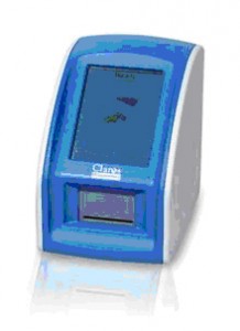
Claros Diagnostics has received regulatory approval from the EU for a new microfluidic point-of-care instrument that measures PSA levels from a finger prick within 15 minutes allowing monitoring of prostrate cancer patients.
 The business end of the machine is a $1 credit card sized, injection moulded, microfluidic cartridge that accepts a small drop of blood and is then inserted into a special reader. Captured proteins are tagged with gold nanoparticles and then developed in a silver solution to form silver plated particles which are easily read by a photodetector. PSA levels are then reported within about 15 minutes of sample input with an accuracy similar to laboratory tests.
Read a related paper on a point-of-care-device for lithium in blood
Microfluidics allows the delivery of rapid results indicate Claros Diagnostics, who aim to make point-of-care PSA monitoring in the doctor’s office a reality. The device recently received EU Regulatory approval and is seeking approval by the U.S. Food and Drug Administration. “Although microfluidics has been a field of scientific endeavour for over 20 years we have not seen full commercialisation of this technology outside the research setting” said Harp Minhas (Editor of the leading journal on micro- and nanofluidics, Lab on a Chip). “However, I have seen evidence that in the next two to three years there are likely to be a flurry of similar point-of-use products that will aim to capture related or overlapping markets.”
Shining light on sperm viability
Optoelectronic tweezers are able to distinguish between live and dead sperm cells, even if they aren’t moving, say US scientists.
To combat this problem, Aaron Ohta at the University of Hawaii and his team have demonstrated that optoelectronic tweezers – which use a combination of light and electric fields to control microscopic objects – can distinguish and sort between live and dead cells, irrespective of mobility.
Link to journal article
Motile and non-motile sperm diagnostic manipulation using optoelectronic tweezers
Aaron T. Ohta, Maurice Garcia, Justin K. Valley, Lia Banie, Hsan-Yin Hsu, Arash Jamshidi, Steven L. Neale, Tom Lue and Ming C. Wu, Lab Chip, 2010, DOI: 10.1039/c0lc00072h
Micropatch detects disease biomarkers in skin
Scientists in Australia have built a microneedle device capable of detecting disease-specific proteins directly from the skin.
 Normally when a clinical sample such as blood is needed to screen a patient for disease, it has to be taken by a specially trained healthcare practitioner using a needle and syringe. The sample is then clotted, centrifuged and stored under controlled conditions ready for analysis.
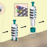
Now Mark Kendall and his colleagues from the University of Queensland, Australia have found an alternative pain-free method which dispenses with invasive needles, specialist training and sample processing. Kendall incorporated a small chip coated with sharp, densely packed microneedles into a patch that can be applied to the skin. The sharp gold-coated silicon needles are less than 1 mm in length and are able to capture and sample protein antibodies directly from the skin.
Link to journal article
Surface-modified microprojection arrays for intradermal biomarker capture, with low non-specific protein binding
Simon R. Corrie, Germain J. P. Fernando, Michael L. Crichton, Marion E. G. Brunck, Chris D. Anderson and Mark A. F. Kendall, Lab Chip, 2010
DOI: 10.1039/c0lc00068j


