Archive for the ‘Hot Articles’ Category

Free access to HOT articles

These HOT articles were recommended by our referees and are free to access for 4 weeks*

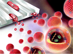
Microfluidic fabrication of cell-derived nanovesicles as endogenous RNA carriers
Wonju Jo, Dayeong Jeong, Junho Kim, Siwoo Cho, Su Chul Jang, Chungmin Han, Ji Yoon Kang, Yong Song Gho and Jaesung Park  
Lab Chip, 2014, Advance Article
DOI: 10.1039/C3LC50993A, Paper

Graphical abstract: Microfluidic fabrication of cell-derived nanovesicles as endogenous RNA carriers
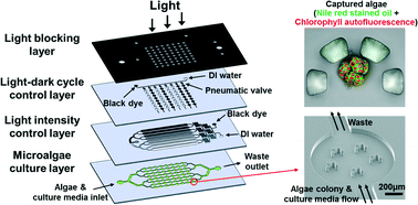
A microfluidic photobioreactor array demonstrating high-throughput screening for microalgal oil production
Hyun Soo Kim, Taylor L. Weiss, Hem R. Thapa, Timothy P. Devarenne and Arum Han  
Lab Chip, 2014, Advance Article
DOI: 10.1039/C3LC51396C, Paper

Graphical abstract: A microfluidic photobioreactor array demonstrating high-throughput screening for microalgal oil production
Mail-order microfluidics: evaluation of stereolithography for the production of microfluidic devices
Anthony K. Au, Wonjae Lee and Albert Folch  
Lab Chip, 2014, Advance Article
DOI: 10.1039/C3LC51360B, Paper

Graphical abstract: Mail-order microfluidics: evaluation of stereolithography for the production of microfluidic devices
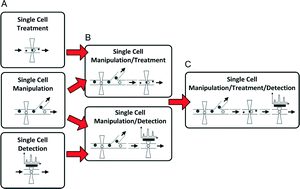
Recent advancements in optofluidics-based single-cell analysis: optical on-chip cellular manipulation, treatment, and property detection
Nien-Tsu Huang, Hua-li Zhang, Meng-Ting Chung, Jung Hwan Seo and Katsuo Kurabayashi  
Lab Chip, 2014, Advance Article
DOI: 10.1039/C3LC51211H, Critical Review

Graphical abstract: Recent advancements in optofluidics-based single-cell analysis: optical on-chip cellular manipulation, treatment, and property detection
Electrokinetically driven reversible banding of colloidal particles near the wall
Necmettin Cevheri and Minami Yoda  
Lab Chip, 2014, Advance Article
DOI: 10.1039/C3LC51341F, Communication

Graphical abstract: Electrokinetically driven reversible banding of colloidal particles near the wall
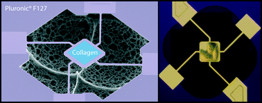
Micro-patterning of mammalian cells on suspended MEMS resonant sensors for long-term growth measurements
Elise A. Corbin, Brian R. Dorvel, Larry J. Millet, William P. King and Rashid Bashir  
Lab Chip, 2014, Advance Article
DOI: 10.1039/C3LC51217G, Technical Innovation

Graphical abstract: Micro-patterning of mammalian cells on suspended MEMS resonant sensors for long-term growth measurements
*Free access to individuals is provided through an RSC Publishing personal account. It’s quick, easy and more importantly – free – to register!

Digg This
Reddit This
Stumble Now!
Share on Facebook
Bookmark this on Delicious
Share on LinkedIn
Bookmark this on Technorati
Post on Twitter
Google Buzz (aka. Google Reader)

Free access to HOT articles!

These HOT articles were recommended by our referees and are free to access for 4 weeks*

Electrostatic potential wells for on-demand drop manipulation in microchannels
Riëlle de Ruiter, Arjen M. Pit, Vitor Martins de Oliveira, Michèl H. G. Duits, Dirk van den Ende and Frieder Mugele  
Lab Chip, 2014,14, 883-891
DOI: 10.1039/C3LC51121A, Paper

Paper-based microfluidics with high resolution, cut on a glass fiber membrane for bioassays
Xueen Fang, Shasha Wei and Jilie Kong  
Lab Chip, 2014,14, 911-915
DOI: 10.1039/C3LC51246K, Paper

The regulation of mobile medical applications
Ali Kemal Yetisen, J. L. Martinez-Hurtado, Fernando da Cruz Vasconcellos, M. C. Emre Simsekler, Muhammad Safwan Akram and Christopher R. Lowe  
Lab Chip, 2014,14, 833-840
DOI: 10.1039/C3LC51235E, Focus 

An automated integrated platform for rapid and sensitive multiplexed protein profiling using human saliva samples
Shuai Nie, W. Hampton Henley, Scott E. Miller, Huaibin Zhang, Kathryn M. Mayer, Patty J. Dennis, Emily A. Oblath, Jean Pierre Alarie, Yue Wu, Frank G. Oppenheim, Frédéric F. Little, Ahmet Z. Uluer, Peidong Wang, J. Michael Ramsey and David R. Walt  
Lab Chip, 2014, Advance Article
DOI: 10.1039/C3LC51303C, Paper

A lab-on-chip cell-based biosensor for label-free sensing of water toxicants
F. Liu, A. N. Nordin, F. Li and I. Voiculescu  
Lab Chip, 2014, Advance Article
DOI: 10.1039/C3LC51085A, Paper

*Free access to individuals is provided through an RSC Publishing personal account. It’s quick, easy and more importantly – free – to register!

Digg This
Reddit This
Stumble Now!
Share on Facebook
Bookmark this on Delicious
Share on LinkedIn
Bookmark this on Technorati
Post on Twitter
Google Buzz (aka. Google Reader)

A microfluidic chip for safekeeping

To accurately diagnose a disease or monitor the effectiveness of a treatment, patient samples (e.g. blood, saliva, or urine) must be analyzed in a central laboratory. After collection, the samples will begin to degrade due to chemical, bacterial, and enzymatic interactions.1 These changes can compromise the integrity of the analyte and introduce errors during analysis. Thus, stabilization techniques are necessary to preserve the diagnostic value of patient samples.

Typically, analyte stabilization is achieved by transporting and storing samples at low temperature using dry ice, refrigerators, freezers, or liquid nitrogen. Unfortunately, low-temperature preservation is costly to implement, requiring substantial infrastructure, specialized equipment, and trained personnel. It becomes particularly expensive for applications involving bio surveillance, long-term sample archiving, and clinical diagnostics in low-resource settings.

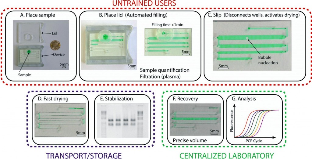
To address this challenge, a research team, led by Prof. Ismagilov at the California Institute of Technology, developed a microfluidic device that can preserve biological samples in the dry state. The device relies on pre-loaded desiccants to rapidly (~30 min) dry samples in a stabilization matrix, protecting the analyte from enzymatic degradation and light or heat activated reactions.2 Importantly, the device is simple to use, allowing minimally trained users to collect and preserve samples without worrying about the precision of the input volume.

The device, based on SlipChip technology,3 was formed by stacking three layers of subassemblies: the top layer has through-holes for sample loading and recovery, the middle layer has channels for sample storage, and the bottom layer houses the desiccants for drying. With the help of a lubricant between each layer, the middle layer can be horizontally moved (slipped), relative to the other layers, allowing the device to be reconfigured into different states.

The device has three states, each with a specific function: loading, drying, and recovery (see figure 1). In the loading state, the loading inlet is connected to the sample storage channel.  An untrained user will place the sample in the inlet and close the lid to create a tight seal. This generates an air pressure that pushes the sample through the channels. To initiate the preservation process, the user will slip the device into the drying state. Here, the sample is disconnected from the inlet and is placed into vapor contact with the desiccant through a porous membrane. At this stage, the device can be stored or transported without the use of low-temperature preservation equipment. Just before analysis in the laboratory, a trained user will use a special tool to slip the device into the recovery state. Here, the sample is disconnected from the desiccant chamber and is connected to the recovery inlets. Using a pipette and water, the samples can be rehydrated and recovered for quantitative analysis.

As a validation, the device was subjected to an accelerated aging test (50oC for 5-weeks) while preserving samples containing control RNA or HIV-1 RNA. Remarkably, the RNA samples stored in the device were indistinguishable from the ones stored in the freezer at -80oC, as tested by electrophoresis and RT-qPCR. In contrast, significant degradation was observed in samples stored in the liquid state at 50oC. These results suggest that the RNA may be stable at room temperature in the device for at least up to 8 months.

In summary, the research team developed and validated a microfluidic device that can preserve biological samples in the dry state, eliminating the need for low-temperature equipment. The device is compact, easy to use, making it compatible for challenging applications such as clinical diagnostics in remote, resource-limited settings. The reduction of cost in transport, sample collection, and storage can open up many new possibilities in health care, diagnostics, and beyond.

To access the full article, click the following link: A microfluidic device for dry sample preservation in remote setting, Stefano Begolo, Feng Shen and Rustem F. Ismagilov.

1.            G. V. Iyengar, K. S. Subramanian and J. R. W. Woittiez, Element Analysis of Biological Samples: Principles and Practice, CRC Press, Boca Raton, New York, 1998.

2.            E. Wan, M. Akana, J. Pons, J. Chen, S. Musone, P. Y. Kwok and W. Liao, Current Issues in Molecular Biology, 2010, 12, 135-142.

3.            W. Du, L. Li, K. P. Nichols and R. F. Ismagilov, Lab on a Chip, 2009, 9, 2286-2292.

Digg This
Reddit This
Stumble Now!
Share on Facebook
Bookmark this on Delicious
Share on LinkedIn
Bookmark this on Technorati
Post on Twitter
Google Buzz (aka. Google Reader)

Free access to HOT articles!

These HOT articles were recommended by our referees and are free to access for 4 weeks*

Antimicrobial susceptibility assays in paper-based portable culture devices
Frédérique Deiss, Maribel E. Funes-Huacca, Jasmin Bal, Katrina F. Tjhung and Ratmir Derda  
Lab Chip, 2014,14, 167-171
DOI: 10.1039/C3LC50887K, Communication
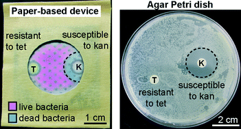
Graphical abstract: Antimicrobial susceptibility assays in paper-based portable culture devices

Cell force measurements in 3D microfabricated environments based on compliant cantilevers
Mattia Marelli, Neha Gadhari, Giovanni Boero, Matthias Chiquet and Jürgen Brugger  
Lab Chip, 2014,14, 286-293
DOI: 10.1039/C3LC51021B, Paper
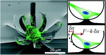
Graphical abstract: Cell force measurements in 3D microfabricated environments based on compliant cantilevers

A differential dielectric affinity glucose sensor
Xian Huang, Charles Leduc, Yann Ravussin, Siqi Li, Erin Davis, Bing Song, Dachao Li, Kexin Xu, Domenico Accili, Qian Wang, Rudolph Leibel and Qiao Lin  
Lab Chip, 2014,14, 294-301
DOI: 10.1039/C3LC51026C, Paper
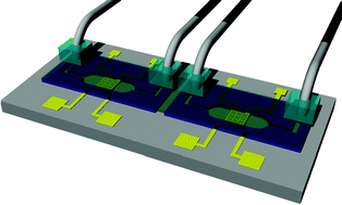
Graphical abstract: A differential dielectric affinity glucose sensor

Microfluidic transwell inserts for generation of tissue culture-friendly gradients in well plates
Christopher G. Sip, Nirveek Bhattacharjee and Albert Folch  
Lab Chip, 2014,14, 302-314
DOI: 10.1039/C3LC51052B, Paper
From themed collection Lab on a Chip Top 10%
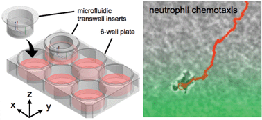
Graphical abstract: Microfluidic transwell inserts for generation of tissue culture-friendly gradients in well plates

Gradient static-strain stimulation in a microfluidic chip for 3D cellular alignment
Hsin-Yi Hsieh, Gulden Camci-Unal, Tsu-Wei Huang, Ronglih Liao, Tsung-Ju Chen, Arghya Paul, Fan-Gang Tseng and Ali Khademhosseini  
Lab Chip, 2014,14, 482-493
DOI: 10.1039/C3LC50884F, Paper
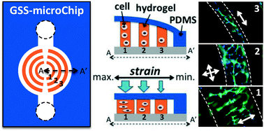
Graphical abstract: Gradient static-strain stimulation in a microfluidic chip for 3D cellular alignment

Biosensor design based on Marangoni flow in an evaporating drop
Joshua R. Trantum, Mark L. Baglia, Zachary E. Eagleton, Raymond L. Mernaugh and Frederick R. Haselton  
Lab Chip, 2014,14, 315-324
DOI: 10.1039/C3LC50991E, Paper
From themed collection Lab on a Chip Top 10%
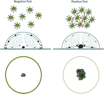
Graphical abstract: Biosensor design based on Marangoni flow in an evaporating drop

Flow of suspensions of carbon nanotubes carrying phase change materials through microchannels and heat transfer enhancement
Sumit Sinha-Ray, Suman Sinha-Ray, Hari Sriram and Alexander L. Yarin  
Lab Chip, 2014,14, 494-508
DOI: 10.1039/C3LC50949D, Paper
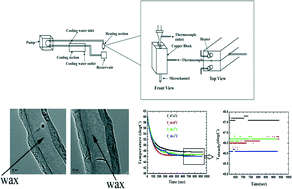
Graphical abstract: Flow of suspensions of carbon nanotubes carrying phase change materials through microchannels and heat transfer enhancement

Micro-scaffold array chip for upgrading cell-based high-throughput drug testing to 3D using benchtop equipment
Xiaokang Li, Xinyong Zhang, Shan Zhao, Jingyu Wang, Gang Liu and Yanan Du  
Lab Chip, 2014,14, 471-481
DOI: 10.1039/C3LC51103K, Paper
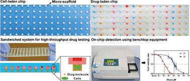
Graphical abstract: Micro-scaffold array chip for upgrading cell-based high-throughput drug testing to 3D using benchtop equipment

Synchronized reinjection and coalescence of droplets in microfluidics
Manhee Lee, Jesse W. Collins, Donald M. Aubrecht, Ralph A. Sperling, Laura Solomon, Jong-Wook Ha, Gi-Ra Yi, David A. Weitz and Vinothan N. Manoharan  
Lab Chip, 2014,14, 509-513
DOI: 10.1039/C3LC51214B, Paper
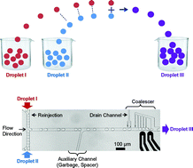
Graphical abstract: Synchronized reinjection and coalescence of droplets in microfluidics

A microfluidic reciprocating intracochlear drug delivery system with reservoir and active dose control
Ernest S. Kim, Erich Gustenhoven, Mark J. Mescher, Erin E. Leary Pararas, Kim A. Smith, Abigail J. Spencer, Vishal Tandon, Jeffrey T. Borenstein and Jason Fiering  
Lab Chip, 2014, Advance Article
DOI: 10.1039/C3LC51105G, Paper
From themed collection Lab on a Chip Top 10%
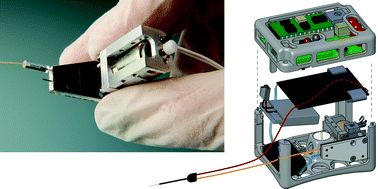
Graphical abstract: A microfluidic reciprocating intracochlear drug delivery system with reservoir and active dose control
Impedance matched channel walls in acoustofluidic systems
Ivo Leibacher, Sebastian Schatzer and Jürg Dual  
Lab Chip, 2014,14, 463-470
DOI: 10.1039/C3LC51109J, Paper
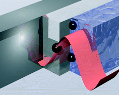
Graphical abstract: Impedance matched channel walls in acoustofluidic systems

Magnetoactive sponges for dynamic control of microfluidic flow patterns in microphysiological systems
Sungmin Hong, Youngmee Jung, Ringo Yen, Hon Fai Chan, Kam W. Leong, George A. Truskey and Xuanhe Zhao  
Lab Chip, 2014,14, 514-521
DOI: 10.1039/C3LC51076J, Paper
From themed collection Lab on a Chip Top 10%
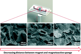
Graphical abstract: Magnetoactive sponges for dynamic control of microfluidic flow patterns in microphysiological systems

The microfluidic post-array device: high throughput production of single emulsion drops
E. Amstad, S. S. Datta and D. A. Weitz  
Lab Chip, 2014, Advance Article
DOI: 10.1039/C3LC51213D, Paper
From themed collection Lab on a Chip Top 10%
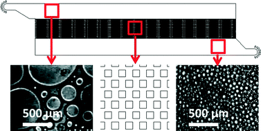
Graphical abstract: The microfluidic post-array device: high throughput production of single emulsion drops

Interdroplet bilayer arrays in millifluidic droplet traps from 3D-printed moulds
Philip H. King, Gareth Jones, Hywel Morgan, Maurits R. R. de Planque and Klaus-Peter Zauner  
Lab Chip, 2014, Advance Article
DOI: 10.1039/C3LC51072G, Paper
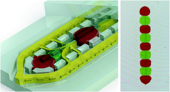
Graphical abstract: Interdroplet bilayer arrays in millifluidic droplet traps from 3D-printed moulds

A 1024-sample serum analyzer chip for cancer diagnostics
Jose L. Garcia-Cordero and Sebastian J. Maerkl  
Lab Chip, 2014, Advance Article
DOI: 10.1039/C3LC51153G, Paper
From themed collection Lab on a Chip Top 10%

Graphical abstract: A 1024-sample serum analyzer chip for cancer diagnostics

Utilization and control of bioactuators across multiple length scales
Vincent Chan, H. Harry Asada and Rashid Bashir  
Lab Chip, 2014, Advance Article
DOI: 10.1039/C3LC50989C, Critical Review
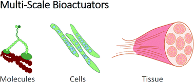
Graphical abstract: Utilization and control of bioactuators across multiple=

Microfabricated perfusable cardiac biowire: a platform that mimics native cardiac bundle
Yun Xiao, Boyang Zhang, Haijiao Liu, Jason W. Miklas, Mark Gagliardi, Aric Pahnke, Nimalan Thavandiran, Yu Sun, Craig Simmons, Gordon Keller and Milica Radisic  
Lab Chip, 2014, Advance Article
DOI: 10.1039/C3LC51123E, Paper
From themed collection Lab on a Chip Top 10%
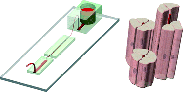
Graphical abstract: Microfabricated perfusable cardiac biowire: a platform that mimics native cardiac bundle
Radiolabelling diverse positron emission tomography (PET) tracers using a single digital microfluidic reactor chip
Supin Chen, Muhammad Rashed Javed, Hee-Kwon Kim, Jack Lei, Mark Lazari, Gaurav J. Shah, R. Michael van Dam, Pei-Yuin Keng and Chang-Jin “CJ” Kim  
Lab Chip, 2014, Advance Article
DOI: 10.1039/C3LC51195B, Paper
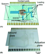
Graphical abstract: Radiolabelling diverse positron emission tomography (PET) tracers using a single digital microfluidic reactor chip

Non-destructive handling of individual chromatin fibers isolated from single cells in a microfluidic device utilizing an optically driven microtool
Hidehiro Oana, Kaori Nishikawa, Hirotada Matsuhara, Ayumu Yamamoto, Takaharu G. Yamamoto, Tokuko Haraguchi, Yasushi Hiraoka and Masao Washizu  
Lab Chip, 2014, Advance Article
DOI: 10.1039/C3LC51111A, Paper
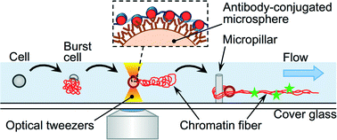
Graphical abstract: Non-destructive handling of individual chromatin fibers isolated from single cells in a microfluidic device utilizing an optically driven microtool

*Free access to individuals is provided through an RSC Publishing personal account. It’s quick, easy and more importantly – free – to register!

Digg This
Reddit This
Stumble Now!
Share on Facebook
Bookmark this on Delicious
Share on LinkedIn
Bookmark this on Technorati
Post on Twitter
Google Buzz (aka. Google Reader)

Re-Writable Microfluidics? New Device Points the Way

Researchers from the University of Tokyo and the CEA-Leti research institute in France have designed a platform that allows channels to be written and rewritten at will in a fluid layer atop a grid of electrodes.

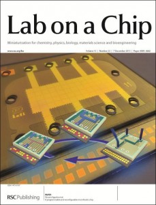
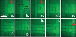
In their recent cover article in Lab on a Chip, Raphael Renaudot and colleagues describe this novel device, which is based on the Electrowetting on Dielectric (EWOD) and Liquid Dielectrophoresis (LDEP) phenomena1. Their goal was to create a flexible, reusable platform that would enable the creation of microfluidic devices without the use of expensive microfabrication techniques. The fluid layer of the device is filled with liquid paraffin, which can easily be solidified and re-melted via thermoelectric cooling and heating (its melting temperature is 35° C.) Water is injected into the paraffin layer, and its flow path is controlled via the underlying electrode layer, which is made up of a grid of 83 electrodes (the small squares seen in the figure). By tuning the interfacial tension between the water and the surface of each electrode via the independently controlled electrode voltages, it is possible to guide a channel (or “finger”) of water along the desired path on the grid (see figure).

After the water is guided into the desired path, the device is cooled, solidifying the paraffin and setting the channels in place. Later, the device can be reheated to erase the existing channels, and a new design can be drawn. The chip can be reused multiple times, reducing waste and making it very useful for prototype testing and low-cost applications. The authors demonstrated two chip designs using the reconfigurable platform: a droplet generator and a device for E. coli confinement within a fluidic cavity.

A water channel is drawn through a paraffin matrix via control of an underlying grid of electrodes (from Figure 2b)

Read this HOT article in Lab on a Chip today!

A Programmable and Reconfigurable Microfluidic Chip, Raphael Renaudot, Vincent Agache, Yves Fouillet, Guillaume Laffite, Emilie Bisceglia, Laurent Jalabert, Momoko Kumemura, Dominique Collard and Hiroyuki Fujita. DOI: 10.1039/c3lc50850a

References:

  1. T. B. Jones and K. L. Wang, Langmuir 2004, 20 (7), 2813–2818.
Digg This
Reddit This
Stumble Now!
Share on Facebook
Bookmark this on Delicious
Share on LinkedIn
Bookmark this on Technorati
Post on Twitter
Google Buzz (aka. Google Reader)

A New Method of Droplet Handling for Ultra-Low Volume Kinetics Studies

In a recent paper in Lab on a Chip, which featured as the Cover Article of Issue 22, Vol 13, researchers from the Ecole Polytechnique, Palaiseau, France describe a micro-droplet based method for measuring reaction kinetics using tiny (20 nL) sample volumes.


When working with precious reagents such as enzymes, minimizing sample volume is of the utmost importance.  Droplet-based microfluidic methods can greatly reduce sample volumes, but still waste considerable amounts of sample in fluid handling processes (e.g. pushing the sample through a channel).  A team led by Marten Vos and Charles Baroud has utilized a unique “rails and anchors” method1 of forming and controlling the movement of droplets to observe the kinetics of a chemical reaction using only 20 nL of each reagent solution.

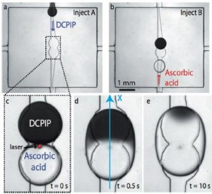
In this scheme, the flow chamber geometry is carefully designed so that when an aqueous solution is injected into the oil-filled chamber, droplets of a reproducible size are released and constrained between the top and bottom surfaces of the chamber.  Then, widening grooves (“rails”) in the bottom of the chamber guide the droplets via surface tension to “anchors” in the center.  (See figure a and b.)  In this way, no additional solution is wasted pushing droplets around.

Droplet-based scheme for observing reaction kinetics (Fig 2 from paper)

The authors used the “rails and anchors” to place two droplets, each containing a different reagent, in proximity, and then used an IR laser pulse to break the surface tension between them (figure c). The interface between the two solutions (in this case, DCPIP and ascorbic acid) can clearly be seen in figure d below.  By tracking the front of the color change that moves across the droplet as the reaction proceeds (figure e), it is possible to calculate the reaction rate (accounting also for the diffusion rates of the reactants).

The researchers also demonstrated the potential of multiplexing their technique by monitoring six different reactions in parallel droplets. They were able to obtain results in good agreement with a commercial stopped-flow spectrometer with greatly reduced cost and time.

Read this HOT article in Lab on a Chip today!

Parallel Measurements of Reaction Kinetics using Ultralow Volumes, Etienne Fradet, Paul Abbyad, Marten H. Vos, and Charles N. Baroud, DOI: 0.1039/c3lc50768h

References:

  1. P. Abbyad et al., Lab on a Chip 2011, 11, 813-821.
Digg This
Reddit This
Stumble Now!
Share on Facebook
Bookmark this on Delicious
Share on LinkedIn
Bookmark this on Technorati
Post on Twitter
Google Buzz (aka. Google Reader)

Free to access HOT articles!

These HOT articles were recommended by our referees and are free to access for 4 weeks*

One-step microfluidic generation of pre-hatching embryo-like core–shell microcapsules for miniaturized 3D culture of pluripotent stem cells
Pranay Agarwal, Shuting Zhao, Peter Bielecki, Wei Rao, Jung Kyu Choi, Yi Zhao, Jianhua Yu, Wujie Zhang and Xiaoming He  
Lab Chip, 2013,13, 4525-4533
DOI: 10.1039/C3LC50678A

A microfluidic approach to synthesizing high-performance microfibers with tunable anhydrous proton conductivity
Mohammad Mahdi Hasani-Sadrabadi, Jules J. VanDersarl, Erfan Dashtimoghadam, Ghasem Bahlakeh, Fatemeh Sadat Majedi, Nassir Mokarram, Arnaud Bertsch, Karl I. Jacob and Philippe Renaud  
Lab Chip, 2013,13, 4549-4553
DOI: 10.1039/C3LC50862E

The microfluidic Kelvin water dropper
Álvaro G. Marín, Wim van Hoeve, Pablo García-Sánchez, Lingling Shui, Yanbo Xie, Marco A. Fontelos,  Jan C. T. Eijkel, Albert van den Berg and Detlef Lohs  
Lab Chip, 2013,13, 4503-4506
DOI: 10.1039/C3LC50832C

A cell rolling cytometer reveals the correlation between mesenchymal stem cell dynamic adhesion and differentiation state
Sungyoung Choi, Oren Levy, Mónica B. Coelho, Joaquim M. S. Cabral, Jeffrey M. Karp and Rohit Karnik  
Lab Chip, 2013, Advance Article
DOI: 10.1039/C3LC50923K

Ultrahigh-throughput sorting of microfluidic drops with flow cytometry
Shaun W. Lim and Adam R. Abate  
Lab Chip, 2013,13, 4563-4572
DOI: 10.1039/C3LC50736J

A programmable and reconfigurable microfluidic chip
Raphael Renaudot, Vincent Agache, Yves Fouillet, Guillaume Laffite, Emilie Bisceglia, Laurent Jalabert, Momoko Kumemura,   Dominique Collard and Hiroyuki Fujita  
Lab Chip, 2013,13, 4517-4524
DOI: 10.1039/C3LC50850A

Digg This
Reddit This
Stumble Now!
Share on Facebook
Bookmark this on Delicious
Share on LinkedIn
Bookmark this on Technorati
Post on Twitter
Google Buzz (aka. Google Reader)

Capillarics – microfluidic circuits

The term ‘microfluidics’ immediately invokes the image of elegant, tiny devices with a small footprint in which fluids are manipulated with ease. It is an image that is most definitely correct. Still, every researcher in the field of microfluidics is aware of ‘the dirty little secret’ of these devices. Namely, that they often require bulky pumps and lots of tubing to be operated.

The requirement for macroscopic equipment to control microfluidic devices is a serious problem. It hinders the development of complex and integrated microfluidic circuits and it prevents the integration of the devices in small portable equipment.

Many researchers in the field are actively developing methods to get rid of all macroscopic equipment to control microfluidic devices. One such method is to build ‘autonomous’ devices, in which the flow is driven only by capillary forces and not by external pumps.

In a recent paper, which featured as the Cover article for Issue 21 of Lab on a Chip, Safavieh and Juncker give a solid overview of the various elements that can be used to autonomously control flow, such as pumps, vents and valves. Moreover, they propose a method called ‘capillarics’ in which autonomous fluidic circuits are designed in a way that is similar to electronic circuits. Finally, they demonstrate the power of their approach by rationally designing a ‘capillaric circuit’ and using it to perform a biochemical assay.

To learn more, read the full HOT article: Capillarics: pre-programmed, self-powered microfluidics circuits built from capillary elements, Roozbeh Safavieh and David Juncker, DOI: 10.1039/C3LC50691F

Digg This
Reddit This
Stumble Now!
Share on Facebook
Bookmark this on Delicious
Share on LinkedIn
Bookmark this on Technorati
Post on Twitter
Google Buzz (aka. Google Reader)

Follow Your Nose: A Micro-aquarium for Probing Bacterial Chemotaxis in Gas Gradients

Many organisms can direct their movements according to their chemical environment. In bacteria, this chemical-directed migration (i.e. chemotaxis) is enabled by receptor-mediated signaling to the bacterial flagellar motors.1 Equipped with this remarkable sensory-motor system, bacteria can measure chemical gradients and swim towards nutritious substances (positive chemotaxis) or flee from cytotoxic chemicals (negative chemotaxis).

Bacterial chemotaxis is critical for various processes including biofilm formation, biomediation, and diseases pathogenesis.2 For example, at the onset of various digestive diseases, bacteria rely on chemotaxis to find a suitable colonization site in the human gastrointestinal tract. Thus, there is great interest in studying the rates and molecular mechanisms in these chemotactic processes.

In the past 10 years, microfluidic gradient-generators have improved the way scientists study bacterial chemotaxis;3 however, these studies have not addressed gas gradients (e.g. O2 and CO2), which are known to be important in bacterial motility.4 Microfluidics devices have enabled the generation of well-controlled chemical gradients and the measurement of bacterial response at high spatiotemporal resolution. Gradients are typically formed by flowing a sample liquid (e.g. aspartate in buffer) and a reference liquid (e.g. buffer) into the device.

A team of researchers at the RIKEN Advanced Science Institute, Japan and Hanyang University, Korea carried out the world’s first microfluidic study of bacterial chemotaxis in gas gradients.5 The microfluidic device (see picture) comprises an isolated chamber (micro-aquarium) for bacterial culture and two bypass channels for sample and reference inputs. In this design, the sample molecules in the channel permeate through the PDMS walls (150 µm thick) and diffuse into the liquid of the micro-aquarium, facilitating the generation of a stable, shear-free gradient. The bacterial response can be monitored optically and quantified via image processing.

Using this device, the research team studied the chemotactic behaviour of Euglena gracilis microbial cells in the presence of CO2 gradients. These studies revealed that the chemotaxis of Euglena cells is rapid and is dependent on CO2 concentration. In particular, negative chemotaxis was observed at high concentrations (>15%) and positive chemotaxis was observed at low concentrations (<15%)— the maximum positive chemotaxis was observed at ~5% CO2, corresponding to the most favourable condition for photosynthesis. Interestingly, the team observed evidence of chemotactic “adaption” when they consecutively switched the sample and reference inputs.

The microfluidic device is also compatible with small molecule liquid samples such as ethanol, H2O2, and culture media. By continuously perfusing culture medium in the bypass channels, the viability of the Euglena cells in the micro-aquarium can be maintained for more than 2 months.

In summary, the research team created a microfluidic gradient-generator that is versatile and straightforward to use. The device is useful for bacterial chemotaxis studies in chemical gradients formed from gas or liquid samples. Furthermore, the device may potentially be useful for other applications including drug screening, toxicity assays, and environmental monitoring.

  1. H. C. Berg, Annual Review of Biochemistry, 2003, 72, 19-54.
  2. T. Ahmed, T. S. Shimizu and R. Stocker, Integrative Biology, 2010, 2, 604-629.
  3. J. Wu, X. Wu and F. Lin, Lab on a Chip, 2013, 13, 2484-2499.
  4. C. Douarche, A. Buguin, H. Salman and A. Libchaber, Physical Review Letters, 2009, 102, 198101.
  5. K. Ozasa, J. Lee, S. Song, M. Hara and M. Maeda, Gas/liquid sensing via chemotaxis of Euglena cells confined in an isolated micro-aquarium, Lab on a Chip, 2013, 13, 4033-4039
Digg This
Reddit This
Stumble Now!
Share on Facebook
Bookmark this on Delicious
Share on LinkedIn
Bookmark this on Technorati
Post on Twitter
Google Buzz (aka. Google Reader)

Molecular Force Assay on a Microfluidic Chip

Researchers in Munich have adapted their molecular force assay technique to a microfluidic system, allowing the strength of protein-DNA interactions to be measured in a chip format.

The measurement of intermolecular forces is a challenging and important task. Two well-known methods, atomic force microscopy and optical trapping, are capable of probing these forces1. However, these instrumentation-intensive techniques are not suitable for every application. A team from the Ludwig-Maximilians-Universität, Germany, headed by Hermann Gaub, a leader in the field of force spectroscopy, has now developed a technique, termed molecular force assay (MFA), to probe the strength of DNA hybridization and the effects of DNA-binding proteins2.

Schematic of the molecular force assay (adapted from Figure 1 of paper)

In MFA, a surface is functionalized with a carefully designed DNA construct, as seen in the left panel of the figure. This surface is brought into contact with another surface which is coated with neutravidin (center). The neutravidin binds the biotin at the end of the DNA construct, so when the two surfaces are separated, the DNA construct rips apart (dehybridization, right). The breakage can happen in one of two ways—if the hybridization of the “probe strand” is stronger than that of the “reference strand”, the reference strand will dehybridize, leaving behind a Cy3 tag. If the hybridization of the reference strand is stronger, the probe strand will dehybridize, leaving both a Cy3 tag and a Cy5 tag behind. Via the resulting fluorescence, it is possible to compare the hybridization strength of the probe strand to that of the known reference. DNA binding proteins which modify the hybridization strength of the probe strand can then be introduced.

In the original formulation of MFA, the surfaces were brought into contact using a piezoelectric device; however, in a recent paper in Lab on a Chip, the researchers have translated their assay into a microfluidic format. A microfluidic chip was used in which the lower layer contains sample wells and the upper layer contains control valves. By pressurizing button valves above each well, the top and bottom surfaces can be brought into contact and then separated. The authors demonstrated the effect of EcoRI binding on probe strand hybridization strength as a proof-of-concept, “paving the way for studies of currently unknown protein-DNA interactions, including those of transcription factors.”

Read this HOT article in Lab on a Chip today!

Protein–DNA force assay in a microfluidic format, Marcus Otten, Philip Wolf, and Hermann E. Gaub. DOI: 10.1039/C3LC50830G

References:

1. K. C. Neuman and A. Nagy, Nature Methods, 2008, 5, 491-505.

2. P. M. D. Severin et al., Lab on a Chip, 2011, 11, 856-862.

Digg This
Reddit This
Stumble Now!
Share on Facebook
Bookmark this on Delicious
Share on LinkedIn
Bookmark this on Technorati
Post on Twitter
Google Buzz (aka. Google Reader)