Archive for the ‘Hot Articles’ Category

Emerging Investigator Series – Kyle Bishop

Kyle Bishop


Introducing Kyle Bishop: Lab on a Chip‘s latest Emerging Investigator

Kyle Bishop received his PhD in Chemical Engineering from Northwestern University under the guidance of Bartosz Grzybowski for work on nanoscale forces in self-assembly. Following his PhD, Dr. Bishop was a post-doctoral fellow with George Whitesides at Harvard University, where he developed new strategies for manipulating flames with electric fields. He started his independent career at Penn State University in the Department of Chemical Engineering. In 2016, Dr. Bishop moved to Columbia University, where he is currently an Associate Professor of Chemical Engineering. Dr. Bishop has been recognized by the 3M Non-tenured Faculty award and the NSF CAREER award. His research seeks to discover, understand, and apply new strategies for organizing and directing colloidal matter through self-assembly and self-organization far-from-equilibrium.


Read Dr Bishop’s article entitled ‘Measurement and mitigation of free convection in microfluidic gradient generators’ and find out more about him in the interview below:

Your recent Emerging Investigator Series paper focuses on the measurement and mitigation of free convection in microfluidic gradient generators. How has your research evolved from your first article to this most recent article?

Our first article in Lab on a Chip focused on harnessing electric potential gradients to power transport and separations within microfluidic systems. Here, we examine how chemical gradients can drive fluid flows as well as motions of colloidal particles, lipid vesicles, and living cells. These topics are linked by our continued interest in harnessing and directing thermodynamic gradients to perform dynamic functions at small scales.

What aspect of your work are you most excited about at the moment?

Currently, we are excited by our pursuit of colloidal “robots” that organise spontaneously in space and time to perform useful functions, which can be rationally encoded within active soft matter.

In your opinion, what is the future of microfluidic gradient generators? Any new applications you foresee for them?

Our interest in microfluidic gradient generators grew from a desire to quantify the motions of lipid vesicles in osmotic gradients (so-called osmophoresis).  These measurements were plagued by undesired gradient-driven flows.  We thought that our efforts to understand and mitigate these flows would be useful to others studying gradient driven motions (e.g., chemotaxis of living cells).

What do you find most challenging about your research?

Staying focused. The world is filled with many micro-mysteries that may pique your curiosity, but time is limited. Picking problems and following through on their solution is an ever-present challenge.

In which upcoming conferences or events may our readers meet you?

Our group regularly attends the AIChE Annual Meeting and the ACS Colloid and Surface Science Symposium.

How do you spend your spare time?

Exploring New York City with my family and thinking about science.

Which profession would you choose if you were not a scientist?

What a horrible thought…perhaps a lawyer as I value evidence-based reasoning and the rule of law (physical or otherwise).

Can you share one piece of career-related advice or wisdom with other early career scientists?

Think big and collaborate often.

Digg This
Reddit This
Stumble Now!
Share on Facebook
Bookmark this on Delicious
Share on LinkedIn
Bookmark this on Technorati
Post on Twitter
Google Buzz (aka. Google Reader)

Quantitative and multiplex microRNA assays from unprocessed cells in isolated nanoliter well arrays

MIT researchers develop a simple new chip that simplifies sample preparation and reduces sample volume for micro-RNA analysis

In recent years, microRNAs (miRNAs) have emerged as potential biomarkers for a range of diseases. These short (~22 nucleotide) sequences of non-coding RNA function by silencing messenger RNA and thus, provide a form of post-transcriptional regulation in the cell. Consequently, their regulation can have a significant impact on cell function. While they are implicated in many diseases, their role in cancer is of particular interest. It is known that multiple miRNAs are dysregulated in tumours compared to healthy tissues and they can be used as biomarkers in cancer diagnosis. Testing for multiple miRNAs provides a better diagnosis, so it is important to be able to test them as a panel. There are many techniques available to test for miRNAs such as gene chips, quantitative RT-PCR, and RNA-seq, but these technologies all have their own shortcomings. What is common to all them is the need for sample preparation, and mainly RNA extraction from the tissue or cells to be tested. In their recent HOT article, Tentori and researchers from the Doyle Group at MIT describe a chip that could overcome this problem and perform multiplexed testing of miRNA all within a minimum volume of just a few nanoliters.

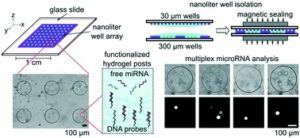
The new chip comprises two glass slides which can be sandwiched together. The bottom slide has an array of 300 µm diameter wells which contain the miRNA sensors and serve to isolate samples into a small reaction volume. The top slide is then used to deliver lysis reagents to the sample. The authors tried a variety of designs for the top plate, but ultimately settled on an array of 30 µm diameter wells. This has two advantages; 1) Reagents are precisely metered and 2) the top and bottom plates can be sandwiched robustly without any need for precision handling or alignment. This last point is really important to Augusto Tentori, a postdoc in the Doyle Group and the lead author of the paper. Tentori wants “to make devices that are simple and robust, so translation is easier.” The miRNA sensors are polyethylene glycol diacrylate (PEGDA) hydrogels that contain complementary DNA probes.  The posts are photopolymerized in the wells and various sizes, shapes, and patterns of posts can be made. Further, a single well can contain a variety of posts, each functionalized with a different DNA probe targeting a different miRNA. In this way, multiplexed assays can be performed with spatial separation. The chip format of the assay is more sensitive than previous formats and can detect miRNA from less than 20 cells.

Tentori is really excited about the prospects of this new chip. His co-authors include pathologists who have been guiding the project to make sure it is clinically useful, and he really wants to see this technology get into the hands of pathologists and diagnostic technicians.

Read the full article by Tentori et al. here “Quantitative and multiplex microRNA assays from unprocessed cells in isolated nanoliter well arrays” that features in Lab on a Chip’HOT article collection

About the webwriter

Darius Rackus (right) is a postdoctoral researcher in the Dittrich Bionalytics Group at ETH Zürich. His research interests are in developing integrated microfluidic tools for healthcare and bioanalysis

Digg This
Reddit This
Stumble Now!
Share on Facebook
Bookmark this on Delicious
Share on LinkedIn
Bookmark this on Technorati
Post on Twitter
Google Buzz (aka. Google Reader)

How soil-worms allow for realistic human physiology studies

Mice, fruit fly, zebra fish easily come to mind when thinking of animal models for human physiology studies – but one animal is often forgotten, although it is as functional as the others. Have you guessed already? We are talking about soil-dwelling worms, aka C. elegans! These animals combine the simplicity of single-cell systems with the complexity of animal models, therefore they can provide significant insights into human disorders. Please take a moment to look at our note below summarizing the key features of C. elegans.

Muscular strength is a good example of human physiology studies  and it relies on calcium-initiated muscle contraction, sarcomere composition and organization, and translocation of actin and myosin molecules. Analysis of such parameters can reveal the formation of muscular dystrophy, a muscle degenerative disorder. However, the measurement of these parameters has been a challenge due to the dependence on random animal-behavior that yields irreproducible results. Recently, researchers from Texas Tech University collaborated with Rutgers University and University of Nottingham to study muscular strength in C. elegans. They achieved to obtain results independent of animal behavior and gait in a miniature system consisting of an elastic micropillar forest (Figure 1a and 1b).

The microfluidic system is made of PDMS, and contains bendable micropillars hanging from the microchannel ceiling. The pillars are bent upon the action of the body muscles when C. elegans crawls through the pillar array. Individual pillar bending events can be quantified using a microscope-camera system and image analysis (Figure 1c). The pillar density is designed to create high mechanical resistance to locomotion, therefore maximum exertable force can be measured independent of animal behavior. Here, maximum exertable force corresponds to the peak force exerted by human quadriceps muscle in a standardized knee extension resistance test.

Figure 1. (a) Image of the microfluidic device with the pillar forest and the ports. (b) Schematic demonstration of the C. elegans strength measurement apparatus. Inset shows a scanning electron microscope picture of the pillars. (c) A sketch of interaction with a pillar by the worm body. The pillar is bent due to the action of the body muscles (shown in red and green). Image from Rahman et al.

The authors of this study explain that animals produce strong forces in highly resistive areas and demonstrate different locomotion regimes based on the body size relative to gap between pillars. Besides the body size, body configuration and behavioral characteristics can be the sources to the magnitude of the force exerted on the pillars. Thanks to the probabilistic nature of the parameters sourcing of the force exerted, a reproducible algorithm can be defined for quantifying muscle strength. Using this strategy, researchers showed for the first time that locomotion between microfluidic pillars comprises of three regimes: non-resistive (worm contacts with 1-2 pillars and doesn’t adjust body posture), moderately resistive (worm contacts with >2 pillars and minimally adjust body posture), and highly resistive (worm contacts with multiple pillars and body posture adjustment is disabled). When operated at highly-resistive regime, the microfluidic system suppresses the animal behavior. This system allows for (1) discriminating between the muscle strength or weakness levels of individual worms of different ages, (2) determining body length decrease and muscular contraction levels led by levamisole treatment, (3) comparing the muscular strength in the wild and mutant C. elegans types. According to the researchers, the future studies can help us to obtain deeper understanding in molecular and cellular circuits of neuromuscular function as well as dissection of degenerative processes in disuse, aging, and disease.

To download the full article for free* click the link below:

NemaFlex: a microfluidics-based technology for standardized measurement of muscular strength of C. elegans
Mizanur Rahman, Jennifer E. Hewitt, Frank Van-Bussel, Hunter Edwards, Jerzy Blawzdziewicz, Nathaniel J. Szewczyk, Monica Driscolld, and Siva A. Vanapalli
Lab Chip, 2018, Lab on a Chip Recent Hot Articles
DOI: 10.1039/c8lc00103k


About the Webwriter

Burcu Gumuscu is a postdoctoral fellow in Herr Lab at UC Berkeley in the United States. Her research interests include development of microfluidic devices for quantitative analysis of proteins from single-cells, next generation sequencing, compartmentalized organ-on-chip studies, and desalination of water on the microscale.

Digg This
Reddit This
Stumble Now!
Share on Facebook
Bookmark this on Delicious
Share on LinkedIn
Bookmark this on Technorati
Post on Twitter
Google Buzz (aka. Google Reader)

What we know about cancer tumors

Cancer tumors are a lot more complex than we think: besides cancer cells, supportive tissue cells, fat, and even immune cells can be found in a tumor. Combined crosstalk in between these cell groups influences the way the tumor develops or responses to drug treatment. On the other hand, the majority of what we know about cancer tumors has been acquired by studying cell ensembles. Recent strides to improve our understanding of cancer revealed that we have long been missing the stochastic interactions and rare events due to ensemble-average measurements. We can unveil how these cell groups work together and how the rare events change the fate of a tumor thanks to single-cell analysis techniques.

Single cells can be identified by extrinsic and intrinsic markers. Extrinsic markers are definitive of genetic and proteomic states of a cell. Flow cytometry and mass spectrometry have been the workhorse of extrinsic marker analysis, where genetic or proteomic materials are often fluorescently labeled for detection. With these techniques, multiplexed analysis of thousands of cells can be employed simultaneously. Intrinsic markers include size, shape, density, optical, mechanical, and electrical properties which do not require labelling. Microfluidic techniques provide with a plethora of different functionalities to sort the cells based on intrinsic markers. Combination of both extrinsic and intrinsic data advances our understanding of how cell heterogeneity is reflected in cell-to-cell variations in tumor development and drug-response. Although many powerful methods are available for determining extrinsic markers, not many techniques can gather information about a panel of different intrinsic markers.

A recent study from Biological Microtechnology and BioMEMS group at MIT represents an important microfluidic approach for the development of multiparameter intrinsic cytometry tool. The approach includes several different microfluidic modules combined with microscope imaging and image processing by machine learning. Separate modules measuring cell size, deformability, and polarization can be combined and organized within the tool (Figure 1). (i) Size module detects the cell size optically in a flow through system. Cell size module is necessary to separate different cell types that can give important cues about disease state. (ii) In deformability module, cells pass through narrow channels, and their transit time defines the deformability. Cell deformability gives cues about cytoskeletal and nuclear changes associated with cancer progression. (iii) In the polarization module, dielectrophoretic force at a fixed frequency is applied on cells driven by opposing hydrodynamic forces. Cells approach coplanar electrodes with different equilibrium positions depending on their polarizability. Cell polarizability allows for distinguishing subtle changes in biological phenotypes. As a proof-of-concept work, drug-induced structural changes in cells were detected for the first time using five different intrinsic markers, including size, deformability, and polarizability at three frequencies. The authors indicate that this powerful tool can further be equipped with visual readout capabilities, such as deterministic lateral displacement array, inertial microfluidics, acoustophoresis, optical techniques.

Figure 1. Multiparameter intrinsic cytometry combines different microfluidic modules on one substrate along with cell tracking to correlate per-cell information across modules for different intrinsic properties including size, polarizability, and deformability.


To download the full article for free* click the link below:

Multiparameter cell-tracking intrinsic cytometry for single-cell characterization
Apichitsopa, A. Jaffe, and J. Voldman
Lab Chip, 2018, Lab on a Chip Recent Hot Articles
DOI: 10.1039/C8LC00240A

*Article free to read until 31st August 2018

About the Webwriter

Burcu Gumuscu is a postdoctoral fellow in Herr Lab at UC Berkeley in the United States. Her research interests include development of microfluidic devices for quantitative analysis of proteins from single-cells, next generation sequencing,compartmentalized organ-on-chip studies, and desalination of water on the microscale.


Digg This
Reddit This
Stumble Now!
Share on Facebook
Bookmark this on Delicious
Share on LinkedIn
Bookmark this on Technorati
Post on Twitter
Google Buzz (aka. Google Reader)

Roll-to-roll PDMS-chips for the masses in molecular diagnostics

PDMS microfluidic devices for molecular diagnostics are now produced at scale using roll-to-roll manufacturing

If there is one material that has enabled microfluidic research in academia, poly(dimethylsiloxane) (PDMS) is surely it. PDMS is cheap and easy to prototype with, and its elastomeric properties have led to complicated structures (e.g. valving) in microfluidic channels. Although it is great for rapid prototyping, there is often a disconnect between the prototype and high throughput manufacturing due to a lack of scalable production methods. Researchers at VTT-Technical Research Centre of Finland and the University of California Berkeley have recently reported a roll-to-roll method for fabricating PDMS microfluidic chips.

In roll-to-roll (R2R) processing—common to the paper industry—long sheets of materials are continuously processed, feeding through rollers and modules with different functionalities. To form R2R microfluidic devices, PDMS was applied to an aluminized paper substrate and then embossed by a heated nickel imprinting cylinder which also cured the PDMS. The devices had good reproducibility and channel depths around 100 µm were achieved. Replication from the nickel master was automated and performed at high throughput of 1.5 m/min. Olli-Heikki Huttunen, one of the authors on the paper, said that “although the process required a lot of fine tuning, it was surprisingly simple.” Like other high-throughput manufacturing techniques (e.g. injection moulding), the nickel tool is quite expensive, but these costs can be overcome by the volume of production.

As a proof-of-principle application, the authors demonstrated nucleic acid detection by loop-mediated isothermal amplification (LAMP). Reagents were spotted and dried in the microchannels using a roll-to-roll compatible dispensing machine, and PDMS lids with vias for fluidic and vacuum connections were formed by a roll-to-roll process (though vias were manually punched) and then bonded manually. Huttunen said that the next steps are to figure out how to manufacture the entire device roll-to-roll, but that it should not be too challenging.

Using aluminized paper as the base substrate for the devices offered a couple advantages. One is that the aluminium dramatically reduced the paper’s autofluorescence. Another advantage was the aluminum reflected back both excitation and emission light, resulting in stronger signals. Results from the test could be read within 20 minutes, suggesting that these devices would be useful for low-cost point-of-care testing.

The challenge for the future, says corresponding author Luke Lee, will be “to learn what the new rules of thinking and design are for roll-to-roll microfluidics in order to solve the problem of mass production in integrated molecular diagnostics for all.” This is an exciting new prospect for both PDMS and the microfluidics community.

To read the full paper for free*, click the link below:

PDMS microfluidic devices for molecular diagnostics are now produced at scale using roll-to-roll manufacturing

*article free to read from 06/06/2018 – 06/07/2018

About the Webwriters

Darius Rackus (Right) is a postdoctoral researcher at the University of Toronto working in the Wheeler Lab. His research interests are in combining sensors with digital microfluidics for healthcare applications.

Digg This
Reddit This
Stumble Now!
Share on Facebook
Bookmark this on Delicious
Share on LinkedIn
Bookmark this on Technorati
Post on Twitter
Google Buzz (aka. Google Reader)

“Cutting edge” technology for cell biology in tape-based devices

Sticker-like devices enable quick, rapid prototyping for cell culture experiments

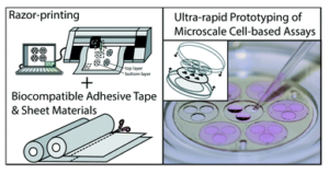
Xurography, or razor-printing, is a low-cost and accessible method for fabricating microfluidic devices. By using a computer controlled razor cutter, sheets of material can be cut precisely to a design. Using adhesive materials, the cut patterns can be used like stickers, and microfluidic devices can then be made by stacking and layering the stickers to create three-dimensional structures. While razor-cut devices might not have the same resolution as soft lithography (150 μm vs. 10-30 μm), their ease of fabrication and rapid turnaround time makes the method very user-friendly and great for rapid prototyping. It is precisely for their ease of use that Jay Warrick (U. Wisconsin) and Maribella Domenech (U. Puerto Rico at Mayagüez) wanted to work with razor-cut microfluidics.

Having access to a very easy fabrication method became a necessity for Domenech. After an electrical fire destroyed her lab and soft lithography equipment in 2016, Domenech was looking for an easy way continue her research while waiting for renovations to be completed. Since she works primarily with undergraduate research students, she needed a fabrication method with a gentle learning curve. “Lithography methods are too difficult to be mastered  within a couple of weeks, but razor-cut devices are easy for anyone to fabricate and use,” says Domenech.

As easy and accessible as any method may be, it won’t gain widespread adoption by a community unless it’s trusted. For biologists, this means trusting that the material is biocompatible and won’t interfere with their experiments. In their recent report, Domenech and Warrick address this challenge and do a service to the community by thoroughly characterizing ARcare 90106, a double-sided adhesive tape for xurography. The tape was compared to polystyrene and PDMS devices, the bread and butter materials of cell biology and microfluidics, respectively. The tape showed good performance across a variety of metrics of cell growth and with a range of cell types. Further, it compared favorably to PDMS in terms of absorption of lipophilic molecules, which means it is less likely to interfere with co-culture experiments where the diffusion of extracellular molecules (e.g., hormones, cytokines, growth factors etc.) is very important.

Easy-made tape-based biocompatible devices open up new opportunities for cell biology. “It’s quite enabling to be able to adhere these devices to so many different types of surfaces,” says Warrick. And because the tape is flexible, it can stick on curved surfaces as well as flat. It also opens up opportunities to integrate new materials with microfluidic devices. Warrick says he’s “often looked at different materials and wished there was an easy way to integrate them. Tape solves this.” In terms of new materials, the team demonstrated the integration of sheets of electrospun collagen within razor-cut microfluidic devices, and co-lead author Yasmín Álvarez-García is currently investigating what other materials could be incorporated. She hopes to expand the current work to include more cell types, perform cell migration studies, and expand the usability of the technique. This will further increase the trustworthiness of the tape’s biocompatibility and lower the barriers for more biologist to get into microfluidics.

To read the full paper for free*, click the link below:

Razor-printed sticker microdevices for cell-based applications

DOI: 10.1039/c7lc00724h (Paper), Lab on a Chip, 2018, 18, 451

__________________

About the Webwriters

Darius Rackus (Right) is a postdoctoral researcher at the University of Toronto working in the Wheeler Lab. His research interests are in combining sensors with digital microfluidics for healthcare applications.


*free access until 28th February 2018

Digg This
Reddit This
Stumble Now!
Share on Facebook
Bookmark this on Delicious
Share on LinkedIn
Bookmark this on Technorati
Post on Twitter
Google Buzz (aka. Google Reader)

Why should we use optofluidics for monitoring marine environment?

Phosphorus is found in natural waters and exerts a major influence on the composition and structure of aquatic ecosystems. It is a crucial nutrient for planktons and algae, which feed fish and other marine organisms. However, human activities may result in excess amount of phosphorus, which, in turn, causes harmful algae to bloom in natural waters. The bloom creates a hostile environment to other forms of marine life by consuming the available oxygen in the sea, and producing toxins. Sea organisms such as fish swim away from the blooms, but the ones that cannot swim, such as shellfish, unfortunately die. We do care about this occurrence as it negatively affects natural life and the economy. There is only one way to interpret the effect of continuously changing phosphorus levels on the strength of the biological pump: real-time monitoring of phosphate levels in the marine environment!

Figure 1. The design of the Fabry-Pérot microcavity, consisting of two parallel mirrors (reflectors) fabricated by coating the surface of the optical fibers with a gold layer. The light is reflected by the mirrors multiple times to enhance the signal. Adapted from Zhu et al., 2017.

Conventional vs. optofluidic monitoring instruments

Conventional phosphate monitoring instruments are mostly used for on-site sampling, then the fresh sample is transported to a laboratory for determining the phosphate level. Laboratories complete one round of analysis in 20 min, often using spectrophotometrical measurement tools. Given the conditions, real-time phosphate monitoring easily becomes laborious, time consuming, and costly. To address this challenge, researchers in Chinese Academy of Sciences, Wuhan University, and The First Institute of Oceanography in China collaborated to develop a portable optofluidic phosphate monitoring tool. However, prototyping an optofluidic marine phosphate detection tool is not straightforward because an absorption cell—a component core to the measurement unit—is simply too big to fit in a microchip. Instead of using a bulky absorption cell, researchers considered integrating a Fabry-Pérot cavity in the microsystem. The Fabry-Pérot cavity consists of two parallel optical fibers with a spacing in between. The cross-sectional surface of each optical fiber is coated with a thin layer of gold to create reflector surfaces (Figure 1) in order to enhance the absorption of phosphate. Shortening the spacing between the reflectors decreases the analysis time from minutes to seconds.


How it works?

In the microchip, filtered water sample and a chromogenic reagent are injected into a curved microchannel. After the chromogenic reaction, the water-soluble components are transported into the optical section (Figure 2). The probe light is sent into the Fabry-Pérot cavity via one of the fibers, bounces between the reflectors multiple times to increase the optical feedback and then analyzed by the detector. The obtained absorbance value, therefore, increases linearly with increasing phosphate concentration. In this microsystem, phosphate detection range is 0.1-100 µmol per liter (400 times greater than the range of a conventional instrument) and detection time is 4 seconds (300 times shorter than detection time of a conventional instrument). The authors of the paper think that this technology can be applied to detect other nutrient levels as well as pH changes in marine environment.


optofluidic phosphate monitoring

Figure 2. A schematic of the optofluidic microchip consisting of two parts: the microfluidics circuit forming the microreactor in the microchannel, and the optical part to provide optical feedback for enhanced absorption analysis. Adapted from Zhu et al., 2017.

To download the full article for free* click the link below:


Optofluidic marine phosphate detection with enhanced absorption using a Fabry–Pérot resonator

M. Zhu, Y. Shi, X. Q. Zhu, Y. Yang, F. H. Jiang, C. J. Sun, W. H. Zhaoc, and X. T. Hanc

Lab Chip, 2017, Lab on a Chip Recent Hot Articles

DOI: 10.1039/C7LC01016H


About the Webwriter

Burcu Gumuscu is a postdoctoral fellow in Herr Lab at UC Berkeley in the United States. Her research interests include development of microfluidic devices for quantitative analysis of proteins from single-cells, next generation sequencing, compartmentalized organ-on-chip studies, and desalination of water on the microscale.

*until 16th February 2018

Digg This
Reddit This
Stumble Now!
Share on Facebook
Bookmark this on Delicious
Share on LinkedIn
Bookmark this on Technorati
Post on Twitter
Google Buzz (aka. Google Reader)

Spotting cancer: one in a billion cells

The human body consists of tens of trillions cells, all of which theoretically should have the same genome. Depending on genetic and environmental factors, some of these cells experience point mutations. Although most of those mutations are cleaned up by DNA repair enzymes, about 0.01% of them stay. A low percentage of the persistent mutations turn out to be ‘cancer’ while others stay recessive. Several genes, including the ones responsible for cell growth cycle, cause the persistent mutations. Uncontrolled growth of cells leads to formation of tumor, which is now prone to experience more mutations due to continuous proliferation. These mutations create heterogeneity among the cell population of a tumor, and eventually some cells leave their original tumor and start a new one in another organ of the body. When a cell leaves its parent tumor, it starts circulating in blood vessels before settling in. Those cells are called circulating tumor cells (CTCs), and around 1-10 CTC can be found in 1 mL of blood (which contains about 1 billion red blood cells, and 1 million white blood cells). Capturing ultra-rare CTCs has enormous implications in early cancer diagnosis. Often times, analysis of at least 10 mL of blood is necessary to capture sufficient CTCs to confirm their presence. The available technology can achieve CTC capturing in about 10 hours leading to the loss of target cells and decay of detection biomarkers.

Microfluidic devices are well known for precise sorting of microscale materials. Parallelizing microscale sorting in microfluidic devices enables high-throughput sample processing. In the light of this principle, David Issadore, Jina Ko, and their fellow researchers at University of Pennsylvania coined CaTCh FISH, a circulating tumor cell fluorescence in situ hybridization platform for rapid detection of CTCs. David Issadore kindly accepted to talk about this exciting lab-on-a-chip device. According to David, “the main strength of the CaTCh FISH is that it preserves the sensitivity and specificity of microfluidic cell sorting and RNA FISH, but through clever engineering allows these normally very slow laboratory-based operations to be performed rapidly and automatically on a chip.”

RNA FISH, high-throughput cell handling

Figure 1. Overview of the CaTCh FISH platform. Whole blood sample is processed in TEMPO step, where magnetic nano particle based cell separation is followed by single-cell RNA analysis (modified from Ko et al., 2017).

Processing a whole blood sample using CaTCh FISH involves three steps (Figure 1). First, white blood cells are labelled with magnetic nanoparticles. Second, whole blood is passed through a magnetic micropore filter to selectively trap magnetically labelled cells. “Our magnetic micropore device rapidly and precisely removes all of the cells that we know are not CTCs”, says David. The operating principle of magnetic micropore filter is based on strong and highly localized microscale field gradients formed at the edge of micropores to enable application of high flow rates. Third, single cell RNA analysis is performed on isolated cells using rapid in-situ hybridization strategy so that CTCs can be identified within the isolated cell population. In this way, targeted CTCs can be isolated from the rest of the cell population regardless of their physical and molecular properties. Analysis of a 10 mL blood sample takes less than an hour.

As a key novelty, the researchers maintained high-throughput processing and high sensitivity at the same time by integrating the FISH technique (hybridization of 20-50 fluorescently-labelled oligonucleotide probes to the target RNA, and subsequent fluorescence-signal based detection to enhance signal-to-noise ratio) in a microfluidic chip. CaTCh FISH has also been tested in patients with pancreatic cancer and detected CTCs in the real patient samples.

“The CaTCH FISH technology can be easily modified to measure other rare cells, for the diagnosis of other cancers or for stem cell research for example, by modifying the RNA FISH probes”, says David. He considers converting the platform to a high-precision hospital-based diagnostic tool and he collaborates with a company in the Bay area for this process. The CaTCh FISH device is poised to have a big impact on the way cancer is diagnosed.

To download the full article for free* click the link below:

A magnetic micropore chip for rapid (<1 hour) unbiased circulating tumor cell isolation and in situ RNA analysis

Jina Ko, Neha Bhagwat, Stephanie S. Yee, Taylor Black, Colleen Redlinger, Janae Romeo, Mark O’Hara, Arjun Raj, Erica L. Carpenter, Ben Z. Stanger and David Issadore

Lab Chip, 2017, Paper

DOI: 10.1039/C7LC00703E


About the Webwriter

Burcu Gumuscu is a postdoctoral fellow in Herr Lab at UC Berkeley in the United States. Her research interests include development of microfluidic devices for quantitative analysis of proteins from single-cells, next generation sequencing, compartmentalized organ-on-chip studies, and desalination of water on the microscale.

*until 5th January 2018

Digg This
Reddit This
Stumble Now!
Share on Facebook
Bookmark this on Delicious
Share on LinkedIn
Bookmark this on Technorati
Post on Twitter
Google Buzz (aka. Google Reader)

Zenith in “artery”

When cutting a finger, thrombocytes and fibrin in the blood make up the blood-clotting mechanism, aka. haemostasis, to stop the blood loss. Another way to trigger this mechanism is having an artery damaged by atherosclerosis, which is often caused by several genetic or acquired factors. In the latter case, thrombosis develops within a vein or artery, obstructing or stopping the blood flow to major organs like the heart and eventually causing heart attack. Considering every year over 14 million lives worldwide are lost to heart attacks, more investigation on this topic is needed without any doubt.

Recently, a research team led by Andries van der Meer published a research article in Lab on a Chip on mimicking arterial thrombosis in 3D vascular structures, representing a major step forward in the development of accurate and faster methods of studying arterial thrombosis without using animals. The authors highlighted the inconvenience of using animal models to predict arterial thrombosis in humans. This is mainly due to fundamental differences between human and animal physiology, the researchers explain. For instance, rodent platelet biology, coagulation dynamics, and shear stress in mice arteries significantly vary between humans and mice.

thrombosis on chip

Figure 1. Three-dimensional models of a healthy and stenotic vessels and thrombosis formation upon blood perfusion through the channels.

The paper uses miniaturized vascular structures mimicking 3D architectures found in both healthy and stenotic blood vessels in-vitro (Figure 1). They combined stereolithography and 3D printing of computed tomography angiography data to construct 3D-printed templates of vessels in PDMS microchips. The 3D printed vessels are then coated with human umbilical vein endothelial cells, forming a monolayer fully covering the surface. In the next step, the artificial vessels are perfused with blood at normal arterial shear rates, allowing a blood clot to form as it would happen in the human body. The 3D printed vessel is clinically more relevant when compared to 2D vessel models, since the realistic flow profiles of blood and even distribution of shear stress across the vessel are of great importance when researching arterial thrombosis. Hugo Albers, the co-first author of the paper explains what led the team to try 3D models: “Other groups have worked on thrombosis-on-a-chip before, but we wanted to incorporate flow profiles that are similar to what one would find in-vivo. So we opt for a round and thus 3D shape. Since the stenotic geometry is an important part of this work, we wanted to find a technique that allowed us to make almost any shape we could come up with. Thus 3D-printing seemed to be the way to go.”

When it comes to defining the challenges in 3D organ-on-chip modeling and fabrication, “we needed to replicate the cellular environment using human endothelial cells and human whole blood to fully mimic the nature of vasculature” says Albers. “Incorporating the shape of vasculature to recreate the flow profiles found in-vivo and recreating the shape of vasculature on a small scale was quite challenging, since the resolution of 3D-printing quickly started to be the limiting factor. Furthermore, we ran into problems related to working with whole blood. We had to figure out how to perfuse small channels with blood without instigating thrombosis outside of the microfluidic channel.” The researchers successfully overcame the challenges mentioned by Albers and mimicked the formation of thrombosis in a stenotic vessel model as seen in Figure 1 (bottom).

The researchers note that the next step involves co-culturing arterial endothelial cells and smooth muscle cells with human umbilical vein endothelial cells or moving to different cell lines such as differentiated human induced pluripotent stem cells. “I think we can also apply the 3D-printing technique to create thrombosis-on-a-chip devices with different geometries, e.g. aneurysms or bifurcated geometries”, says Albers.

To download the full article for free* click the link below:

Mimicking arterial thrombosis in a 3D-printed microfluidic in vitro vascular model based on computed tomography angiography data

Pedro F. Costa, Hugo J. Albers, John E. A. Linssen, Heleen H. T. Middelkamp, Linda van der Hout, Robert Passier, Albert van den Berg, Jos Malda and Andries D. van der Meer

Lab Chip, 2017, Paper

DOI: 10.1039/C7LC00202E

This paper is included in our Organ-, Body- and Disease-on-a-Chip Thematic Collection. To read other articles in the collection, visit – rsc.li/organonachip

About the Webwriter

Burcu Gumuscu is a postdoctoral fellow in Herr Lab at UC Berkeley in the United States. Her research interests include development of microfluidic devices for quantitative analysis of proteins from single-cells, next generation sequencing, compartmentalized organ-on-chip studies, and desalination of water on the microscale.

Digg This
Reddit This
Stumble Now!
Share on Facebook
Bookmark this on Delicious
Share on LinkedIn
Bookmark this on Technorati
Post on Twitter
Google Buzz (aka. Google Reader)

Droplets droplets everywhere: optimising genetic circuits using cell-free extracts and droplet microfluidics

The design and engineering of biological systems — a field known as Synthetic Biology —has been heralded by many to be the defining technology of the 21st century. Just as electronics and computers have changed almost every facet of our day-to-day lives, the consequences of engineering microbes might also be as far reaching. Engineered bugs are already being developed to produce pharmaceuticals and fuels, monitor their environment, detect trace chemicals, degrade plastic waste, and perform computing operations.

Microbes, however, are considerably more complex — and hence less predictable — than electronic components. For this reason, one of the best strategies scientists have for engineering them is to use a brute force approach: try out as many different biodesigns as possible and see what works best.

Doing this with microbes is tricky. Designing and constructing a single engineered bacteria is labour intensive and can take weeks. To circumvent this, researchers have turned to cell-free systems: a collection of biomolecules that can reproduce biochemical features of interest, for example the synthesis of proteins from a genetic circuit. This greatly simplifies things as after all you are now merely handling what is essentially a sack of chemicals.

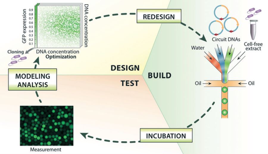
This is where it really gets interesting. As opposed to testing out different genetic circuits one-by-one in a test tube, Hori et al developed a microfluidic device which forms millions of droplets an hour – each droplet acts as a mini test tube, each containing a slightly different version of the circuit. The circuit was made up of three strands of DNA which interact with one another in a non-linear way to produce a Green Fluorescent Protein. The fact that it is non-linear means predicting how they interact is difficult and makes the tactic of trying out different combinations more appealing. Using microfluidics, the authors created a generic library by combining different ratios of these three components. By incubating the droplets and monitoring protein production levels, they were able to map out the parameter space to see which genetic circuit from their library worked best.

Although simply a proof-of-concept, this droplet microfluidic approach has shown to be incredibly powerful, and in addition to being used in genetic circuit design has the potential to rapidly explore large parameter spaces in other areas of chemistry and biology.


To download the full article for free* click the link below:

Cell-free extract based optimization of biomolecular circuits with droplet microfluidics
Yutaka Hori, Chaitanya Kantak, Richard M. Murray and Adam R. Abate
Lab Chip, 2017
DOI: 10.1039/C7LC00552K

*Free to access until 8th September 2017.


About the Webwriter

Yuval Elani is an EPSRC Research Fellow in the Department of Chemistry at Imperial College London. His research involves using microfluidics in bottom-up synthetic biology for biomembrane construction, artificial cell assembly, and for exploring bionterfaces between living and synthetic microsystems.

Digg This
Reddit This
Stumble Now!
Share on Facebook
Bookmark this on Delicious
Share on LinkedIn
Bookmark this on Technorati
Post on Twitter
Google Buzz (aka. Google Reader)