Archive for the ‘Cover articles’ Category

LOC Issue 8 now online: Art in Science

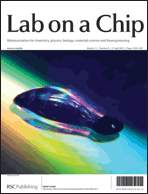
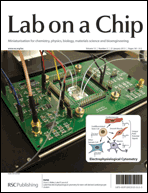
The winner of the 2012 Art in Science Award presented at October’s MicroTas meeting was Yi Zhang, from Johns Hopkins University, USA and the striking winning image is featured on the outside front cover of Issue 8!

Read what’s in the issue here

A description of the winning image and advice on what makes a worthy Art in Science submission are discussed in the editorial by Michael Gaitan from NIST, USA, and Harp Minhas, Editor of LOC, who were part of the award selection committee.

The Art in Science of microTAS
Michael Gaitan and Harp Minhas
DOI: 10.1039/C3LC90026F
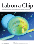

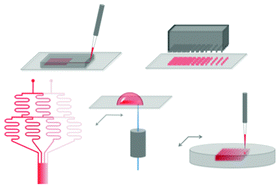
In keeping with the artistry of the front cover, work from Shoji Takeuchi et al. in Japan is featured on the inside front cover of Issue 8. In this paper, they address the problem of how to exchange the solution within a droplet for a new solution, for example to introduce membrane proteins or to wash-out applied chemicals during ion channel analysis. They use microfluidic channels to carry out solution exchange by droplet contact in under 20 seconds with the membrane still intact. Cover articles are free to access for 6 weeks*!

Droplet-based lipid bilayer system integrated with microfluidic channels for solution exchange
Yutaro Tsuji, Ryuji Kawano, Toshihisa Osaki, Koki Kamiya, Norihisa Miki and Shoji Takeuchi
DOI: 10.1039/C3LC41359D


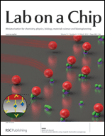
The back cover features the laboratory of Sergey Shevkoplyas at Tulane University, USA. The communication by this laboratory describes their simple point-of-care test for sickle cell disease using characteristic patterns made by blood samples on paper, which can even differentiate between sickle cell disease and those healthy people with sickle cell traits.

A simple, rapid, low-cost diagnostic test for sickle cell disease
Xiaoxi Yang, Julie Kanter, Nathaniel Z. Piety, Melody S. Benton, Seth M. Vignes and Sergey S. Shevkoplyas
DOI: 10.1039/C3LC41302K


This issue also includes plenty of HOT research and a focus article from Tony Huang on the more unconventional applications of microfluidics:

Unconventional microfluidics: expanding the discipline
Ahmad Ahsan Nawaz, Xiaole Mao, Zackary S. Stratton and Tony Jun Huang
DOI: 10.1039/C3LC90023A

Programmable parylene-C bonding layer fluorescence for storing information on microfluidic chips
Ata Tuna Ciftlik, Diego Gabriel Dupouy and Martin A. M. Gijs
DOI: 10.1039/C3LC41280F

Engineering of functional, perfusable 3D microvascular networks on a chip
Sudong Kim, Hyunjae Lee, Minhwan Chung and Noo Li Jeon
DOI: 10.1039/C3LC41320A

In situ synthesis of silver nanoparticle decorated vertical nanowalls in a microfluidic device for ultrasensitive in-channel SERS sensing
Joseph Parisi, Liang Su and Yu Lei
DOI: 10.1039/C3LC41249K

Digg This
Reddit This
Stumble Now!
Share on Facebook
Bookmark this on Delicious
Share on LinkedIn
Bookmark this on Technorati
Post on Twitter
Google Buzz (aka. Google Reader)

Issue 7 online! The latest HOT miniaturisation research and critical reviews on microfabricated organ systems, nucleic acid amplification and microfluidics in IVF

As highlighted on the outside front cover, physicists, engineers and chemists at the University of Illinois , USA, describe a new laser-based label-free resonant optical biosensor with high resolution and high sensitivity. The difference with this sensor is that they use a photonic crystal resonant reflector surface to introduce optical gain to the sensor from an external source via simulated emission. All of our cover articles are free to access for 6 weeks,* so you can read the full work now:

External cavity laser biosensor
Chun Ge, Meng Lu, Sherine George, Timothy A. Flood, Clark Wagner, Jie Zheng, Anusha Pokhriyal, J. Gary Eden, Paul J. Hergenrother and Brian T. Cunningham
DOI: 10.1039/C3LC41330F

On the inside front cover, researchers at California Institute of Technology, USA, and LeukoDx Inc., Israel, present a point-of-care test for leukocyte counting using a microflow cytometer and fluorescent dye. This method eliminated excessive dilution and sheath flow, giving a minimal needed sample volume.

Four-part leukocyte differential count based on sheathless microflow cytometer and fluorescent dye assay
Wendian Shi, Luke Guo, Harvey Kasdan and Yu-Chong Tai
DOI: 10.1039/C3LC41059E

Work from Tino Frank and Savaş Tay at ETH Zurich is featured on the inside back cover. To improve studies of cell signalling in vitro, this article introduces a simple cell culture platform that can produce programmable diffusion-based gradients using microfluidics based on modifying flow over time. Read it here:

Flow-switching allows independently programmable, extremely stable, high-throughput diffusion-based gradients
Tino Frank and Savaş Tay
DOI: 10.1039/C3LC41076E

The outside back cover highlights the work of Sung Gap Im at KAIST, South Korea. This article is about a doubly cross-linked nano-adhesive system (DCNA) and the team demonstrate fabrication of microfluidic devices with flexible and rigid substrates with high strength and stability. The flexible devices could be manipulated without delamination occurring.

A doubly cross-linked nano-adhesive for the reliable sealing of flexible microfluidic devices
Jae Bem You, Kyoung-Ik Min, Bora Lee, Dong-Pyo Kim and Sung Gap Im
DOI: 10.1039/C2LC41266G


In addition to the primary microfluidics research and high number of HOT articles in issue 7, there are also three critical reviews:

Microfabricated mammalian organ systems and their integration into models of whole animals and humans
Jong H. Sung, Mandy B. Esch, Jean-Matthieu Prot, Christopher J. Long, Alec Smith, James J. Hickman and Michael L. Shuler
DOI: 10.1039/C3LC41017J

Thinking big by thinking small: application of microfluidic technology to improve ART
J. E. Swain, D. Lai, S. Takayama and G. D. Smith
DOI: 10.1039/C3LC41290C

Nucleic acid amplification using microfluidic systems
Chen-Min Chang, Wen-Hsin Chang, Chih-Hung Wang, Jung-Hao Wang, John D. Mai and Gwo-Bin Lee
DOI: 10.1039/C3LC41097H

View the whole issue here

*Free access to individuals is provided through an RSC Publishing personal account. Registration is quick, free and simple


Digg This
Reddit This
Stumble Now!
Share on Facebook
Bookmark this on Delicious
Share on LinkedIn
Bookmark this on Technorati
Post on Twitter
Google Buzz (aka. Google Reader)

HOT review: Forming the wiring of the nervous system – discovering the mechanisms of axon guidance through new technologies

Today’s HOT article is a review article that was featured on the front cover of our Neuroengineering themed issue.

In this review, Santiago Costantino et al. at the University of Montreal and McGill University, Canada, critically discuss the most recent insights into axon guidance that have been enabled by developments in methods and technologies for engineered cell substrates.

This review includes:

  1. Extracellular cues and growth cones
  2. Historical background
  3. The stripe assay in discovery of guidance cues and signalling mechanisms
  4. Microcontact printing – development from the stripe assay
  5. Insights via microfluidics
  6. Studies using laser-assisted printing and patterning 3D hydrogels

The focus is on the biological insights into how axons recognise and interpret external signals in order to reach their synaptic target that have come out of using these technologies, rather than the development of the technology itself. This cover article is still free to access for another couple of weeks*, read it now by clicking the link below:

Engineered cell culture substrates for axon guidance studies: moving beyond proof of concept
Joannie Roy, Timothy E. Kennedy and Santiago Costantino
DOI: 10.1039/C2LC41002H

*Free access to individuals is provided through an RSC Publishing personal account. Registration is quick, free and simple

Digg This
Reddit This
Stumble Now!
Share on Facebook
Bookmark this on Delicious
Share on LinkedIn
Bookmark this on Technorati
Post on Twitter
Google Buzz (aka. Google Reader)

Welcome to Lab on a Chip Issue 6! A microrobot, adaptive coatings, protein detection, flow cytometry, a critical review and the final Acoustofluidics paper

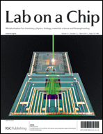
On the outside front cover is a Technical Innovation from Oliver Hayden, Michael Helou and co-workers in Germany describing a magnetic time-of-flight flow cytometry workflow for point-of-care tests, integrating sample preparation with the use of superparamagnetic labels:

Time-of-flight magnetic flow cytometry in whole blood with integrated sample preparation
Michael Helou, Mathias Reisbeck, Sandro F. Tedde, Lukas Richter, Ludwig Bär, Jacobus J. Bosch, Roland H. Stauber, Eckhard Quandt and Oliver Hayden
DOI: 10.1039/C3LC41310A


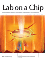
The inside front cover highlights work by Martin Gijs’s team at EPFL, Switzerland, on a microfluidic method for ultra-sensitive protein detection in serum using magnetic beads:

Attomolar protein detection using a magnetic bead surface coverage assay
H. Cumhur Tekin, Matteo Cornaglia and Martin A. M. Gijs
DOI: 10.1039/C3LC41285G


Mengsu Yang and co-workers at the City University of Hong Kong are advertised on the back cover, which illustrates their research on monitoring of intracellular calcium signals after mechanical stimulation using a single integrated microfluidic device:

Microfluidics study of intracellular calcium response to mechanical stimulation on single suspension cells
Tao Xu, Wanqing Yue, Cheuk-Wing Li, Xinsheng Yao and Mengsu Yang
DOI: 10.1039/C3LC40880A


Remember, all of our cover articles are free to access for 6 weeks!*

*Free access to individuals is provided through an RSC Publishing personal account. Registration is quick, free and simple


The final paper in our highly interesting Acoustofluidics series of tutorial articles is published in Issue 6. It points out the ability to combine acoustic forces with others for microfluidic applications:

Acoustofluidics 23: acoustic manipulation combined with other force fields
Peter Glynne-Jones and Martyn Hill
DOI: 10.1039/C3LC41369A

Issue 6 also includes a Critical Review on sample preparation for cell analysis technology from Dino Di Carlo et al. at the University of California, USA:

Microfluidic sample preparation for diagnostic cytopathology
Albert J. Mach, Oladunni B. Adeyiga and Dino Di Carlo
DOI: 10.1039/C2LC41104K

HOT articles in Issue 6 include:

On-chip microrobot for investigating the response of aquatic microorganisms to mechanical stimulation
Tomohiro Kawahara, Masakuni Sugita, Masaya Hagiwara, Fumihito Arai, Hiroyuki Kawano, Ikuko Shihira-Ishikawa and Atsushi Miyawaki
DOI: 10.1039/C2LC41190C

Dynamic pH mapping in microfluidic devices by integrating adaptive coatings based on polyaniline with colorimetric imaging techniques
Larisa Florea, Cormac Fay, Emer Lahiff, Thomas Phelan, Noel E. O’Connor, Brian Corcoran, Dermot Diamond and Fernando Benito-Lopez
DOI: 10.1039/C2LC41065F

For more fascinating Lab on a Chip content, read the full issue here

Digg This
Reddit This
Stumble Now!
Share on Facebook
Bookmark this on Delicious
Share on LinkedIn
Bookmark this on Technorati
Post on Twitter
Google Buzz (aka. Google Reader)

Constant stretching of DNA in a microfluidic device for higher throughput Genome Sequence Scanning

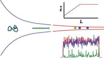
This HOT article by Robert Meltzer et al. at Pathogenetix Inc., USA, describes new geometries for the stretching funnel in Genome Sequence Scanning that increase DNA throughput by 30 times.

Genome Sequence Scanning (GSS) detects sequence-specific fluorescent tags on DNA fragments. It is used to identify bacteria by first lysing open the bacteria cell to release the DNA, then using a restriction endonuclease enzyme to digest the DNA into smaller fragments. Fluorescent tags are added that recognise specific repeated elements present in all bacterial genomes. GSS characterises the bacterial genome by the spatial distribution of the tags.

genome sequence scanning, GSSThe detection is done in a continuous flow microfluidic device with confocal microscopy. In order to carry out the spatial recognition of the fluorescent tags along the length of the DNA fragment, it needs to be stretched out into a linear form using a funnel. High molecule throughput is important as the detection confidence of this technology relies on observing as many tags as possible in the specified experimental period.

The team looks at the relationship between the funnel taper shape and related parameters, such as fluid velocity and fragment length, to improve the current designs and increase throughput. Their new geometries are able to keep the tension applied to the DNA constant during the detection process. Because DNA fragments come in various lengths, a very important goal is to maximise the range of lengths that can be stretched effectively with the funnel. The influence of channel etch depth on fluid flow, and therefore throughput, is also considered.

You can learn more about the work of Pathogenetix on Genome Sequence Scanning by visiting their website. This HOT article, which was featured on the cover of Issue 2, is free to access for the next 4 weeks* and you can read it by clicking the link below:

High-throughput genome scanning in constant tension fluidic funnels
Joshua W. Griffis, Ekaterina Protozanova, Douglas B. Cameron and Robert H. Meltzer
DOI: 10.1039/C2LC40943G#

*Free access to individuals is provided through an RSC Publishing personal account. Registration is quick, free and simple


Keep up to date with the latest Lab on a Chip news and blogs by following us on Twitter! @LabonaChip

Digg This
Reddit This
Stumble Now!
Share on Facebook
Bookmark this on Delicious
Share on LinkedIn
Bookmark this on Technorati
Post on Twitter
Google Buzz (aka. Google Reader)

Lab on a Chip Issue 5 is online! Towards point-of-care biosensors, an RNAi screening method and a review of microfluidics for liposome production

The front cover of Issue 5 illustrates work from Peter Ertl and colleagues at AIT, Austria, in which they combine organic photodiodes and electrode structures in a lab on a chip to develop the ability to quickly determine cell number to standardise microfluidic cell culture and simultaneously measure cell adhesion and interactions.

Standardization of microfluidic cell cultures using integrated organic photodiodes and electrode arrays
Verena Charwat, Michaela Purtscher, Sandro F. Tedde, Oliver Hayden and Peter Ertl
DOI: 10.1039/C2LC40965H


Research by scientists at the National Research Council and McGill University, Canada, features on the striking inside front cover. Teodor Veres and co-workers take a step towards the development of point-of-care nanoplasmonic biosensors with their all-polymeric transmission surface plasmon resonance sensor. It uses a simple and widely available technique and they demonstrate its application in sensing glycoprotein sCD44 at picomolar to nanomolar concentrations.

All-thermoplastic nanoplasmonic microfluidic device for transmission SPR biosensing
Lidija Malic, Keith Morton, Liviu Clime and Teodor Veres
DOI: 10.1039/C2LC41123G


The back cover advert highlights research from the groups of Anup Singh and Oscar Negrete at Sandia National Laboratories, USA, on their advantageous microfluidic RNA interference screening method, which they validate using vesicular stomatitis virus and Rift Valley fever virus.

Microfluidic platforms for RNA interference screening of virus–host interactions
Benjamin R. Schudel, Brooke Harmon, Vinay V. Abhyankar, Benjamin W. Pruitt, Oscar A. Negrete and Anup K. Singh
DOI: 10.1039/C2LC41165B


This issue also features Research highlights from Ali Khademhosseini on biomimetic honeycombs, microfluidic vessel chips and BioMEMS, and a critical review written by Andrew deMello and Dirk van Swaay at ETH Zurich on the microfluidic technologies available for producing liposomes:

Research highlights
Gulden Camci-Unal, Šeila Selimović, Mehmet R. Dokmeci and Ali Khademhosseini
DOI: 10.1039/C3LC90002A

Microfluidic methods for forming liposomes
Dirk van Swaay and Andrew deMello
DOI: 10.1039/C2LC41121K


Find the issue in full here

Digg This
Reddit This
Stumble Now!
Share on Facebook
Bookmark this on Delicious
Share on LinkedIn
Bookmark this on Technorati
Post on Twitter
Google Buzz (aka. Google Reader)

LOC issue 4 – the Neuroengineering 2013 themed issue is online now!


Lab on a Chip’s 2013 Neuroengineering themed issue is introduced by guest editors David T. Eddington and Justin Williams, who also contributed the outside front cover design. This is followed by profiles of all of the contributors to the Neuroengineering themed issue.

Neuroengineering
David T. Eddington and Justin Williams
DOI: 10.1039/C3LC90003G


As well as plenty of cutting-edge primary research into lab on a chip technology for neuroengineering, Issue 4 includes two Critical Reviews.

A critical review of the development of engineered cell culture substrates and techniques for investigating axon development guidance by Santiago Costantino et al. at University of Montreal and McGill University, Canada, is highlighted on the inside front cover. They look at the opportunities that are now opening up due to these new technological developments, the biological insights that can now be gained and the breakthroughs waiting to happen in the near future.

Engineered cell culture substrates for axon guidance studies: moving beyond proof of concept
Joannie Roy, Timothy E. Kennedy and Santiago Costantino
DOI: 10.1039/C2LC41002H


The second critical review from Noo Li Jeon et al. at Seoul National University, Korea, and The Salk Institute, USA, summarises the most recent technological developments of BioMEMs devices and their application to neuroscience research. They look at platforms for disease culture, modelling disease in vitro, neuron electrophysiology and stem cell biology.

Advances in microfluidics-based experimental methods for neuroscience research
Jae Woo Park, Hyung Joon Kim, Myeong Woo Kang and Noo Li Jeon
DOI: 10.1039/C2LC41081H


The HOT articles in this issue include:

Integration of pre-aligned liquid metal electrodes for neural stimulation within a user-friendly microfluidic platform
Nicholas Hallfors, Asif Khan, Michael D. Dickey and Anne Marion Taylor
DOI: 10.1039/C2LC40954B

Inherent amplitude demodulation of an AC-EWOD (electrowetting on dielectric) droplet
Myung Gon Yoon, Sang Hyun Byun and Sung Kwon Cho
DOI: 10.1039/C2LC41043E

One-step polymer surface modification for minimizing drug, protein, and DNA adsorption in microanalytical systems
Esben Kjær Unmack Larsen and Niels B. Larsen
DOI: 10.1039/C2LC40750G


To learn all about the exciting advances happening in applying microtechnology to neuroengineering, read the full issue here

Digg This
Reddit This
Stumble Now!
Share on Facebook
Bookmark this on Delicious
Share on LinkedIn
Bookmark this on Technorati
Post on Twitter
Google Buzz (aka. Google Reader)

Microdevices in cell biology: not one, but three HOT articles!

microfluidic x-ray cellsBritta Weinhausen and Sarah Köster study hydrated eukaryotic cells device using X-rays in a new microfluidic device in their Technical Innovation, free to access for 4 weeks*.

The team at Georg-August-Universität Göttinghen, Germany, overcome some of the limitations of previous work to create a new type of device suitable for studying hydrated cells, with the possibility of applying it to living cells.

They use UV-curable glue (NOA 81) allowing sophisticated channel structures to be defined, two thin Kapton foils, with advantageous properties for X-ray studies, and a Si3N4 membrane window, upon which cells can be grown and fixed before the device is closed with a second membrane. Despite the small difference in electron density between the cellular material and water, they successfully produce X-ray dark-field images:

Microfluidic devices for X-ray studies on hydrated cells
Britta Weinhausen and Sarah Köster
DOI: 10.1039/C2LC41014A


cytometryAs mentioned in the LOC issue 2 blog, Luke Lee et al. at the University of California, Berkeley and Stanford University, featured on the front cover with their work on sorting stem cells using a cell cytometer based on the electrophysiological response to stimulus. As a HOT article that was featured on the cover, this article has been free to access* for 6 weeks from mid-December so get your skates on to read it in full in the next week:

Label-free electrophysiological cytometry for stem cell-derived cardiomyocyte clusters
Frank B. Myers, Christopher K. Zarins, Oscar J. Abilez and Luke P. Lee
DOI: 10.1039/C2LC40905D


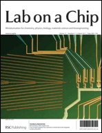
electroporationIn this Technical Innovation, Rashid Bashir led a team of researchers from the USA, Sweden and Singapore in using silicon nanowires for rapid lysis of single cells by ultra-localized electroporation to release cell components for analysis. They integrate label-free magnetic manipulation to correctly position cells with the use of field effect transistors to apply an electric field for cell lysis.

Their method eliminates the use of microfluidic trapping techniques, transparent substrates for optical tweezing and high voltages, amongst other requirements for previous devices.

This work is featured on the inside front cover of Issue 3, so it’s currently free to access for 6 weeks*. Do have a read of the article here:

Ultra-localized single cell electroporation using silicon nanowires
Nima Jokilaakso, Eric Salm, Aaron Chen, Larry Millet, Carlos Duarte Guevara, Brian Dorvel, Bobby Reddy, Amelie Eriksson Karlstrom, Yu Chen, Hongmiao Ji, Yu Chen, Ratnasingham Sooryakumar and Rashid Bashir
DOI: 10.1039/C2LC40837F

*Free access to individuals is provided through an RSC Publishing personal account. Registration is quick, free and simple

Digg This
Reddit This
Stumble Now!
Share on Facebook
Bookmark this on Delicious
Share on LinkedIn
Bookmark this on Technorati
Post on Twitter
Google Buzz (aka. Google Reader)

LOC issue 2 – sorting stem cell-derived cardiomyocyte clusters, cross talk between cancer and immune cells and Genome Sequence Scanning

Welcome to Issue 2 of Lab on a Chip! This issue features the winner of the Lab on a Chip Pioneers of Miniaturisation Lectureship, Andrew deMello, on the back cover. Read more about this award and others given at MicroTAS here.

On the front cover of issue 2 is featured work from the group of Luke Lee at the University of California, Berkeley, in conjunction with colleagues at Stanford University.

In the paper, the authors use a ‘non-genetic, label-free cell cytometry method based on electrophysiological response to stimulus’ to sort undifferentiated stem cells from heterogeneous stem cell progeny.

The cell cytometer can identify human induced pluripotent stem cell-derived cardiomyocyte clusters from their extracellular field potential signals – these stem cells can then be used for various stem cell therapies.

Label-free electrophysiological cytometry for stem cell-derived cardiomyocyte clusters
Frank B. Myers, Christopher K. Zarins, Oscar J. Abilez and Luke P. Lee
DOI: 10.1039/C2LC40905D

Work from the Italian National Research Council – Institute for Photonics and Nanotechnologies and the Istituto Superiore di Sanità is featured on the inside front cover.

The groups, led by Fabrizio Mattei and Luca Businaro, have used an on-chip co-culture system to investigate interactions between cancer cells and a host’s immune system.

Cross talk between cancer and immune cells: exploring complex dynamics in a microfluidic environment
Luca Businaro, Adele De Ninno, Giovanna Schiavoni, Valeria Lucarini, Gabriele Ciasca, Annamaria Gerardino, Filippo Belardelli, Lucia Gabriele and Fabrizio Mattei
DOI: 10.1039/C2LC40887B

On the inside back cover, work from Robert Meltzer and co-workers at Pathogenetix, Inc. is featured.

In their paper, they present a novel compound funnel design for use with Genome Sequence Scanning (GSS) technology, which improves molecule throughput.

High-throughput genome scanning in constant tension fluidic funnels
Joshua W. Griffis, Ekaterina Protozanova, Douglas B. Cameron and Robert H. Meltzer
DOI: 10.1039/C2LC40943G

As with all our cover articles these are free to access for 6 weeks (following a simple registration for an RSC Publishing account).

Other HOT articles featured in the issue include:

Benchtop fabrication of microfluidic systems based on curable polymers with improved solvent compatibility
Michinao Hashimoto, Robert Langer and Daniel S. Kohane
DOI: 10.1039/C2LC40888K

Microfluidic assisted self-assembly of chitosan based nanoparticles as drug delivery agents
Fatemeh Sadat Majedi, Mohammad Mahdi Hasani-Sadrabadi, Shahriar Hojjati Emami, Mohammad Ali Shokrgozar, Jules John VanDersarl, Erfan Dashtimoghadam, Arnaud Bertsch and Philippe Renaud
DOI: 10.1039/C2LC41045A

Microfluidic devices for X-ray studies on hydrated cells
Britta Weinhausen and Sarah Köster
DOI: 10.1039/C2LC41014A

For even more exciting microfluidics research, read the full issue here.

Digg This
Reddit This
Stumble Now!
Share on Facebook
Bookmark this on Delicious
Share on LinkedIn
Bookmark this on Technorati
Post on Twitter
Google Buzz (aka. Google Reader)

Issue 22 – Focus on Scandinavia

Issue 22 of Lab on a Chip features work from Scandinavia – read the editorial from the Guest Editors Professors Thomas Laurell and Jörg Kutter here. The issue features 8 articles from Scandinavian authors, and profiles of the athours included can be found here.

The front cover features work from Jörg Kutter and co-workers at the Technical University of Denmark, reporting on the optical properties of gold nanoparticle probes, used as sensors for environmental contaminants.

Gold nanoparticle-based optical microfluidic sensors for analysis of environmental pollutants
Josiane P. Lafleur, Silja Senkbeil, Thomas G. Jensen and Jörg P. Kutter
DOI: 10.1039/C2LC40543A

Artwork linked to research from Vincent Aimez and colleagues from the Université de Sherbrooke can be seen on the inside front cover of the issue – they have developed a microfluidic device to monitor the radioactivity concentration in the blood of rats and mice in real time.

Blood compatible microfluidic system for pharmacokinetic studies in small animals
Laurence Convert, Frédérique Girard Baril, Vincent Boisselle, Jean-François Pratte, Réjean Fontaine, Roger Lecomte, Paul G. Charette and Vincent Aimez
DOI: 10.1039/C2LC40550D

The back cover of the issue features work from Samir Iqbal and co-workers at the University of Texas and Lehigh University. The team designed a Hele-Shaw device with aptamer functionalized glass beads to isolate cancer cells from a cellular mixture.

Capture, isolation and release of cancer cells with aptamer-functionalized glass bead array
Yuan Wan, Yaling Liu, Peter B. Allen, Waseem Asghar, M. Arif Iftakher Mahmood, Jifu Tan, Holli Duhon, Young-tae Kim, Andrew D. Ellington and Samir M. Iqbal
DOI: 10.1039/C2LC21251J

Other HOT papers in the issue include:

Two-hundredfold volume concentration of dilute cell and particle suspensions using chip integrated multistage acoustophoresis
Maria Nordin and Thomas Laurell
DOI: 10.1039/C2LC40629B

Inertial microfluidics in parallel channels for high-throughput applications
Jonas Hansson, J. Mikael Karlsson, Tommy Haraldsson, Hjalmar Brismar, W. van der Wijngaart and Aman Russom
DOI: 10.1039/C2LC40241F

Low cost integration of 3D-electrode structures into microfluidic devices by replica molding
Benjamin Mustin and Boris Stoeber
DOI: 10.1039/C2LC40728K

A microfluidic device with removable packaging for the real time visualisation of intracellular effects of nanosecond electrical pulses on adherent cells
C. Dalmay, M. A. De Menorval, O. Français, L. M. Mir and B. Le Pioufle
DOI: 10.1039/C2LC40857K

Programmable microfluidic synthesis of spectrally encoded microspheres
R. E. Gerver, R. Gómez-Sjöberg, B. C. Baxter, K. S. Thorn, P. M. Fordyce, C. A. Diaz-Botia, B. A. Helms and J. L. DeRisi
DOI: 10.1039/C2LC40699C

Digg This
Reddit This
Stumble Now!
Share on Facebook
Bookmark this on Delicious
Share on LinkedIn
Bookmark this on Technorati
Post on Twitter
Google Buzz (aka. Google Reader)