Archive for the ‘Chemistry World Highlights’ Category

Unravelling chromosomes

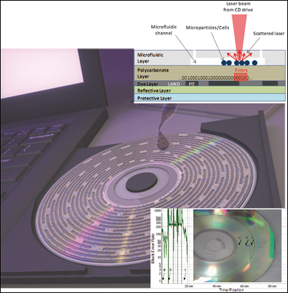
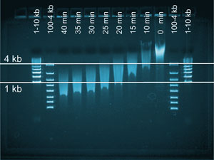
Chromosomes were extracted by washing cell extracts with a protein digester in a microfluidic trap

Danish scientists have used a micro device to isolate centimetre-long portions of human DNA to help study the genetic make-up of diseased cells.

Rodolphe Marie at the Technical University of Denmark, Kongens Lyngby, and colleagues made the device to isolate chromosomes from cell extract samples and manipulate them in such a way that the strands remain intact.

Being able to sequence DNA accurately is a challenge, but Marie’s technique allows the DNA to remain intact so that the gene sequence can be read in one go. The device is made from silicon and consists of an isolation zone in which cell samples are trapped and washed to obtain chromosomes.

Read Catherine Bacon’s Chemistry World article online here or go straight to the HOT Lab on a Chip paper:

A device for extraction, manipulation and stretching of DNA from single human chromosomes
Kristian H. Rasmussen, Rodolphe Marie, Jacob M. Lange, Winnie E. Svendsen, Anders Kristensen and Kalim U. Mir
Lab Chip, 2011, Advance Article
DOI: 10.1039/c0lc00603c

Digg This
Reddit This
Stumble Now!
Share on Facebook
Bookmark this on Delicious
Share on LinkedIn
Bookmark this on Technorati
Post on Twitter
Google Buzz (aka. Google Reader)

Diagnosing diseases with CDs

Cells in the microfluidic channels interfere with the laser beam in a standard CD drive and cause errors in reading and decoding the digital data previously burned on the CD

A digital compact disc integrated with a microfluidic device to analyse cells has been developed by scientists in the US. The disc can be inserted into a standard computer disc drive for analysis and could be used to diagnose diseases.

Gang Logan Liu and coworkers at the University of Illinois at Urbana-Champaign built a layer onto a CD that converts biological information into digital information to detect the presence of cells and their size and number. Blood cell counting and sizing are often standard medical practices in the diagnosis of diseases such as leukemia, anemia and Aids.

The team made the device by burning an audio file consisting of a repeating sequence of binary numbers onto a CD’s surface. On top of this, they added a microfluidic layer with a channel. They injected small fluid samples of cells into the channel and focused the laser beam from a CD drive onto the disc’s data layer, which reads the binary code. Any particles on the layer interfere with the laser beam, interrupting the code and altering the data readout in graphs of error against time. The size of the response correlates with the cells’ shape, concentration and optical density.

Read the full Chemistry World article online here or go straight to the Lab on a Chip paper:

Microparticle and cell counting with digital microfluidic compact disc using standard CD drive
Syed M. Imaad, Nathan Lord, Gulsim Kulsharova and Gang Logan Liu
Lab Chip, 2011, Advance Article
DOI: 10.1039/c0lc00451k

Digg This
Reddit This
Stumble Now!
Share on Facebook
Bookmark this on Delicious
Share on LinkedIn
Bookmark this on Technorati
Post on Twitter
Google Buzz (aka. Google Reader)

Mapping brain networks

The ring structure of the neuron networks was prepared by stamping proteins onto a glass substrate and growing neurons on the proteins

US scientists have created a model of the ring-shaped networks of neurons in the brain, which could help researchers to understand small changes within diseased brain cells.

Henry Zeringue, and colleagues at the University of Pittsburgh, developed the model to enable the study of brain circuitry in an in vitro system instead of using brain slices. In neurological diseases, such as Alzheimer’s disease, subtle changes in this activity take years, or even decades, to become clinically detectable. Most studies only explore the electrical properties of a single neuron or the connection between two neurons. Until now, there hasn’t been a good model to study network activity in vitro.

Zeringue and his team are considering  using their platform to characterise network-level changes based on small experimental perturbations, like changes in network geometry or expression of mutant proteins in 10 per cent of the neurons. ‘Deciphering the relationship between the connectivity of the neurons and the activity in the ring culture will be challenging and unravelling the connections between the neurons may still prove to be a daunting task,’ he concludes.

Read Carl Saxton’s Chemistry World article online here or go straight to the Lab on a Chip paper:

Ring-shaped neuronal networks: a platform to study persistent activity
Ashwin Vishwanathan, Guo-Qiang Bi and Henry C. Zeringue,
Lab Chip, 2011, 11, 1081
DOI: 10.1039/c0lc00450b

Digg This
Reddit This
Stumble Now!
Share on Facebook
Bookmark this on Delicious
Share on LinkedIn
Bookmark this on Technorati
Post on Twitter
Google Buzz (aka. Google Reader)

Remote powered lab on a chip

The chip consists of a printed integrated circuit and a microfluidic device, powered wirelessly by a palm sized RFID reader

A team of US scientists has developed the first lab on a chip device to be powered remotely.

Wen Qiao at the University of California, San Diego, made a microfluidic chip that can be powered with a commercially available radio frequency transmitter for electrophoresis experiments.

Qiao’s team made the chip by printing a circuit onto a plastic sheet. Within the circuit, they placed a chamber containing microwells.

The device is cheap to produce and simple to use and can be used in the same way as a microscope slide, with the RFID transmitter mounted next to a microscope stage and a camera to capture images of the moving nanoparticles. Qiao says that the chip will ‘greatly simplify the operation of the device for pathologists and clinicians whose training and practices have been mostly on optical microscopes, with limited experience with sophisticated electronic instruments.’

Check out the full Chemistry World story online here or read the Lab on a Chip article:

Wirelessly powered microfluidic dielectrophoresis devices using printable RF circuits
Wen Qiao, Gyoujin Cho and Yu-Hwa Lo
Lab Chip, 2011, Advance Article
DOI: 10.1039/c0lc00457j

Digg This
Reddit This
Stumble Now!
Share on Facebook
Bookmark this on Delicious
Share on LinkedIn
Bookmark this on Technorati
Post on Twitter
Google Buzz (aka. Google Reader)

Crime scene DNA testing on the move

A microfluidic chip that can come up with a DNA profile in less than three hours has been designed by US scientists for use at crime scenes.

With current techniques, forensic scientists have to wait up to eight hours to get results. Using microchips to speed up the process has been investigated but integrating all of the profiling steps in one device has remained elusive until now. Richard Mathies from the University of California, Berkeley, and colleagues, in collaboration with the US Department of Justice, have produced a portable method to test DNA at a crime scene that integrates all of the steps in one device.

Andy Hopwood, an expert in DNA analysis techniques from the UK’s Forensic Science Service, believes that the work is ‘without a doubt a very exciting and significant development toward the total integration of the DNA-based human identification process onto a single microchip’.

Read Holly Sheahan’s Chemistry World article online here or go straight to the HOT Lab on a Chip paper:

Integrated DNA purification, PCR, sample cleanup, and capillary electrophoresis microchip for forensic human identification
Peng Liu, Xiujun Li, Susan A. Greenspoon, James R. Scherer and Richard A. Mathies
Lab Chip, 2011, 11, 1041
DOI: 10.1039/c0lc00533a

Digg This
Reddit This
Stumble Now!
Share on Facebook
Bookmark this on Delicious
Share on LinkedIn
Bookmark this on Technorati
Post on Twitter
Google Buzz (aka. Google Reader)

Chips make short work of RNA synthesis

Chinese scientists have developed a much easier way to make the short strands of RNA that are an essential tool in understanding what genes do.

Short interfering ribonucleic acids (siRNAs) were first discovered in 1999, and found to interfere with the expression of specific genes, giving them a key role in controlling the molecular machinery in living organisms. Though initially identified in plants, they were later found in animals too, and this spurred an interest in using them as tools to investigate what specific genes do in the body.

One type of siRNAs, endoribonuclease-prepared siRNAs (esiRNAs), has recently attracted attention because of their greater specificity and their cost effectiveness. Jianzhong Xi and colleagues at Peking University have now demonstrated a lab on a chip method that makes large scale manufacture of esiRNAs much easier.

The chip consists of 96 pins. Each pin has a polymer bead at its end in which a number of DNA probes are immobilised, allowing hundreds of esiRNA products to be manipulated at the same time

The chip consists of 96 pins. Each pin has a polymer bead at its end in which a number of DNA probes are immobilised, allowing hundreds of esiRNA products to be manipulated at the same time.

Read Catherine Bacon’s Chemistry World article online here or go straight to the HOT Lab on a Chip paper:

A polyacrylamide microbead-integrated chip for the large-scale manufacture of ready-to-use esiRNA
Huang Huang, Qing Chang, Changhong Sun, Shenyi Yin, Juan Li and Jianzhong Jeff Xi
Lab Chip, 2011, 11, Advance Article
DOI: 10.1039/C0LC00564A

Digg This
Reddit This
Stumble Now!
Share on Facebook
Bookmark this on Delicious
Share on LinkedIn
Bookmark this on Technorati
Post on Twitter
Google Buzz (aka. Google Reader)

Microfluidic pinball

A device to deposit polymer layers on oil droplets has been made by researchers from Singapore, who say that their design is faster and

Oil droplets are guided through three liquid streams - two polymers and a washing station - by micropillars arranged in a zigzag fashion

more efficient than conventional deposition techniques and it achieves the highest number of polymer layers reported so far using microfluidics. The device could be used to encapsulate drugs for delivery or be used to create capsules for biosensing.

Dieter Trau from the National University of Singapore and colleagues have used a method that they call ‘microfluidic pinball’ to guide oil droplets through channels created by rows of micropillars, like a pinball machine. The rows are orientated in a zigzag fashion across three liquid streams – two polymers and a washing station. The angles created by the rows, as well as the flow rates in the streams, determine how long droplets stay in each channel.

The droplets are guided along the rows to travel repeatedly through the three streams. The team found that six polyelectrolyte layers could be deposited on a droplet in under three minutes – they were able to see the layers with fluorescence spectroscopy. Atomic force microscopy revealed the thickness of each layer to be approximately 2.8nm.

Read Jennifer Newton’s Chemistry World article online here or go straight to the HOT Lab on a Chip paper:


A microfluidic pinball for on-chip generation of Layer-by-Layer polyelectrolyte microcapsules

Chaitanya Kantak, Sebastian Beyer, Levent Yobas, Tushar Bansal and Dieter Trau,
Lab Chip, 2011
DOI: 10.1039/c0lc00381f

Digg This
Reddit This
Stumble Now!
Share on Facebook
Bookmark this on Delicious
Share on LinkedIn
Bookmark this on Technorati
Post on Twitter
Google Buzz (aka. Google Reader)

Ferrofluids help you see better

US researchers have used ferrofluids as liquid pistons that could be used to make adjustable liquid lenses with nearly perfect spherical interfaces for applications such as an optometrist’s phoropter. A phoropter measures the way light is focused in the eye and is used to determine prescriptions for glasses and contact lenses.

Ferrofluids are colloidal solutions of ferromagnetic nanoparticles suspended in a dispersing liquid. Ferrofluid droplets can be manipulated by a magnetic field, so they could be used in systems that need precise control, such as optics, drug delivery, and electronic devices.

Amir Hirsa and colleagues from Rensselaer Polytechnic Institute in Troy, New York, have made such a device by filling three of four holes in a substrate with ferrofluid; the ferrofluid’s surface tension allows droplets to protrude from either side of the substrate. They filled the fourth hole with 1-methylnaphthalene, a compound used as a liquid lens. They sealed the system and filled it with water, producing two chambers, with the substrate as the separator and the ferrofluid and 1-methylnaphthalene being the only connections between them.

As a magnetic field was applied to the device, the ferrofluid moved further into one chamber, pushing the 1-methylnaphthalene liquid lens and changing its curvature

Read the full story here

Link to journal article
Electromagnetic liquid pistons for capillarity-based pumping 
Bernard A. Malouin Jr, Michael J. Vogel, Joseph D. Olles, Lili Cheng and Amir H. Hirsa,
Lab Chip, 2011, DOI: 10.1039/c0lc00397b

Digg This
Reddit This
Stumble Now!
Share on Facebook
Bookmark this on Delicious
Share on LinkedIn
Bookmark this on Technorati
Post on Twitter
Google Buzz (aka. Google Reader)

Lung implant is a breath of fresh air

Artificial lung technology could reduce the death rate for patients awaiting a lung transplant, say US scientists.

Advanced lung disease is characterised by an inability to remove carbon dioxide from the blood and reduced oxygen uptake efficiency. A shortage of donors can mean long delays and high mortality rates for those awaiting a transplant. The only technology available to aid sufferers during this time is based in intensive care units, hindering quality of life.

Now, Joseph Vacanti and coworkers at Massachusetts General Hospital, Boston, have developed a device that achieves the CO2/O2 gas exchange that, when implanted in the body, could allow patients more freedom when awaiting a transplant. Their design is a microfluidic branched vascular network through which blood flows, separated from a gas-filled chamber by a silicone membrane less than 10um thick. The network is formed by casting polydimethylsiloxane, a biocompatible polymer, on a micro machined mould.

A device that achieves carbon dioxide/oxygen gas exchange could allow patients more freedom when awaiting a lung transplant

Read the full story here

Link to journal article
Lung assist device technology with physiologic blood flow developed on a tissue engineered scaffold platform
David M. Hoganson, Howard I. Pryor II, Erik K. Bassett, Ira D. Spool and Joseph P. Vacanti, Lab Chip, 2011
DOI: 10.1039/c0lc00158a

Digg This
Reddit This
Stumble Now!
Share on Facebook
Bookmark this on Delicious
Share on LinkedIn
Bookmark this on Technorati
Post on Twitter
Google Buzz (aka. Google Reader)

Medicine gets smart

Korean scientists have developed a fast and simple mobile phone-based device to test urine samples for common diseases in developing countries. This could provide a cheap, painless solution for detecting disease in remote areas.

Dae-Sik Lee at the Electronics and Telecommunications Research Institute and colleagues developed a pocket-sized urinalysis colorimetric reader capable of sending data wirelessly via a smart phone. To take urine samples, the team used a commercially available 10-parameter urinalysis paper strip that detects glucose, protein, bilirubin, urobilinogen, ketones, nitrite, pH, specific gravity, erythrocytes and leukocytes.

Lee’s team tested the device on 1000 human urine samples and the results were comparable with those given by hospital equipment, demonstrating reliable glucose and protein analysis. It uses a colorimetric multidetection diode comprising LEDs, photodiodes and an optical splitter, which reads the colour intensity changes on the paper strips.

In remote areas of the developing world early detection and prevention of disease is rare. Portable lab on a chip devices have been developed to help combat this but many are not up to the standards required for use in such environments, as they are often expensive with high power consumption and take a long time to produce results, whereas Lee’s device can produce readings within six seconds.

The simple and easy to use handheld device could prove vital for patients in remote areas of the developing world

Read the full story here

Link to journal article
A simple and smart telemedicine device for developing regions: a pocket-sized colorimetric reader
Dae-Sik Lee, Byoung Goo Jeon, Chunhwa Ihm, Je-Kyun Park and Mun Yeon Jung, Lab Chip, 2011
DOI: 10.1039/c0lc00209g

Digg This
Reddit This
Stumble Now!
Share on Facebook
Bookmark this on Delicious
Share on LinkedIn
Bookmark this on Technorati
Post on Twitter
Google Buzz (aka. Google Reader)