Author Archive

New speedy method for the production of monodisperse droplets!

A group of scientists at IMTEK, University of Freiburg have developed a new method for the production of monodisperse droplets. Previous methods, such as T-junctions and flow focusing require several channels, containing either the disperse phase (which will form the droplet) or the continuous phase (which will surround the droplet), with the droplets forming at a constriction point in the tubing. Extremely precise control of flow rate is therefore required in order to achieve consistent droplet diameters. These methods also have substantial dead volumes due to sample material remaining in the tubing at the end of the process.

An alternative method is step emulsification (as highlighted recently in a Lab on a Chip HOT article). This only requires one channel, containing both phases, and the droplet formation is caused by a change in capillary pressure. The droplet size depends on the nozzle, rather than on pressure and flow rate, so this method is less sensitive to fluctuations than the methods mentioned previously. The main limitation of step emulsification is the relatively low throughput due to droplet accumulation at the nozzle. This publication reports the use of centrifugal force in order to solve this problem.

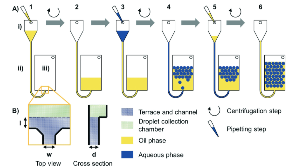
Workflow of centrifual step emulsification without dead volume

By spinning the whole system, the disperse phase (water) is overpressured relative to the continuous phase (oil), resulting in the droplet being forced away from the nozzle by the centrifugal gravitational field (step 3 to 4 above). In order to avoid sample material being wasted as dead volume, an additional aliquot of oil is added in order to push the last few droplets of water out of the nozzle (step 5 to 6 above).

Medium throughput monodisperse droplet formation

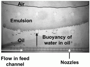
The authors found that droplet diameter is controlled by the nozzle geometry, while rate of formation is controlled by spinning frequency. In light of these findings, they were able to increase droplet production rate from less than 1 droplet per second, to greater than 500 droplets per second, while maintaining monodispersity. They were also able to set up multiple nozzles in parallel (as seen in the microscopic image), all feed by a larger channel, to further increase throughput.

In order to demonstrate the potential applications of this new method, the authors performed digital droplet recombinase polymerase amplification (ddRPA) of L. monocytogenes (a potential contaminant during food production). They found that the number of copies measured with ddRPA was consistent with those measured with digital droplet PCR, and the overall processing was 30 minutes, compared with 2 hourlis for ddPCR.

ddRPA is just one small example of how this new technique can be used – there are a huge range of potential applications where formation of monodisperse particles are a requirement and hopefully we will see this new method being adopted!

To download the full article for free click the link below:

Centrifugal step emulsification applied for absolute quantification of nucleic acids by digital droplet RPA
Friedrich Schuler, Frank Schwemmer, Martin Trotter, Simon Wadle, Roland Zengerle, Felix von Stetten and Nils Paust
DOI: 10.1039/C5LC00291E

Digg This
Reddit This
Stumble Now!
Share on Facebook
Bookmark this on Delicious
Share on LinkedIn
Bookmark this on Technorati
Post on Twitter
Google Buzz (aka. Google Reader)

Direct measurement of the tensile strength of water using an optofluidic chip

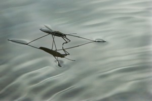
As children, you may remember being fascinated by pond skaters and their ability to walk on water. This is due to water’s high surface tension and there are numerous other ways in which this property is vital to many biological functions. It is also an important factor to take into account when it comes to engineering, and therefore it is essential that there is an accurate and straight forward method for measuring surface tension.

To measure this property, the pressure has to be reduced to such an extent that it causes the water to rupture and form vapour cavities. These vapour cavities must be a result of homogeneous nucleation alone and not heterogeneous nucleation (so should occur spontaneously and randomly, rather than due to nucleation sites). The pressure at which cavitation occurs in termed the tensile strength.

Previous methods have shown a large discrepancy in results, due to the requirement of large volumes of water leading to heterogeneous nucleation. More recently, the mineral inclusion method has overcome this, however has other limitations, such as the requirement of an autoclave. Alternatively, microfluidics allows the use small volumes of water to ensure homogenous nucleation, but this method is limited to low-viscosity liquids.

Graphical Abstract

Dr. Liu Ai Lin and co-workers, at NTU, Singapore, have reported the direct measurement of water’s tensile strength using an optofluidic chip. Their method relies on an infrared laser that is focussed into a microchannel partially filled with water. The laser pulse results in the formation and recombination of plasma, which in turn produces a bubble, causing a spherical shock wave. The reflection of the shock wave on the air-water interface generates a negative pressure and, if this is larger than the tensile strength, the water ruptures, causing nucleation of vapour bubbles near the interface. The pressure value can be attained by both measuring the spreading of the shock wave over time and the displacement of the water-air interface.

By imaging the microchannel at increasing standoff distances (defined in the diagram above), the distance and pressure at which water no longer ruptures can be found. This can be directly converted to a value for the tensile strength.

Graphical Abstract

This work provides a simple, low-cost method for calculating tensile strength that can easily allow rapid testing of a wide range of samples. In order to demonstrate this, the authors also measured the tensile strength of glycerol, a highly viscous fluid.

To download the full article for free* click the link below:

Water’s tensile strength measured using an optofluidic chip
Z. G. Li,  S. Xiong,  L. K. Chin,  K. Ando,  J. B. Zhang and  A. Q. Liu
DOI: 10.1039/ C5LC00048C

*Access is free through a registered RSC personal publishing account

Digg This
Reddit This
Stumble Now!
Share on Facebook
Bookmark this on Delicious
Share on LinkedIn
Bookmark this on Technorati
Post on Twitter
Google Buzz (aka. Google Reader)

Textile Weaving: a new cheaper alternative to electrochemical sensors

Electrochemical sensors are widely used as analytical tools. They are disposable, cheap to make, and small – making them ideal for many applications.

The current method for commercial electrochemical sensors uses screen printing onto plastic or ceramic surfaces to generate the circuit elements, which requires specialised fabrication equipment. This screen printing method also leads to wastage of electrode inks and reagents. One way of avoiding these issues is using paper microfluidics – Professor George Whitesides is a well-known name in this field.

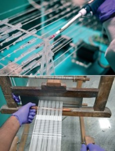
Dendukuri and coworkers, from Achira Labs, Bangalore, have come at this problem from a different angle. They have developed an alternative approach using textile weaving. Instead of screen printing, they coat their yarn with the required reagents in a way that results in no wastage, as shown in the photo. They use silk as their material which is biodegradable, unlike the plastics usually used. It is also easily processed – initially silk is hydrophobic but it can be made hydrophobic by degumming. This can be achieved by simply boiling the yarn.

To make the sensors, electrode yarns are prepared by coating in conductive inks and reagents, and then woven into the fabric. Large numbers of sensors can be woven as patches on the fabric, which are then stuck onto an adhesive backing and laminated, leaving a window for application of the sample and for contact with a reader.To demonstrate this effectiveness of this new method, the authors developed glucose and haemoglobin sensors. The glucose sensors were found to have a clinically acceptable performance, according to FDA criteria, while the haemoglobin sensors were able to detect physiologically relevant concentrations. Multiplexed sensors capable of detecting more than one analyte were easily prepared by adding an additional electrode.

One of the most pleasing aspects of this new method, is its potential in the developing world, where weaving is still widely used. In addition, the cost of manufacture was calculated as less than 20 USD per 1000 sensors and this could even lower on scale up.

To download the full article for free* click the link below:

Woven electrochemical fabric-based test sensors (WEFTS): a new class of multiplexed electrochemical sensors
Tripurari Choudhary, G. P. Rajamanickam and Dhananjaya Dendukuri
DOI: 10.1039/C5LC00041F

*Access is free until 30.04.2015 through a registered RSC Personal Publishing Account

Digg This
Reddit This
Stumble Now!
Share on Facebook
Bookmark this on Delicious
Share on LinkedIn
Bookmark this on Technorati
Post on Twitter
Google Buzz (aka. Google Reader)

Scientists can quickly detect waterborne pathogens using a smartphone!

More than half a billion people have to survive with unimproved water, as providing safe drinking water is still a problem in many parts of the developing world. Of the waterborne pathogens, Giardia lambia (G. lambia) is one of the most common intestinal parasites that are difficult to remove using traditional water purification methods. Current methods for their detection take up to two days and require analysis laboratories with trained specialists and expensive equipment. Because of this there is an ongoing effort to design low-cost and field-portable methods that can rapidly analyse large volumes of water.

Ozcan and co-workers at UCLA have developed a method for the detection of G. lambia cysts in water using a light weight attachment to a smartphone. The attachment consists of a fluorescence microscope, aligned to the smartphone camera, and a disposable water sample cassette that can hold 20 mL of water. The whole test can be carried out in just 1 hour, from taking the sample from the source, to receiving the total number of cysts detected in the sample.

The process is relatively simple, with the test sample first being fluorescently labelled and then filtered through a membrane that traps the G. lambia cysts. A fluorescence image is taken and wirelessly transmitted to servers using an app designed by the group. Digital analysis is carried out using a machine learning algorithm that can specifically recognise the cysts over other fluorescent micro-objects. The results of this analysis are then transmitted back to the phone and displayed on the app.

The group were able to achieve an impressive limit of detection of 12 cysts per 10 mL of sample, citing several factors that led to this limit. They have put forward a number of suggestions for how they hope to further improve their system, so it will be interesting to hear more from this group.

To download the full article for free* click the link below:

Rapid imaging, detection and quantification of Giardia lamblia cysts using mobile-phone based fluorescent microscopy and machine learning
Hatice Ceylan Koydemir, Zoltan Gorocs, Derek Tseng, Bingen Cortazar, Steve Feng, Raymond Yan Lok Chan, Jordi Burbano, Euan McLeod and Aydogan Ozcan
DOI: 10.1039/C4LC01358A

*Access is free until 31st March 2015 through a publishing personal account. It’s quick, easy and free to register.

About the web writer

Claire Weston is currently studying for a PhD at Imperial College London, focussing on developing novel photoswitches and photoswitchable inhibitors.

Digg This
Reddit This
Stumble Now!
Share on Facebook
Bookmark this on Delicious
Share on LinkedIn
Bookmark this on Technorati
Post on Twitter
Google Buzz (aka. Google Reader)