Author Archive

Our current HOT articles on cell sorting, demulsification kinetics, modelling microvalves and materials for chips

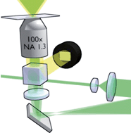
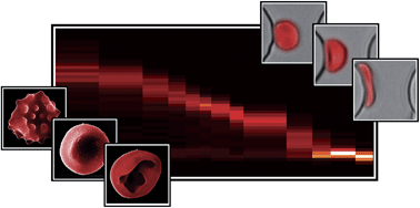
C2LC21083E graphical abstractJason P. Beech and colleagues at Lund University use shape and deformability to sort cells, as well as the more usual parameter of size in this microfluidic device:

Sorting cells by size, shape and deformability
Jason P. Beech, Stefan H. Holm, Karl Adolfsson and Jonas O. Tegenfeldt
DOI: 10.1039/C2LC21083E

Thomas Krebs and colleagues at Wageningen University present the results of experiments studying droplet coalescence in a dense layer of emulsion droplets using microfluidic circuits:

A microfluidic method to study demulsification kinetics
Thomas Krebs, Karin Schroen and Remko Boom
DOI: 10.1039/C2LC20930F

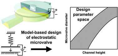
C2LC21133E graphical abstractPaul Kenis et al. present an analytical model to guide the design of electrostatic microvalves that can be integrated into microfluidic chips:

Design considerations for electrostatic microvalves with applications in poly(dimethylsiloxane)-based microfluidics
Amit V. Desai, Joshua D. Tice, Christopher A. Apblett and Paul J. A. Kenis
DOI: 10.1039/C2LC21133E

David Beebe et al. discuss the use of PDMS and polystyrene by researchers working at the interface of microfluidics and cell biology research:

Engineers are from PDMS-land, Biologists are from Polystyrenia
Erwin Berthier, Edmond W. K. Young and David Beebe
DOI: 10.1039/C2LC20982A

These HOT articles are free to access for the next four weeks (following a simple registration for individual users), so why not take a look?

Digg This
Reddit This
Stumble Now!
Share on Facebook
Bookmark this on Delicious
Share on LinkedIn
Bookmark this on Technorati
Post on Twitter
Google Buzz (aka. Google Reader)

Pressurising red blood cells for information

Scientists in Canada have developed a method to study the changes in red blood cells caused by the most common malaria parasite Plasmodium falciparum.

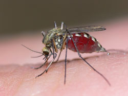
Mosquito

The malaria parasite is spread by mosquitos. © Shutterstock

Malaria causes approximately one million deaths each year and there is a lot of research surrounding the disease’s biomechanics, aimed at diagnosing patients and developing treatments. The parasite, spread by mosquitoes, infects red blood cells and reduces their ability to deform. Measuring the deformability of an infected red blood cell can provide vital information about the disease’s mechanism and response to treatment.

Current methods to measure deformability are complicated or not sensitive enough, but Hongshen Ma at the University of British Columbia, Vancouver, and colleagues, have designed an accurate and simple microfluidic device for this purpose. The device consists of two layers that control the cells so that only a single cell is introduced into a funnel containing a series of different sized constrictions. The pressure required to push the cell through a constriction is measured precisely and used to calculate the deformability.

The team used their device to show that the deformability of uninfected red blood cells can be distinguished from cells at various stages of infection.

Abhishek Jain at Boston University, US, is an expert in biomedical devices and comments that ‘the device is elegant in the sense that it can be easily scaled up for diagnosis and high throughput drug testing for not only malaria but other pathologies like sickle cell anaemia’.

‘We hope our technique will provide a useful biomechanical assay for the development of new drugs,’ says Ma, who adds that ‘the ability to easily measure the deformability of red blood cells will help researchers study the mechanism of the disease and investigate complex challenges, such as drug resistance.’

Ma’s team plans to further test their device and use it to study the mechanisms of drug resistance.

Microfluidic biomechanical assay for red blood cells parasitized by Plasmodium falciparum
Quan Guo , Sarah J. Reiling , Petra Rohrbach and Hongshen Ma
Lab Chip, 2012, Advance Article
DOI: 10.1039/C2LC20857A

Original article published at Chemistry World

Digg This
Reddit This
Stumble Now!
Share on Facebook
Bookmark this on Delicious
Share on LinkedIn
Bookmark this on Technorati
Post on Twitter
Google Buzz (aka. Google Reader)

Issue 5 just published including hot articles on sensors for microgas chromatography

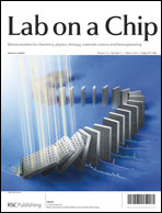
Issue 5 front coverIssue 5 is now available online and on the outside front cover we have a hot article on the batch fabrication of disposable SERS arrays using screen printing with silver nanoparticle-containing ink from Lu-Lu Qu et al.

Batch fabrication of disposable screen printed SERS arrays
Lu-Lu Qu,  Da-Wei Li,  Jin-Qun Xue,  Wen-Lei Zhai,  John S. Fossey and Yi-Tao Long
DOI: 10.1039/C2LC20926H

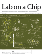
On the inside front cover is another hot article, this time from Andrew Griffiths et al. who have developed an ultrahigh-throughput system which combines droplet PCR and IVTT to provide a completely in vitro screen.

Issue 5 inside front cover

A completely in vitro ultrahigh-throughput droplet-based microfluidic screening system for protein engineering and directed evolution
Ali Fallah-Araghi,  Jean-Christophe Baret,  Michael Ryckelynck and Andrew D. Griffiths
DOI: 10.1039/C2LC21035E

Plus you can read the latest Research Highlights from Ali Khademhosseini – shrink-film for cell culture, optically adjustable microfluidic chips and protein profiling with microgels – or the latest in our Acoustofluidics series on measurement techniques for the characterization of ultrasonic particle manipulation devices.

The issue also features several hot articles which will be free to access* for 4 weeks:

Rapid, sensitive, and multiplexed on-chip optical sensors for micro-gas chromatography
Karthik Reddy, Yunbo Guo, Jing Liu, Wonsuk Lee, Maung Kyaw Khaing Oo and Xudong Fan
DOI: 10.1039/C2LC20922E

Microfluidic single-cell cultivation chip with controllable immobilization and selective release of yeast cells
Zhen Zhu, Olivier Frey, Diana Silvia Ottoz, Fabian Rudolf and Andreas Hierlemann
DOI: 10.1039/C2LC20911J

Dual-electrode microfluidic cell for characterizing electrocatalysts
Ioana Dumitrescu, David F. Yancey and Richard M. Crooks
DOI: 10.1039/C2LC21181E

* Following a simple registration for individual users

Digg This
Reddit This
Stumble Now!
Share on Facebook
Bookmark this on Delicious
Share on LinkedIn
Bookmark this on Technorati
Post on Twitter
Google Buzz (aka. Google Reader)

Have you seen our collection of review articles? Reviews on holographic optical tweezers to microfluidics for the food industry and optoelectrofluidics

During 2011 we published a number of topical reviews on a wide range of topics by expert researchers in their fields.  We’ve collected some of them below but take a look here for the whole list, we hope you’ll find something interesting in your area.

Graphical abstract for C0LC00526FHolographic optical tweezers and their relevance to lab on chip devices
Miles Padgett and Roberto Di Leonardo

Lab-on-a-chip based immunosensor principles and technologies for the detection of cardiac biomarkers: a review
Mazher-Iqbal Mohammed and Marc P. Y. Desmulliez

Nanomanipulation using near field photonics
David Erickson, Xavier Serey, Yih-Fan Chen and Sudeep Mandal

Microfluidics for food, agriculture and biosystems industries
Suresh Neethirajan, Isao Kobayashi, Mitsutoshi Nakajima, Dan Wu, Saravanan Nandagopal and Francis Lin

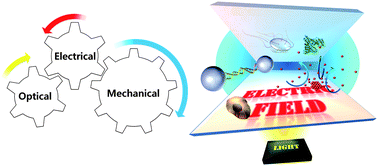
Graphical abstract for C0LC00117AOptoelectrofluidic platforms for chemistry and biology
Hyundoo Hwang and Je-Kyun Park

Disposable microfluidic substrates: Transitioning from the research laboratory into the clinic
Jason S. Kuo and Daniel T. Chiu

Miniaturized isothermal nucleic acid amplification, a review
Peter J. Asiello and Antje J. Baeumner

If you have an idea for a review article that hasn’t been covered and you would like to see included, contact the Editorial Office – we’d love to hear from you.

Digg This
Reddit This
Stumble Now!
Share on Facebook
Bookmark this on Delicious
Share on LinkedIn
Bookmark this on Technorati
Post on Twitter
Google Buzz (aka. Google Reader)

Top ten most accessed articles in November

This month sees the following articles in Lab on a Chip that are in the top ten most accessed:

A low cost point-of-care viscous sample preparation device for molecular diagnosis in the developing world; an example of microfluidic origami
A. V. Govindarajan, S. Ramachandran, G. D. Vigil, P. Yager and K. F. Böhringer
Lab Chip, 2012, 12, 174-181
DOI: 10.1039/C1LC20622B

A new method of UV-patternable hydrophobization of micro- and nanofluidic networks
Rerngchai Arayanarakool, Lingling Shui, Albert van den Berg and Jan C. T. Eijkel
Lab Chip, 2011, 11, 4260-4266
DOI: 10.1039/C1LC20716D

Highly-integrated lab-on-chip system for point-of-care multiparameter analysis
Soeren Schumacher, Jörg Nestler, Thomas Otto, Michael Wegener, Eva Ehrentreich-Förster, Dirk Michel, Kai Wunderlich, Silke Palzer, Kai Sohn, Achim Weber, Matthias Burgard, Andrzej Grzesiak, Andreas Teichert, Albrecht Brandenburg, Birgit Koger, Jörg Albers, Eric Nebling and Frank F. Bier
Lab Chip, 2012, 12, 464-473
DOI: 10.1039/C1LC20693A

Surfactants in droplet-based microfluidics
Jean-Christophe Baret
Lab Chip, 2012, 12, 422-433
DOI: 10.1039/C1LC20582J

Integrated separation of blood plasma from whole blood for microfluidic paper-based analytical devices
Xiaoxi Yang, Omid Forouzan, Theodore P. Brown and Sergey S. Shevkoplyas
Lab Chip, 2012, 12, 274-280
DOI: 10.1039/C1LC20803A

Photolithographic surface micromachining of polydimethylsiloxane (PDMS)
Weiqiang Chen, Raymond H. W. Lam and Jianping Fu
Lab Chip, 2012, 12, 391-395
DOI: 10.1039/C1LC20721K

A digital microfluidic platform for primary cell culture and analysis
Suthan Srigunapalan, Irwin A. Eydelnant, Craig A. Simmons and Aaron R. Wheeler
Lab Chip, 2012, 12, 369-375
DOI: 10.1039/C1LC20844F

Simultaneous high speed optical and impedance analysis of single particles with a microfluidic cytometer
David Barat, Daniel Spencer, Giuseppe Benazzi, Matthew Charles Mowlem and Hywel Morgan
Lab Chip, 2012, 12, 118-126
DOI: 10.1039/C1LC20785G

Ensembles of engineered cardiac tissues for physiological and pharmacological study: Heart on a chip
Anna Grosberg, Patrick W. Alford, Megan L. McCain and Kevin Kit Parker
Lab Chip, 2011, 11, 4165-4173
DOI: 10.1039/C1LC20557A

Fuel cell-powered microfluidic platform for lab-on-a-chip applications
Juan Pablo Esquivel, Marc Castellarnau, Tobias Senn, Bernd Löchel, Josep Samitier and Neus Sabaté
Lab Chip, 2012, 12, 74-79
DOI: 10.1039/C1LC20426B

Why not take a look at the articles today and blog your thoughts and comments below.

Fancy submitting an article to Lab on a Chip? Then why not submit to us today or alternatively email us your suggestions.
Digg This
Reddit This
Stumble Now!
Share on Facebook
Bookmark this on Delicious
Share on LinkedIn
Bookmark this on Technorati
Post on Twitter
Google Buzz (aka. Google Reader)

Staining tissue samples at the microscale

A vertical microfluidic probe developed by researchers in Switzerland can create a range of immunohistochemistry staining conditions on a single tissue sample.

Immunohistochemistry is a process of detecting antibody biomarkers and is regularly used to reveal abnormal cells in tissue sections. Pathologists conducting immunohistochemistry tests often work with very limited samples for which they don’t know the optimal staining conditions. Under-staining can give a false negative, but over-staining causes loss of contrast and can generate false positives.

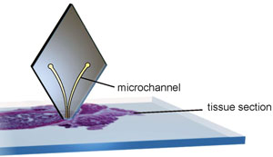
Diamond-shaped probe used to analyse tissue samples

The diamond-shaped probe is positioned above the tissue sample and scans horizontally across it. The microchannels inject and aspirate liquids at a distance of 1-30µm from the tissue section

Now, a microfluidic device developed by Emmanuel Delamarche and colleagues at the IBM Zurich Research Laboratory, Rüschlikon, offers local staining of tissue sections, which allows a range of staining conditions to be used on one section. The microfluidic probe is positioned vertically above the tissue section and scans horizontally across it. The head of the probe has two apertures at its apex. The liquid is injected onto the tissue section from one aperture and aspirated at the second. ‘Instead of incubating the entire tissue section, the probe can scan it with a variable speed so as to vary locally the incubation time. This is a simple trick, but it should give an optimal staining contrast at least on one spot of the sample,’ explains Delamarche. Tissue sections are prepared for staining in the conventional fashion and post-staining processing is also unchanged.

Delamarche and the team have shown proof of concept with their current research, but in the future, they hope it will receive clinical validation. In addition, the probe could be used for fundamental research. ‘Developing a novel tissue staining method to detect various biomarkers is critical to obtain a more accurate and sophisticated understanding of drug discovery and clinical pathology,’ explains Je-Kyun Park, an expert in bioengineering at the Korea Advanced Institute of Science and Technology, South Korea. Using the microfluidic probe to scan multiple locations with different conditions could help analyse tissue microarrays.

Micro-immunohistochemistry using a microfluidic probe
Robert D. Lovchik, Govind V. Kaigala, Marios Georgiadis and Emmanuel Delamarche
Lab Chip, 2012, Advance Article
DOI: 10.1039/C2LC21016A

Original article published at Chemistry World

Digg This
Reddit This
Stumble Now!
Share on Facebook
Bookmark this on Delicious
Share on LinkedIn
Bookmark this on Technorati
Post on Twitter
Google Buzz (aka. Google Reader)

LOC article on a new device for antibiotic susceptibility testing is mentioned in the press

The recent LOC article from Douglas Weibel and team at the University of Wisconsin-Madison has been highlighted on MedGadget. The article describes a new portable self-loading technology for determining minimum inhibitory concentration values, vital in clinical bacteriology for determining whether an organism is reported susceptible or resistant.

Congratulations to Douglas Weibel and team!

You can read the MedGadget article online here or go straight to the Lab on a Chip paper:

A self-loading microfluidic device for determining the minimum inhibitory concentration of antibiotics
Nate J. Cira, Jack Y. Ho, Megan E. Dueck and Douglas B. Weibel
Lab Chip, 2012, Advance Article
DOI: 10.1039/C2LC20887C

Digg This
Reddit This
Stumble Now!
Share on Facebook
Bookmark this on Delicious
Share on LinkedIn
Bookmark this on Technorati
Post on Twitter
Google Buzz (aka. Google Reader)

New YouTube videos

View the new videos on the Lab on a Chip YouTube site below:

Uniform mixing in paper-based microfluidic systems using surface acoustic waves

Digg This
Reddit This
Stumble Now!
Share on Facebook
Bookmark this on Delicious
Share on LinkedIn
Bookmark this on Technorati
Post on Twitter
Google Buzz (aka. Google Reader)

New YouTube videos

View the new videos on the Lab on a Chip YouTube site below:

A robust diffusion-based gradient generator for dynamic cell assays

Toward active-matrix lab-on-a-chip: programmable electrofluidic control enabled by arrayed oxide thin film transistors

Reconfigurable virtual electrowetting channels

Digg This
Reddit This
Stumble Now!
Share on Facebook
Bookmark this on Delicious
Share on LinkedIn
Bookmark this on Technorati
Post on Twitter
Google Buzz (aka. Google Reader)

Top ten most accessed articles in October

This month sees the following articles in Lab on a Chip that are in the top ten most accessed:

Rapid prototyping polymers for microfluidic devices and high pressure injections
Elodie Sollier, Coleman Murray, Pietro Maoddi and Dino Di Carlo
Lab Chip, 2011, 11, 3752-3765
DOI: 10.1039/C1LC20514E

Droplet microfluidics—a tool for protein engineering and analysis
Haakan N. Joensson and Helene Andersson-Svahn
Lab Chip, 2011, 11, 4144-4147
DOI: 10.1039/C1LC90102H

Surfactants in droplet-based microfluidics
Jean-Christophe Baret
Lab Chip, 2012, Advance Article
DOI: 10.1039/C1LC20582J

Bubbles navigating through networks of microchannels
Wonjae Choi, Michinao Hashimoto, Audrey K. Ellerbee, Xin Chen, Kyle J. M. Bishop, Piotr Garstecki, Howard A. Stone and George M. Whitesides
Lab Chip, 2011, 11, 3970-3978
DOI: 10.1039/C1LC20444K

Integration of intra- and extravasation in one cell-based microfluidic chip for the study of cancer metastasis
Min Kyeong Shin, Sung Kyu Kim and Hyungil Jung
Lab Chip, 2011, 11, 3880-3887
DOI: 10.1039/C1LC20671K

Microfluidic static droplet arrays with tuneable gradients in material composition
Meng Sun, Swastika S. Bithi and Siva A. Vanapalli
Lab Chip, 2011, 11, 3949-3952
DOI: 10.1039/C1LC20709A

Controlled viable release of selectively captured label-free cells in microchannels
Umut Atakan Gurkan, Tarini Anand, Huseyin Tas, David Elkan, Altug Akay, Hasan Onur Keles and Utkan Demirci
Lab Chip, 2011, 11, 3979-3989
DOI: 10.1039/C1LC20487D

Droplet formation via flow-through microdevices in Raman and surface enhanced Raman spectroscopy—concepts and applications
Anne März, Thomas Henkel, Dana Cialla, Michael Schmitt and Jürgen Popp
Lab Chip, 2011, 11, 3584-3592
DOI: 10.1039/C1LC20638A

1-Million droplet array with wide-field fluorescence imaging for digital PCR
Andrew C. Hatch, Jeffrey S. Fisher, Armando R. Tovar, Albert T. Hsieh, Robert Lin, Stephen L. Pentoney, David L. Yang and Abraham P. Lee
Lab Chip, 2011, 11, 3838-3845
DOI: 10.1039/C1LC20561G

Large-scale plasmonic microarrays for label-free high-throughput screening
Tsung-Yao Chang, Min Huang, Ahmet Ali Yanik, Hsin-Yu Tsai, Peng Shi, Serap Aksu, Mehmet Fatih Yanik and Hatice Altug
Lab Chip, 2011, 11, 3596-3602
DOI: 10.1039/C1LC20475K

Why not take a look at the articles today and blog your thoughts and comments below.

Fancy submitting an article to Lab on a Chip? Then why not submit to us today or alternatively email us your suggestions.

Digg This
Reddit This
Stumble Now!
Share on Facebook
Bookmark this on Delicious
Share on LinkedIn
Bookmark this on Technorati
Post on Twitter
Google Buzz (aka. Google Reader)