Author Archive

SLAS 2018


SLAS will host SLAS2018, the seventh Annual International SLAS International Conference and Exhibition, in San Diego, California, from Feb. 3-7, 2018.


Through a unique combination of education, access to innovative technologies and intelligent peer networking, SLAS2018 delivers unmatched value for professionals and students looking to discover the latest life sciences technologies and how they can be applied to drive research objectives. SLAS 2018 invites research scientists, engineers, academics and business leaders to submit abstracts for presentation.

SLAS is a global community of more than 20,000 life sciences professionals—from academia, government and industry—collectively focused on leveraging the power of technology to achieve scientific objectives. Showcase your research on this global stage by presenting at SLAS2018


Key deadlines:

18th December: Early-Bird Registration Discount

Monday, January 22, 2018 (Final Poster Abstract Submission Deadline)

Digg This
Reddit This
Stumble Now!
Share on Facebook
Bookmark this on Delicious
Share on LinkedIn
Bookmark this on Technorati
Post on Twitter
Google Buzz (aka. Google Reader)

Emerging Investigator Series – Ashleigh Theberge

We are delighted to introduce our latest Lab on a Chip Emerging Investigator, Ashleigh Theberge!

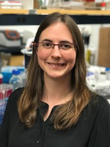
Ashleigh Theberge is an Assistant Professor of Chemistry at the University of Washington and Adjunct Assistant Professor of Urology at the University of Washington School of Medicine. She received her BA in Chemistry at Williams College and her PhD in Chemistry at the University of Cambridge, UK with Wilhelm Huck. During her graduate work, she was a Visiting Scientist with Andrew Griffiths at the Université de Strasbourg, France. Her graduate research focused on droplet-based microfluidics for chemical synthesis and analysis. She completed her postdoctoral fellowship in Biomedical Engineering and Urology with David Beebe, William Ricke, and Wade Bushman at the University of Wisconsin-Madison. In 2014, she was awarded an NIH K Career Development Award from the NIDDK in Urology. She joined the faculty at the University of Washington in 2016. Her group develops new microscale culture and analysis methods to study cell-cell, cell-extracellular matrix, and host-microbe interactions, with a focus on inflammation in urologic diseases and asthma.

Read Ashleigh’s Emerging Investigator series paper “Upgrading well plates using open microfluidic patterning” and find out more about her in the interview below:

Your recent Emerging Investigator Series paper focuses on upgrading well plates using microfluidic patterning. How has your research evolved from your first article to this most recent article? 

My research has changed dramatically since my first article, which focused on chemical synthesis in droplet-based microfluidics. The common thread in both articles is manipulation of fluids on the microscale, but almost everything else is different! While my graduate work focused on chemical applications of microfluidics in conventional closed microfluidic channels, I now work on methods for studying cell signaling in biological systems using primarily open microfluidic channels. For example in this most recent article, the “walls” of the “channel” are comprised of the floor of the well plate, a plastic insert, and two free air-liquid interfaces.

What aspect of your work are you most excited about at the moment?

I am excited about our group’s fundamental work advancing fluid manipulation in open microfluidics using capillary flow. On the applications side, I am excited to be exploring signaling in ever-more complex cellular systems, including “multikingdom” systems where we study the chemical signals exchanged between host and microbe. I am also happy to work with a fantastic group of students who are taking our group in new directions both biologically and technologically.

In your opinion, what is the biggest advantage of the presented patterning device over the current systems?

Our method enables biologists to easily “upgrade” any existing assay developed in conventional well plates to a co- or multiculture assay. Well plates are the most common cultureware used for cell-based assays, and our device is a simple plastic insert that fits within the well and enables the user to pipette hydrogels (such as collagen, Matrigel, or synthetic gels) to make biocompatible partitions that enable segregated cell culture and maintain soluble factor signaling through the gel wall. Our device enables biologists to use a familiar off-the-shelf platform (well plates) and culture cells on surfaces that have been optimized by decades of industry experience with tissue culture treated surfaces. Since our device uses “open microfluidics,” it does not require bonding of multiple layers and can be fabricated in a single step via injection molding to scale up production.

What do you find most challenging about your research?

A challenge is the interdisciplinary of our work; but this is also what I love about it. Much of our work involves new methods to isolate and study chemical signals from complex cultures – falling under the category of analytical chemistry or bioengineering. But we also focus heavily on disease mechanisms, delving deeply into biology, and our work in the fundamentals of open microfluidics is at the interface of physics and chemistry. Our work also touches on applied aspects of microscale device fabrication, traditionally falling under mechanical engineering, such as our recent paper in press at Lab on a Chip entitled “Fundamentals of rapid injection molding for microfluidic cell-based assays.”

In which upcoming conferences or events may our readers meet you?

I regularly attend microTAS and the Gordon Research Conference on Microfluidics. I also plan to attend the IEEE EMBS Micro and Nanoengineering in Medicine Conference and biological conferences related to our group’s focus areas (microbiome, urologic research, etc.).

How do you spend your spare time?

I try to spend spare time outdoors and enjoy sailing, open water swimming, and climbing. I also like to travel – both to explore different cultures and also wilderness such as the Yukon Territory, Patagonia, and national forest land nearby in Washington.

Which profession would you choose if you were not a scientist?

I would enjoy having a leadership role in a company or organization. I like brainstorming ideas to develop a vision for large projects and working in teams toward a common goal – be it a product, a discovery, or a process. I decided to be a scientist because the scientific method provides an opportunity to rigorously explore questions, but there are many complex problems outside of science that require exciting new strategies and the cohesive effort of many minds to solve.

Can you share one piece of career-related advice or wisdom with other early career scientists?

One strategy that has been helpful for me is to be persistent in looking for opportunities to support projects that I care about. As an undergraduate researcher, I designed my own project that required augmenting our department’s gas chromatograph. I was able to secure the funds by talking with different offices on campus (dean’s office initiatives, student project grants, etc.); I was turned down many times but even those experiences helped me to refine my explanation of the project to have a more compelling conversation with the next person I approached. As an undergraduate summer intern working for Merck, my department did not have funds to send me to a conference to present my work, so I asked the human resources department to fund my trip as a way to promote the intern program as well as present my science; they agreed. In graduate school, I set up an international collaboration in order to develop the expertise required to pursue a multidisciplinary project that I had designed. So for early career scientists, I think it is key to remember that there are many opportunities (even if they are not always obvious at first), and facing road blocks or being turned down several times doesn’t preclude a successful outcome.

Digg This
Reddit This
Stumble Now!
Share on Facebook
Bookmark this on Delicious
Share on LinkedIn
Bookmark this on Technorati
Post on Twitter
Google Buzz (aka. Google Reader)

1st UK Microfluidics for Analytical Chemistry Conference

1st UK Microfluidics for Analytical Chemistry Conference will be held on Thursday 1 February 2018 at the National Oceanography Centre in Southampton, UK.


This one day conference aims to bring together UK researchers developing and applying microfluidic systems for chemical and biochemical analysis. The meeting will cover all types of devices (lab-on-chip, digital microfluidics, paper microfluidics, total analytical systems etc.) and concern application to all forms of molecular analysis (biomedical, environmental, forensic, quality control etc.)

It will provide an opportunity to discuss recent developments in the field and develop future research opportunities as part of an overall aim to nurture and promote the UK microfluidic and analytical communities. This will also offer PhD students and early career researchers a chance to present their work.

Sessions will feature presentations by invited speakers, including keynote presentations from Nicole Pamme (Advisory Board Member for Lab on a Chip) and Joshua Edel (Advisory Board Member for Analyst), in addition to presentations selected from submission of abstracts. There will also be a chance to view the posters and exhibition, with additional networking possible during a wine reception at the end of the event.

Key deadlines:

Abstract deadline – 3rd December, 2018

Earlybird registration deadline -17th December, 2018

Standard Registration deadline – 12th January, 2018


To register, please click here and for more information, please visit the Conference website here.

Digg This
Reddit This
Stumble Now!
Share on Facebook
Bookmark this on Delicious
Share on LinkedIn
Bookmark this on Technorati
Post on Twitter
Google Buzz (aka. Google Reader)

MicroTAS “Late News” Posters

This year’s 21st International Conference on Miniaturized Systems for Chemistry and Life Sciences (more commonly known as MicroTAS) was held in Savannah, Georgia. As with previous MicroTAS conferences, the event brought together the international microfluidics and lab-on-a-chip community for an outstanding week of talks and posters. Rather than starting with a talk, MicroTAS 2017 opened with a conversation with George Whitesides moderated by Thomas Laurell, who asked questions pre-selected from the conference attendees. Whitesides, one of the pioneers of microfluidics, provided salient and humorous takes on the past, present, and future of our field. Two of Whitesides’ memorable takeaways were: (1) keep things simple, and (2) make sure you can answer the question “who cares?” The spirit of open discussion that started at the beginning of the conference continued through the many oral presentations and poster sessions at the conference. While the talks and posters represented the usual gamut of microfluidic technologies, 3D printing made a big splash this year. There was a 3D printing session, with a great keynote from Greg Nordin, as well as posters and companies featuring 3D-printed microfluidics. The sense of community was also palpable during many ferry rides to the Savannah International Trade & Convention Centre, the student trivia night, female faculty mixer event, and the conference ending banquet (and unofficial after party!).

In addition to all the great talks, we’ve highlighted some of our favourite “Late News” posters for our readers:


Rapid Extraction and Concentration of Magnetic Particles from Whole Blood with Microfluidic Magnetic Ratcheting, 

Oladunni Adeyiga, Coleman Murray, Dino Di Carlo

Magnetic particles are a useful tool for extracting and concentrating target analytes. Conventionally, pipetting, centrifuge tubes, and magnetic racks are used, but this approach is prone to loss of particles and errors from pipetting. Dunni presented a new microfluidic device that concentrates magnetic particles using magnetic ratcheting. A rotating permanent magnet was used to induce magnetic fields in permalloy micropillars embedded within the devices, which could then move magnetic particles along like on a conveyor belt. At the end of the process, the particles are concentrated into a drop within an immiscible fluid. While demonstrated as a standalone device, this tool could also function as a pre-concentrator in an integrated microfluidic device.


High Density, Reversible 3D Printed Microfluidic Interconnects, 

Hua Gong, Adam T. Woolley, Greg P. Nordin

Earlier this year, Lab on a Chip published a paper describing the first 3D printed microchannels that truly had microscale dimensions. You can read the blog post summarizing the work here. It was nice to see this poster from Hua demonstrating the capabilities of their custom 3D printer, including printing complex valves. Hua explained how their 3D printer has a very small print area, and so one of the challenges to overcome was how to fit all the interconnects needed to control the valves into such a small space. This was achieved by printing a world-to-chip interface that connects tubing to the smaller, more densely packed inlets and outlets on the microfluidic chip.


3D-printing, Ink Casting and Lamination (3-D PICL): A Rapid, Robust, and Cost Effective Process Technology Toward The Fabrication of Microfluidic and Biological Devices, 

Tariq Ausaf, Avra Kundu, and Swaminathan Rajaraman

Tariq, Avra, and Swami presented an application of 3D-printing aimed at developing microelectrode arrays, microneedles, and microfluidic chips without cleanroom facilities. This work was motivated by a desire for disposable microelectrode arrays that can be developed from concept to functional prototype in < 24 hours. The authors integrated stereolithographic 3D-printing to form the device body, selective ink casting to define conductive traces, lamination of an insulating layer, and micromachining of electrodes and connecting layers. Combining multiple benchtop fabrication techniques could increase the functionality of the developed microdevices and could speed up the transition from prototype to final product in a cost effective manner.


An Automated Modular Microsystem For Enzymatic Digestion With Gut-On-A-Chip Applications
Pim de Haan, Margaryta A. Ianovska, Klaus Mathwig, Hans Bouwmeester and Elisabeth Verpoorte.

What is the primary function of the gastrointestinal tract? Digestion. However, most human gut-on-a-chip models tend to focus on cultured cells in one region of the gut (typically the small intestine) and do not model the initial digestive processes that take place in the mouth, stomach, and small intestine. Pim’s work is focused on developing bioreactors to study the conversion of food into chyme, as it moves from the mouth, through the stomach, and into the small intestine. This requires accurate modelling and realization of the vast pH differences that take place from one region of the gut to the next and verifying enzyme activity within his gut-on-a-chip model. Future work will focus on integration of this upstream digestion model with the downstream study of absorption of nutrients across an intestinal cell layer.


__________

About the Webwriters

Darius Rackus (Right) is a postdoctoral researcher at the University of Toronto working in the Wheeler Lab. His research interests are in combining sensors with digital microfluidics for healthcare applications.


Ayokunle Olanrewaju (Left) is an industrial postdoctoral fellow at McGill University working in the Juncker lab (dj.lab.mcgill.ca). He is excited about projects that use engineering design to effect real world change, especially in healthcare. Currently, he builds portable and self-powered microchips that rapidly detect bacteria in urine.


Digg This
Reddit This
Stumble Now!
Share on Facebook
Bookmark this on Delicious
Share on LinkedIn
Bookmark this on Technorati
Post on Twitter
Google Buzz (aka. Google Reader)

Magnetic separation of circulating tumor cells…but it’s not what you think

Cancers typically originate in one organ yet can spread to distant regions of the body forming secondary tumours called metastases. This happens as cells from the primary tumour migrate into the circulatory system and then travel to other organs. These cells, which are a very rare population within the circulatory system, are termed circulating tumor cells (CTCs). Because of their role in cancer biology, they have garnered a lot of interest lately. Their detection and isolation present several analytical challenges. For one, they are the proverbial “needle in a haystack”, with counts on the order of one CTC for every billion blood cells. This has traditionally led to a paradox: these rare cells are best handled in microscale systems but the world-to-chip mismatch limits microfluidic devices from rapidly processing the large (> 5 mL) samples necessary. Second, recent studies have revealed CTCs to be very heterogeneous populations, limiting the use of surface markers for labelling and capturing a broad range of CTCs. Because there is much still to learn about CTCs, there’s also an interest in recovering viable CTCs for further analysis. In their recent report, Zhao et al. demonstrate a microfluidic device capable of enriching CTCs using magnetic separation. But it’s not that typical magnetophoretic separations you may be familiar with!

Magnetic separation of circulating tumor cells - nanoparticles

Rather than using magnetic particles to bind to surface antigens and eventually separate out CTCs, they capitalize on a phenomenon known as “negative magnetophoresis”. Cells are suspended within a uniformly magnetic medium and application of a non-uniform magnetic field results in a magnetic buoyant force. (This is akin to how negative dielectrophoresis exerts a force on particles in a non-uniform electric field.) The advantage of this method is that the “working principle applies to every non-magnetic material,” according to Prof. Leidong Mao. “Naturally,” he thought “it could apply to CTC enrichment.” However, despite previous work separating different cell populations with negative magnetophoresis, moving to CTC enrichment is not so straightforward. CTC enrichment is the most challenging separation. “All previous applications in our group are with cells at high concentrations,” mentioned Prof. Mao. The main challenge in developing the chip was trying to preserve the characteristics of an “ideal” CTC enrichment device; one that could process a significant amount of blood quickly, have a high recovery rate of CTCs, give reasonable purity of isolated CTCs, and retain cell integrity and viability for further analysis.

With this method, heterogeneous populations of CTCs can be enriched as selection is size dependent rather than based on expression of certain surface markers. This also avoids the costs associated with traditional magnetic labelling – typically used to label and deplete the millions of white blood cells. The device is capable of working at flow rates of 5-7 mL/hr, which is what is necessary to process an entire blood sample and can achieve high recovery rates (>90%). While the authors report purities that appear low (10-12%), they are working on improving purity. One strategy they suggest in their report is to follow the route of the iChip and combine size based separations with magnetic WBC depletion.

To read the full paper for free*, click the link below:

Label-free ferrohydrodynamic cell separation of circulating tumor cells
DOI: 10.1039/C7LC00680B (Paper) Lab Chip, 2017, 17, 3097-3111

—————-

About the Webwriter

Darius Rackus is a postdoctoral researcher at the University of Toronto working in the Wheeler Lab. His research interests are in combining sensors with digital microfluidics for healthcare applications.

*free to access until 14th December 2017


Digg This
Reddit This
Stumble Now!
Share on Facebook
Bookmark this on Delicious
Share on LinkedIn
Bookmark this on Technorati
Post on Twitter
Google Buzz (aka. Google Reader)