Author Archive

Introducing Editorial Board Member Holger Becker

For the third post in the Introducing series, here we’re very happy to introduce you to Editorial Board member Holger Becker and his research vision, including the development of lab on a chip technology to marketable products:

Dr Holger Becker is co-founder and CSO of microfluidic ChipShop GmbH. He obtained physics degrees from the University of Western Australia/Perth and the University of Heidelberg in 1990 and 1991 respectively. He started to work on miniaturized systems for chemical analysis during his PhD thesis at the Institute for Applied Physics at Heidelberg University, where he obtained his PhD on miniaturized chemical surface acoustic wave (SAW) sensors in 1995. Between 1995 and 1997 he was a Research Associate at the Department of Chemistry at Imperial College in London with Prof. Andreas Manz. In 1998 he joined Jenoptik Mikrotechnik GmbH where he was responsible for the realisation of a polymer-based microfabrication production line. Since then, he founded and led several companies in the field of microsystem technologies in medicine and the life sciences, for which he was nominated for the German Founder’s Prize in 2004. He lead the Industry Group of the German Physical Society between 2004 and 2009, and is the current chair of the SPIE ‘‘Microfluidics, BioMEMS and Medical Microsystems’’ conference as well as co-chair for MicroTAS 2013. Besides serving on the Editorial Board of “Lab-on-a-Chip”, he is a member of the General Advisory Board of MANCEF (Micro and Nanotechnology Commercialization Education Foundation), the expert panel on “Security Research” of the Federal Ministry of Economics and Technology as well as several other advisory boards and is acting as a regular reviewer of project proposals on a national and international level.


RESEARCH VISION: As lab-on-a-chip technologies make tremendous progress on their transition from a purely scientific topic to a commercially usable enabling technology, our work in industry concentrates on three main fields: In the area of the design of microfluidic structures, a clear trend towards fully integrated devices, i.e. devices which can perform a complete analytical or diagnostic process from sample input to result output, can be observed. We have over the years developed a microfluidic toolbox which allows a rapid development and validation of such integrated devices. The second field is the development of commercially viable back-end processing technologies. In higher volume production, these processes such as heterogeneous integration of sensors, filters or membranes, assembly, bonding, reagent storage or surface modifications, can make up to 80% of the overall manufacturing cost of a microfluidic device and many solutions which are used by the academic community cannot be scaled to higher volume manufacturing. For a commercial success however, manufacturing cost play a decisive role and research into these processes is therefore vital for the industry. The third field is the adoption of application cases onto a microfluidic platform. In order to run in a miniaturised format, existing protocols and assays have to be modified with respect to reagent composition, volumes, flow rates, timing and other parameters. We have therefore established an application lab with possibilities for processes like biomolecule deposition, reagent lyophilisation, cell culture or real-time PCR to name just a few. In our experience, a successful commercialization of a microfluidic system needs to address all the issues mentioned above in addition to a thorough business planning. It is nice to see that more and more microfluidics-enabled products are making it onto the market.
Digg This
Reddit This
Stumble Now!
Share on Facebook
Bookmark this on Delicious
Share on LinkedIn
Bookmark this on Technorati
Post on Twitter
Google Buzz (aka. Google Reader)

Free software tool for quantitative droplet analysis

Prof. Amar Basu from Wayne State University, USA, uses a tutorial video to explain his new freely available software tool Droplet Morphometry and Velocimetry (DMV):

A common problem for researchers in droplet microfluidics is the quantitative analysis of droplets.  Developments in the droplet microfluidics literature have shown that there is a great deal of relevant information encoded into a droplet’s physical characteristics, such its size, shape, velocity and trajectory.   Indeed, several techniques utilize such characteristics as measurements for label-free assays.  Currently however, much of the video or image analysis must be done manually. 

Droplet Morphometry and Velocimetry (DMV) is a software tool which uses image processing techniques to track droplets in digital videos, providing quantitative, time-resolved, label-free measurements.  DMV tracks 20 different parameters, including size, shape, trajectory, velocity, pixel statistics and nearest neighbor spacing.    Our recent Lab on a Chip paper provides details on DMV performance and how it can be used to analyze common droplet operations and systems reported by industry and academic labs, including: droplet generators, splitting and merging devices, cell encapsulation efficiency, serial dilutions, emulsion packing, size distributions and sorting efficiency.

Droplet Morphometry and Velocimetry (DMV): A video processing software for time-resolved, label-free tracking of droplet parameters
A. S. Basu
DOI:10.1039/C3LC50074H

DMV is available free of charge to researchers in the droplet microfluidics community.  Currently >25 labs worldwide have requested the software.  To obtain the software, please feel free to contact Prof. Amar Basu at abasu@eng.wayne.edu.   Accompanying the software are a video training tutorial, installation tutorial and a playlist of videos showing the application of DMV in various operations.

This example video of DMV applied to drop splitting is one of eight available application example videos:

We welcome your feedback and new applications of DMV!  Updates will be posted to our website at http://microfluidics.wayne.edu.

DMV video tutorial (58 minutes, HD): http://www.youtube.com/watch?v=XOaYX4Zk9mA

DMV Installation tutorial (4 minutes, HD): http://www.youtube.com/watch?v=lDpAIn2MsAQ

Digg This
Reddit This
Stumble Now!
Share on Facebook
Bookmark this on Delicious
Share on LinkedIn
Bookmark this on Technorati
Post on Twitter
Google Buzz (aka. Google Reader)

Introducing Editoral Board member Helene Andersson Svahn

In the second in our Introducing series, we’re very pleased to introduce Editorial Board member Helene Andersson Svahn to readers of the blog!

Helene Andersson Svahn

Helene Andersson Svahn received her M. Sc. in Molecular Biotechnology from Uppsala University and her Ph. D. in Electrical Engineering at the Royal Institute of Technology in 2001. In 2002-2005 Dr Andersson Svahn was Marketing Director at Silex Microsystems. In 2003 she was selected as member of the ‘TR100: Innovators under 35 who will create the future’ at MIT in USA. In 2005-2008 she was professor in Applied BIOMEMS at MESA+ Research Institute in Holland. In 2006 Prof Andersson Svahn was awarded a prestigious research fellowship from the Royal Swedish Academy of Sciences. In 2011-2012 she was the President of the Young Academy of Sweden and she is also a member of The Royal Swedish Academy of Sciences. Currently she is heading the Nanobiotechnology division at the Royal Institute of Technology in Sweden and CEO of the startup company Picovitro AB (part time). Her main research focus is micro- and nano-fluidic devices for biotech and medical applications.

RESEARCH VISION: “The Nanobiotechnology group at The Royal Institute of Technology was initiated in 2005 and consists today of approximately 20 people with a wide variety of backgrounds such as electrical engineering, medicine, biotechnology, chemistry and physics creating a very dynamic and interdisciplinary environment. The Nanobiotechnology group is focusing on interdisciplinary research with a focus to combine nanotechnology and microfluidics with various biotechnology and medical applications. In 2013 the research group moved to the Science for Life Laboratory, which is a new national resource center in Sweden devoted to high-throughput bioscience with a focus on health and environment. The aim is that SciLifeLab will become the leading technology-driven national life science center in Europe. By moving into the SciLifeLab, my research group will have closer contacts with biological expertise which in combination with our cutting edge nanotechnology tools can help to maximize the output of these tools. For the future I believe that it is critical for the microfluidics field to develop a common language and understanding with biologists to enable us to shoulder biologically complex and technically demanding challenges.”
Digg This
Reddit This
Stumble Now!
Share on Facebook
Bookmark this on Delicious
Share on LinkedIn
Bookmark this on Technorati
Post on Twitter
Google Buzz (aka. Google Reader)

HOT article: Low cost, miniaturised, thermoplastic SPR biosensor

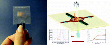
SPR biosensorAs featured on the bright inside front cover of Issue 5, this HOT article from Teodor Veres and colleagues at the National Research Council and McGill University, Canada, steps towards low cost point-of-care sensors for disease diagnosis.

The team present their all-polymeric nanoplasmonic microfluidic (NMF) transmission surface plasmon resonance (SPR) biosensor. SPR, involving light stimulated electron oscillation, is advantageous for sensing as it means label-free, real-time detection with high throughput and automation. The device is miniaturised with a view to small, point-of-care applications. The approach involves nanostructures called nanogratings for transmission SPR, which gives a more stable response. The signal can be turned by altering their characteristics and they are easily fabricated en mass.

Thermoplastic materials present an advantage over traditional PDMS for such miniaturised SPR devices as they are more mechanically robust, inert, transparent and crucially viable for large scale production and commercial applications.  The novel aspect of this work is that the nanostructured surface and the microchannels are incorporated into one substrate quickly and at low cost. Thermoplastic valves are used in large numbers for the first time for a multiplex detection scheme.

They demonstrate its application in sensing glycoprotein sCD44 at picomolar to nanomolar concentrations. Further work by the group is focused on integrating this device with a CCD spectrometer.

See the design and performance results in the full paper, now available free for 4 weeks*:

All-thermoplastic nanoplasmonic microfluidic device for transmission SPR biosensing
Lidija Malic, Keith Morton, Liviu Clime and Teodor Veres
DOI: 10.1039/C2LC41123G

Digg This
Reddit This
Stumble Now!
Share on Facebook
Bookmark this on Delicious
Share on LinkedIn
Bookmark this on Technorati
Post on Twitter
Google Buzz (aka. Google Reader)

LOC Issue 9 online! Micronanofabrication, neurotransmitters, SAW-controlled droplets and pharmaceutical screening

Abraham LeeThe packed Issue 9 begins with an editorial celebrating over three decades since the birth of microfluidics by Lab on a Chip Associate Editor Abraham Lee.

Submit your work to his editorial office today at http://mc.manuscriptcentral.com/lc!

The third decade of microfluidics
Abraham Lee
DOI: 10.1039/C3LC90031B


An urgent communication from Lothar Schmid and Thomas Franke at University of Augsburg, Germany, and Harvard University, USA, is featured on the outside front cover, in which a surface acoustic wave is applied to control droplet size in real time:

SAW-controlled drop size for flow focusing
Lothar Schmid and Thomas Franke
DOI: 10.1039/C3LC41233D

The inside front cover illustrates the work of researchers in New Zealand and Beijing led by Wenhui Wang who investigate locomotion metrics and muscular forces of C. elegans in one microfluidic assay:

On-chip analysis of C. elegans muscular forces and locomotion patterns in microstructured environments
Shazlina Johari, Volker Nock, Maan M. Alkaisi and Wenhui Wang
DOI: 10.1039/C3LC41403E


Issue 9 includes one Tutorial Review on using microfluidics to study neurotransmitters from Callie Croushore and Jonathan Sweedler at University of Illinois at Urbana-Champaign, USA, and one Critical Review discussing micronanofabrication techniques:

Microfluidic systems for studying neurotransmitters and neurotransmission
Callie A. Croushore and Jonathan V. Sweedler
DOI: 10.1039/C3LC41334A

Fabrication and multifunction integration of microfluidic chips by femtosecond laser direct writing
Bin-Bin Xu, Yong-Lai Zhang, Hong Xia, Wen-Fei Dong, Hong Ding and Hong-Bo Sun
DOI: 10.1039/C3LC50160D

The high quality research published in Issue 9 includes a HOT article from Paul Kenis et al. again at the University of Illinois at Urbana-Champaign, USA, which describes a new microfluidic platform for the screening of salt forms of pharmaceuticals:

Microfluidic Platform for Evaporation-based Salt Screening of Pharmaceutical Parent compounds
Sachit Goyal, Michael R. Thorson, Cassandra L. Schneider, Geoff G. Z. Zhang, Yuchuan Gong and Paul J. A. Kenis
DOI: 10.1039/C3LC41271G

View all of the articles in Issue 9 here

Digg This
Reddit This
Stumble Now!
Share on Facebook
Bookmark this on Delicious
Share on LinkedIn
Bookmark this on Technorati
Post on Twitter
Google Buzz (aka. Google Reader)

Introducing Editorial Board Member Yoon-Kyoung Cho

In the first of a short series of blog posts introducing the newest members of the Lab on a Chip Editorial Board, here we are very pleased to welcome Yoon-Kyoung Cho:

Yoon-Kyoung Cho received her B.S. and M.S. in Chemical Engineering from POSTECH in South Korea in 1992 and 1994, respectively. She continued her studies in the USA at the University of Illinois at Urbana-Champaign (UIUC) where she received her Ph.D. in Materials Science and Engineering in 1999 under the supervision of Prof. Steve Granick. Following her graduate work at UIUC, she returned to her homeland of South Korea and joined Samsung Advanced Institute of Technology (SAIT) as a senior research scientist. During her nine years at SAIT, she was involved in the research and development of a wide range of lab-on-a-chip technologies for biomedical applications, several of which have made it to the commercial marketplace.

In 2008, she returned to academia as an assistant professor in the school of Nano-Bioscience and Chemical Engineering at the Ulsan National Institute for Science and Technology (UNIST), South Korea and was promoted to associate professor in 2010. Since 2009, she has been the chair of her department and the director of an ambitious and prestigious program, the World Class University (WCU) program, geared to perform international research with leading scientists at UNIST. Her current research interests include novel micro/nano fluidic devices for advanced diagnostics, environmental monitoring, and cell biology. She has had a prolific career in academia and industry, publishing more than 38 scientific papers and 107 registered patents to date. 

Below, Professor Cho shares her views on Lab on a Chip, and the research areas she is working in currently:

RESEARCH VISION: ““Lab on a Chip” is an emerging research area where new discoveries and innovations are realized through multidisciplinary thinking and miniaturization to solve today’s most challenging problems in human health, energy and environment. Beyond the classic definition of a device that can integrates multiple laboratory functions on a small sized chip, Lab on a Chip has advanced the fundamental understanding of biological systems, broadened the basic knowledge on the molecular interactions in nano-scales, and translated into innovative designs and engineering of novel materials, devices and processes in order to provide paradigm-shifting solutions to the complex issues in chemistry, physics, biology and bioengineering. It is expected that there will be more and more examples of Lab on a Chip that go beyond chip-scale test devices and provide real impact in clinics and industry. My research group, under the title of “Integrated Nano-Biotechnology Lab”, in the school of Nano-Bioscience and Chemical Engineering at UNIST, is focused on the development of Lab on a Chip systems with fundamental understanding of bio-molecular interactions and fluidic behavior in micro/nano scales and its smart implementation by utilizing various engineering tools. Current research interests include novel micro/nano fluidic devices for advanced biomedical diagnostics, environmental monitoring and cell biology; e.g., fully integrated lab-on-a-disc for bioanalysis, biosensors using novel nano-materials, and cell chips for the investigation of cell to cell communication in cancer.”
Digg This
Reddit This
Stumble Now!
Share on Facebook
Bookmark this on Delicious
Share on LinkedIn
Bookmark this on Technorati
Post on Twitter
Google Buzz (aka. Google Reader)

Focus writer Tony Huang recognised for outstanding research

Tony Huang, writer of a series of Lab on a Chip Focus review articles, has been awarded a 2013 Faculty Scholar Medal by his institution, Penn State University. This Medal is awarded in recognition of his outstanding research in the area of microfluidic engineering, in particular acoustofluidics. His most recent work on optoacoustic tweezers is available to read in Lab on a Chip:

Optoacoustic tweezers: a programmable, localized cell concentrator based on opto-thermally generated, acoustically activated, surface bubbles
Yuliang Xie, Chenglong Zhao, Yanhui Zhao, Sixing Li, Joseph Rufo, Shikuan Yang, Feng Guo and Tony Jun Huang
DOI: 10.1039/C3LC00043E

Tony continues to write a series of varied and fascinating Focus articles published in Lab on a Chip on a regular basis, with topics from the unconventional uses of microfluidic technologies and applications of acoustofluidics through to imaging, circulating tumour cells, diagnostics and mechanical biomarkers:

Unconventional microfluidics: expanding the discipline
Ahmad Ahsan Nawaz, Xiaole Mao, Zackary S. Stratton and Tony Jun Huang
DOI: 10.1039/C3LC90023A

Probing circulating tumor cells in microfluidics
Peng Li, Zackary S. Stratton, Ming Dao, Jerome Ritz and Tony Jun Huang
DOI: 10.1039/C2LC90148J

Optofluidic imaging: now and beyond
Yanhui Zhao, Zackary S. Stratton, Feng Guo, Michael Ian Lapsley, Chung Yu Chan, Sz-Chin Steven Lin and Tony Jun Huang
DOI: 10.1039/C2LC90127G

Exploiting mechanical biomarkers in microfluidics
Xiaole Mao and Tony Jun Huang
DOI: 10.1039/C2LC90100E

Digg This
Reddit This
Stumble Now!
Share on Facebook
Bookmark this on Delicious
Share on LinkedIn
Bookmark this on Technorati
Post on Twitter
Google Buzz (aka. Google Reader)

HOT article: No separate demodulator for oscillating droplet manipulation

Electrowetting on dielectric-coated electrodes (EWOD) enables manipulation of individual droplets. Rather than direct current, AC-EWOD is used to additionally oscillate droplets for mixing and transport.

For wireless AC-EWOD, magnetic induction with a high frequency signal is needed, however this is above the dynamic range for droplet oscillation and an amplitude demodulator is currently needed in front of the EWOD chip to reduce this below 1 kHz.

In today’s HOT article, collaborators from Gangneung-Wonju National University, Republic of Korea, and University of Pittsburgh, USA, show theoretically and experimentally that an EWOD-actuated droplet actually has the ability to demodulate a high frequency amplitude modulation frequency itself due to the contact angle and does not require external artificial demodulation.

droplet, demodulationThe team point out that this phenomenon is unique in that this is the first physical entity to show visible demodulation behaviour. They briefly show that this demodulating functionality is not applicable with frequency modulation signals. Further investigation will focus on the possible range of signal that can be applied and successfully transmitted to the droplet and recovered by the inherent demodulation.

Read the theoretical and experimental explanation in full, as this HOT article is free to access for four weeks*:

Inherent amplitude demodulation of an AC-EWOD (electrowetting on dielectric) droplet
Myung Gon Yoon, Sang Hyun Byun and Sung Kwon Cho
DOI: 10.1039/C2LC41043E

*Free access to individuals is provided through an RSC Publishing personal account. Registration is quick, free and simple

Digg This
Reddit This
Stumble Now!
Share on Facebook
Bookmark this on Delicious
Share on LinkedIn
Bookmark this on Technorati
Post on Twitter
Google Buzz (aka. Google Reader)

LOC Issue 8 now online: Art in Science

The winner of the 2012 Art in Science Award presented at October’s MicroTas meeting was Yi Zhang, from Johns Hopkins University, USA and the striking winning image is featured on the outside front cover of Issue 8!

Read what’s in the issue here

A description of the winning image and advice on what makes a worthy Art in Science submission are discussed in the editorial by Michael Gaitan from NIST, USA, and Harp Minhas, Editor of LOC, who were part of the award selection committee.

The Art in Science of microTAS
Michael Gaitan and Harp Minhas
DOI: 10.1039/C3LC90026F


In keeping with the artistry of the front cover, work from Shoji Takeuchi et al. in Japan is featured on the inside front cover of Issue 8. In this paper, they address the problem of how to exchange the solution within a droplet for a new solution, for example to introduce membrane proteins or to wash-out applied chemicals during ion channel analysis. They use microfluidic channels to carry out solution exchange by droplet contact in under 20 seconds with the membrane still intact. Cover articles are free to access for 6 weeks*!

Droplet-based lipid bilayer system integrated with microfluidic channels for solution exchange
Yutaro Tsuji, Ryuji Kawano, Toshihisa Osaki, Koki Kamiya, Norihisa Miki and Shoji Takeuchi
DOI: 10.1039/C3LC41359D


The back cover features the laboratory of Sergey Shevkoplyas at Tulane University, USA. The communication by this laboratory describes their simple point-of-care test for sickle cell disease using characteristic patterns made by blood samples on paper, which can even differentiate between sickle cell disease and those healthy people with sickle cell traits.
A simple, rapid, low-cost diagnostic test for sickle cell disease
Xiaoxi Yang, Julie Kanter, Nathaniel Z. Piety, Melody S. Benton, Seth M. Vignes and Sergey S. Shevkoplyas
DOI: 10.1039/C3LC41302K


This issue also includes plenty of HOT research and a focus article from Tony Huang on the more unconventional applications of microfluidics:

Unconventional microfluidics: expanding the discipline
Ahmad Ahsan Nawaz, Xiaole Mao, Zackary S. Stratton and Tony Jun Huang
DOI: 10.1039/C3LC90023A

Programmable parylene-C bonding layer fluorescence for storing information on microfluidic chips
Ata Tuna Ciftlik, Diego Gabriel Dupouy and Martin A. M. Gijs
DOI: 10.1039/C3LC41280F

Engineering of functional, perfusable 3D microvascular networks on a chip
Sudong Kim, Hyunjae Lee, Minhwan Chung and Noo Li Jeon
DOI: 10.1039/C3LC41320A

In situ synthesis of silver nanoparticle decorated vertical nanowalls in a microfluidic device for ultrasensitive in-channel SERS sensing
Joseph Parisi, Liang Su and Yu Lei
DOI: 10.1039/C3LC41249K

Digg This
Reddit This
Stumble Now!
Share on Facebook
Bookmark this on Delicious
Share on LinkedIn
Bookmark this on Technorati
Post on Twitter
Google Buzz (aka. Google Reader)

Innovative cell culture and analysis microdevices

Here we have two HOT articles for you to read, both of which are free to access for the next 4 weeks*:

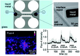
Researchers at North Carolina State University, USA, integrate microelectrodes into a neuron culture platform creating a single device with which cells can be electrically stimulated and their behaviour recorded simultaneously for scientific experiments. They do this using gallium and its alloys, which are liquid at room temperature and flow into microchannels to create the microelectrodes. This integrated biocompatible device avoids the complication of manually aligning the electrodes and provides subcellular accuracy.

Integration of pre-aligned liquid metal electrodes for neural stimulation within a user-friendly microfluidic platform
Nicholas Hallfors, Asif Khan, Michael D. Dickey and Anne Marion
DOI: 10.1039/C2LC40954B

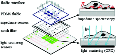
As featured on the outside front cover of Issue 5, HOT article number two is from a team led by Peter Ertl at the Austrian Institute of Technology, Austria, and Siemens AG, Germany. They combine two methods of cell analysis into one miniaturised device for dual-parameter analysis of cell cultures. The first technique is electrical impedance spectroscopy, normally used in analysing adherent cell cultures. The second is optical light scattering, normally used in flow cytometry. The combination of the two to detect light scattering from adherent cells enables rapid identification of cell number variations during culture, assessment of cell adhesion and interactions in physiological conditions. Applications are envisaged in cell–drug interaction assays and cytotoxicity screening. It also identifies intracellular granularity, which indicates apoptosis.

Standardization of microfluidic cell cultures using integrated organic photodiodes and electrode arrays
Verena Charwat, Michaela Purtscher, Sandro F. Tedde, Oliver Hayden and Peter Ertl
DOI: 10.1039/C2LC40965H

*Free access to individuals is provided through an RSC Publishing personal account. Registration is quick, free and simple

Digg This
Reddit This
Stumble Now!
Share on Facebook
Bookmark this on Delicious
Share on LinkedIn
Bookmark this on Technorati
Post on Twitter
Google Buzz (aka. Google Reader)