Archive for November, 2018

Emerging Investigator Series – Jerome Charmet

Jérôme Charmet received the Diplôme d’Ingénieur in Microtechnology Engineering from HES-SO Arc in Switzerland in 1998, the M.Sc. degree in Biomedical Engineering from the University of Bern, Switzerland, in 2010, and the PhD degree from the University of Cambridge in 2015. Overall, he worked for more than 10 years in both industrial and academic positions, including Intel Corporation, the National Centre for Sensor Research of Dublin City University in Ireland, the Microtechnology Institute of HES-SO Arc in Switzerland and the Centre for Misfolding Diseases of the University of Cambridge, UK. He joined the University of Warwick as an Assistant Professor in 2016 where he is developing integrated microfluidic platforms to study complex fluids and biological environments with applications in diagnosis, monitoring and drug screening/discovery. Read more about his group research here.

Read his Emerging Investigator article “Resolving protein mixtures using microfluidic diffusional sizing combined with synchrotron radiation circular dichroism” and read about him in the interview below:

Your recent Emerging Investigator Series paper focuses on protein mixtures using microfluidic diffusional sizing combined with synchrotron radiation circular dichroism. How has your research evolved from your first article to this most recent article?

It has evolved quite a lot, in fact it was not even directly related to microfluidics!

What aspect of your work are you most excited about at the moment?

I have started to explore organs-on-a-chip platforms with some colleagues biologists and I find it quite fascinating. But to be honest, every aspects of my work is exciting. I have a great team and collaborators I really enjoy to work with on a daily basis!

In your opinion, what applications can your current approach be used for?

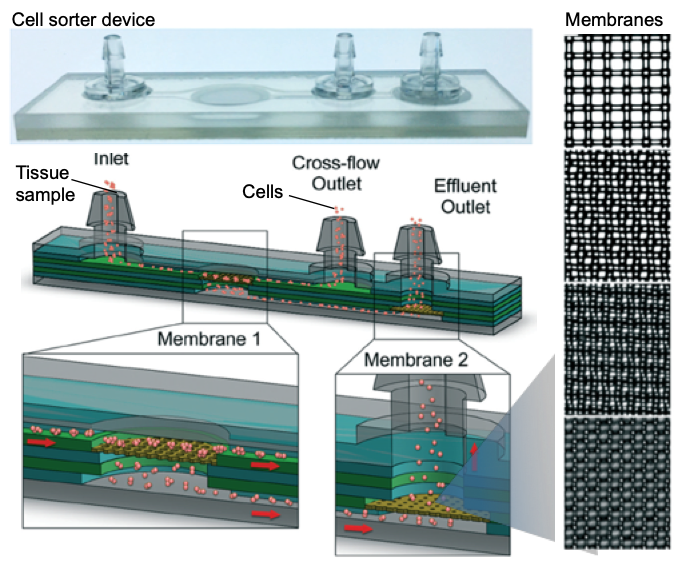
In the manuscript we have used diffusional sizing to resolve the secondary structure of a complex mixture of proteins using synchrotron radiation circular dichroism, but the approach can be applied to other biomolecules with other bulk measurement techniques. We are taking advantage of laminar flow to separate the mixture into “controllable” fractions. By measuring the mixture and the different fractions, we can retrieve information about each component in the mixture.

What do you find most challenging about your research?

It’s multidisciplinary nature.. but it is also one of the most rewarding (when it works).

In which upcoming conferences or events may our readers meet you?

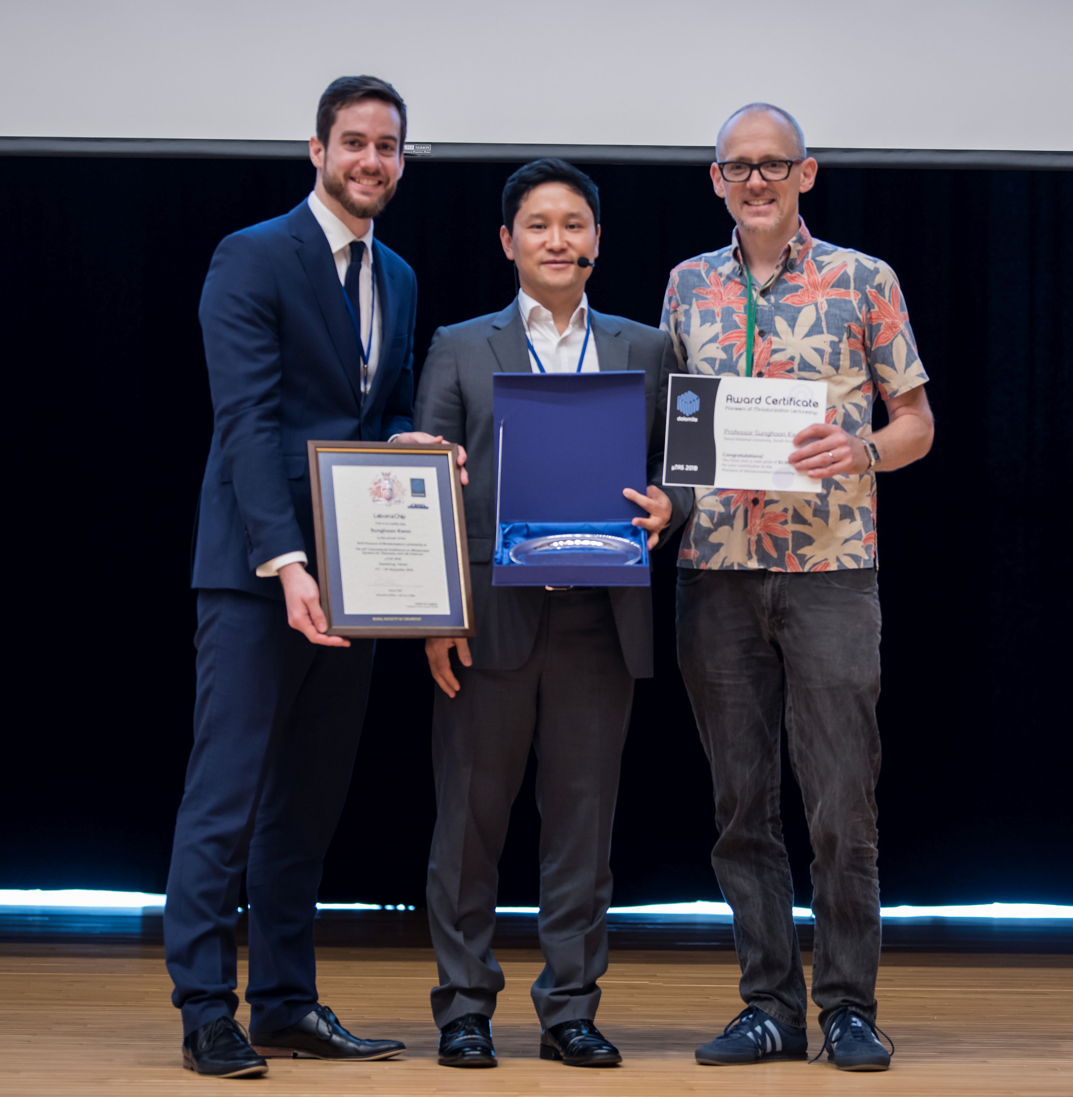
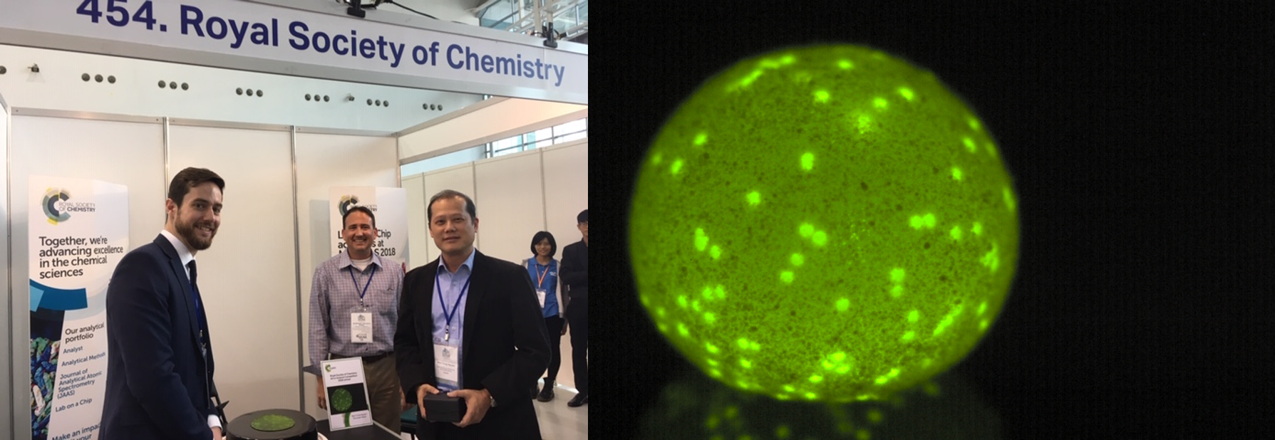
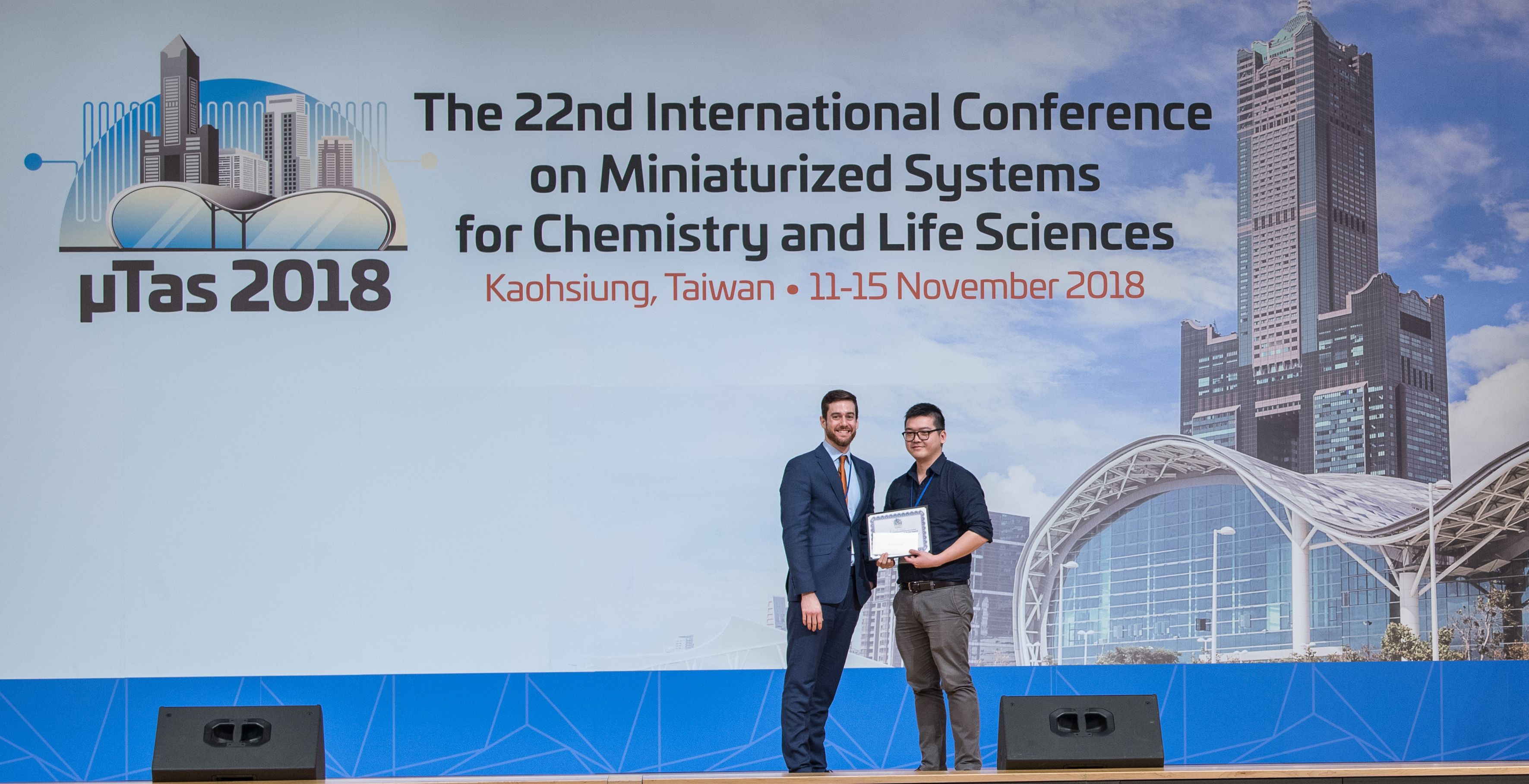
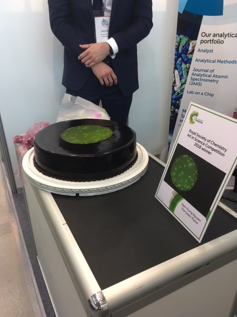
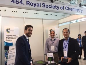
I’m just back from MicroTAS 2018 in Kaohsiung (Taiwan). Next, I will be attending the 8th Annual UK and Ireland Early Career Blood Brain Barrier Symposium 2018 in Oxford.

How do you spend your spare time?

Hiking, running … and these days spending as much time as possible with my 1 year old son.

Which profession would you choose if you were not a scientist?

I got to work with art conservator-restorers (…for some reason) and it is something I would definitely enjoy.

Can you share one piece of career-related advice or wisdom with other early career scientists?

It will sound a bit cheesy, but I will say “believe in your own ideas and importantly, find the right environment to develop them”.

Digg This
Reddit This
Stumble Now!
Share on Facebook
Bookmark this on Delicious
Share on LinkedIn
Bookmark this on Technorati
Post on Twitter
Google Buzz (aka. Google Reader)

Lab on a Chip Thematic Collections

We’ve brought together all of our latest Lab on a Chip Article Collections, Themed Issues, and Editor’s Choice collections to enable you to easily navigate to content most relevant to you. We hope you enjoy reading the papers in these collections!

Ongoing Collections

Thematic Collections

Digg This
Reddit This
Stumble Now!
Share on Facebook
Bookmark this on Delicious
Share on LinkedIn
Bookmark this on Technorati
Post on Twitter
Google Buzz (aka. Google Reader)

Lab on a Chip presents prestigious prizes at MicroTAS 2018

The µTAS 2018 Conference was held during 11th-15th November in Kaohsiung, Taiwan.  Simon Neil, Executive Editor of Lab on a Chip, attended the conference and announced the prestigious Lab on a Chip awards which include the Pioneers of Miniaturization Lectureship (in partnership with Dolomite Microfluidics), the Widmer Young Researcher Poster Prize and the Art in Science competition (in partnership with NIST). All three competitions received many fantastic submissions and we are delighted to present the winners, below:

“Pioneers of Miniaturization” Lectureship

Professor Sunghoon Kwon (Seoul National University) won the 13th “Pioneers of Miniaturization” Lectureship, sponsored by Dolomite and Lab on a Chip. The “Pioneers of Miniaturization” Lectureship rewards early to mid-career scientists who have made extraordinary or outstanding contributions to the understanding or development of miniaturised systems. Professor Sunghoon Kwon received a certificate, a monetary award and delivered a short lecture titled “Miniaturization for Personalised Medicine” at the conference.


Left to right: Simon Neil (Lab on a Chip), Sunghoon Kwon (winner) and Mark Gilligan (Dolomite)


Art in Science Competition

Lab on a Chip Executive Editor Simon Neil and Greg Cooksey from the National Institute of Standards Technology (NIST) presented the Art in Science award and a cake featuring the winning image at the Royal Society of Chemistry booth to Nam-Trung Nguyen for his entry “The Green Planet”. This award aims to highlight the aesthetic value in scientific illustrations while still conveying scientific merit.


Left to right: Simon Neil (Lab on a Chip), Greg Cooksey (NIST) and winner, Nam-Trung Nguyen with the personalised cake, and the winning image ‘The Green Planet’: an image of a floating liquid marble, decorated with green fluorescent beads. The image was taken with a colour USB camera. The liquid marble is made of a water droplet containing green fluorescent beads and coated with Teflon powder.


Widmer Young Researcher Poster Prize

The Widmer Young Researcher Poster Prize was awarded to Richard Cheng from the University of Toronto for his poster on “In Situ Delivery And Patterning Of Skin Cell Containing Biomaterial Sheet Using A Microfluidic Bioprinter”.


Simon Neil (left) with Richard Cheng (winner)


Congratulations to all the winners at the conference, we look forward to seeing you at µTAS 2019 in Basel, Switzerland! 


Digg This
Reddit This
Stumble Now!
Share on Facebook
Bookmark this on Delicious
Share on LinkedIn
Bookmark this on Technorati
Post on Twitter
Google Buzz (aka. Google Reader)

Art in Science Competition Winner Announced at MicroTAS 2018

Lab on a Chip and the National Institute of Standards Technology (NIST) presented the Art in Science award at the µTAS 2018 Conference on 14 November 2018 at the Royal Society of Chemistry booth. The award highlights the aesthetic value in scientific illustrations while still conveying scientific merit. The competition received many fantastic submissions this year which were judged by Simon Neil, Lab on a Chip Executive Editor, Greg Cooksey, NIST representative and Manabu TokeshiLab on a Chip Associate Editor .

Simon Neil and Greg Cooksey announced the winner of the competition was Nam-Trung Nguyen with his entry “The Green Planet” and presented Dr Nguyen with his award and certificate and a cake featuring the winning image.

The Green Planet 

Nam-Trung Nguyen, Griffith University, Australia

The Green Planet


The runners up are:


Magnetic Artificial Cilia with a Brush-shaped Cap 

Shuaizhong Zhang, Eindhoven University of Technology, Netherlands

magnetic artificial cilia with a brush-shaped cap


Embracing Chaos

Samantha Byrnes, Intellectual Ventures Laboratory, USA


A big thank you to all the contributors this year!

Digg This
Reddit This
Stumble Now!
Share on Facebook
Bookmark this on Delicious
Share on LinkedIn
Bookmark this on Technorati
Post on Twitter
Google Buzz (aka. Google Reader)

Emerging Investigator Series – Weian Zhao

Dr. Weian Zhao is an Associate Professor at the Sue and Bill Gross Stem Cell Research Center, Chao Family Comprehensive Cancer Center, Department of Biomedical Engineering, and Department of Pharmaceutical Sciences at University of California, Irvine. Dr. Zhao is also the co-founder of Velox Biosystems Inc, Baylx Inc, and Amberstone Biosciences LLC, start-up companies that aim to develop technologies for rapid diagnosis, stem cell therapy, and drug discovery, respectively. Dr. Zhao’s research aims to 1) elucidate and eventually control the fate of transplanted stem cells and immune cells to treat cancer and autoimmune diseases, and 2) develop novel miniaturized devices for early diagnosis and monitoring for conditions including sepsis, antibiotic resistance and cancer. Dr. Zhao has received several awards including the MIT’s Technology Review TR35 Award: the world’s top 35 innovators under the age of 35 and NIH Director’s New Innovator Award. Dr. Zhao completed his BSc and MSc degrees in Chemistry at Shandong University and then obtained his PhD in Chemistry at McMaster University in 2008. During 2008-2011, Dr. Zhao was a Human Frontier Science Program (HFSP) Postdoctoral Fellow at Harvard Medical School, Brigham and Women’s Hospital and MIT.

Read Dr Zhao’s Emerging Investigator article “Functional TCR T cell screening using single-cell droplet microfluidics” and read more about him in the interview below:

Your recent Emerging Investigator Series paper focuses on functional TCR T cell screening using single-cell droplet microfluidics. How has your research evolved from your first article to this most recent article?

I was trained as a colloidal and surface chemist. My first paper back when I was a PhD student was about building nanostructures using DNA as a template. Over the years, my research has evolved towards addressing immediate, major unmet need in biology and medicine; this is achieved by developing new tools and technologies, often using an interdisciplinary and collaborative approach, as exemplified by this new paper.

What aspect of your work are you most excited about at the moment?

It is the perspective that what we do in research can potentially solve real-world problems and help the patients in a short term. Like this new paper illustrated, our single-cell system holds great potential to accelerate development of cancer immunotherapeutics. There is a great and urgent demand in the clinic for many more of this type of promising drugs that can benefit patients who do not have lots of time to wait.

In your opinion, how can droplet microfluidic technologies contribute to immune-screening and immuno-therapy and personalised medicine in general?

Droplet microfluidic technologies and other exciting single-cell systems being developed in our field can contribute to immune-screening, immuno-therapy and personalised medicine by significantly reducing the time needed for the discovery phase. For example, conventional screening platforms for immunologic agents such as antibodies or T cells, usually take months to years to obtain a therapeutic candidate whereas the new single-cell platforms can potentially do this in the matter of days to weeks. In this new paper, this ability is enabled by directly interrogating the “functions” of individual cell clones in greater depth by using single-cell analysis. As biological samples are heterogeneous, this key information is often lost in conventional, population based studies.

What do you find most challenging about your research?

It is always the people. I take time to identify and build the right team that, once in place, can almost conquer any challenges in research itself. I also wish I could spend more time to do research rather than writing grants.

In which upcoming conferences or events may our readers meet you?

IEEE EMBS Micro & Nanotechnology in Medicine 2018, Physical Science of Cancer (GRS) 2019, and PEGS 2019

How do you spend your spare time?

Mostly with the family, and watching Manchester United games.

Which profession would you choose if you were not a scientist?

Probably try to become a football manager in the English Premier League. There is a lot of similarity in running a research laboratory and running a football club, isn’t there?

Can you share one piece of career-related advice or wisdom with other early career scientists?

Ask for help and mentorship from people who have succeeded in what you are trying to do. Try to accelerate your progress through collaboration and partnership.

Digg This
Reddit This
Stumble Now!
Share on Facebook
Bookmark this on Delicious
Share on LinkedIn
Bookmark this on Technorati
Post on Twitter
Google Buzz (aka. Google Reader)

Simple microfluidic cell sorter device to replace manual tissue dissociation protocols

Even a tiny group of cells has the ability to populate a tumor in tissues. Determining cellular diversity and identifying these small cell groups gains importance when it comes to selection of treatment strategies. Tissue samples taken from patients are required to be dissociated into single-cell suspensions, therefore identification can be efficiently done at single-cell level using a powerful suite of technologies including flow cytometry, mass cytometry, and single cell RNA sequencing. However, breaking a tissue down to a single-cell suspension is not an easy task. The old-school way is to cut the tissue sample into small pieces with a blade and mechanically dissociated by vigorous shaking after the application of proteolytic enzymes. Large aggregates are removed by filtering the suspension through a strainer. This technique significantly increases the sample loss, drops the speed of the process and is not ideal for immediate downstream analysis.

In this month’s Lab on a Chip HOT article series, a group of researchers led by Dr Jered Haun at University of California Irvine presented a novel and simple approach that improves the quality of single-cell suspensions obtained from tissue samples using microfluidics. Jeremy Lombardo, a co-author of this article, explains that “the goal of this work was to fully replace manual intensive tissue dissociation protocols by using microfluidic devices.” The developed tool is a microdevice consisting of two nylon membranes, one with 25-50 µm mesh, and the other with 10-15 µm mesh, attached to micron-sized pores and microchannels. The device is made of laser-micromachined hard plastic (PET, aka. polyethylene terephthalate), which enables operation at high flow rates (>10 mL/min) when compared to PDMS (a silicon-based organic material). Also, the chip has multiple layers for connecting nylon mesh membranes at different levels (Figure 1).

cell sorter

Figure 1. Microfluidic cell sorter device for tissue  samples. The sketch shows the inner layers, consisting of two membranes for operating the device in direct or tangential filtration modes. Membrane mesh size can be adjusted to the cell size. Micrographs on the right show lattice network with several pore sizes used in this work. Pore sizes are (top to bottom) 50, 25, 15, 10 µm diameter.

Working principle of the cell sorter device

The inlet of the device connects to a microporous membrane to introduce tissue samples. The Sample passing through the membrane exits through the effluent outlet. It is also possible to direct a portion of the sample along the surface of the membrane that is connected to the cross-flow outlet. The device is either operated in a direct filtration regime to maximize sample recovery and processing speed, or in a tangential filtration regime to sweep larger tissue fragments and cell aggregates away to prevent clogging.

While the researchers initially hypothesized that under pressure-driven flow, cell and tissue aggregates might disaggregate as they pass through the membranes of the device, they were pleasantly surprised by the drastic level of single cell increases seen in the initial testing of these devices, says Jeremy Lombardo and adds “The hardest part in developing and testing this device was to find a combination of membrane pore sizes that could best dissociate cell aggregates and tissue without compromising cell viability. Thorough testing of various pore sizes and combinations were ultimately carried out with both cell line and murine tissue models before we settled on the final 50 and 15 μm pore sizes.”

Advantages, challenges and the future

The authors summarized the advantages of this platform for Lab on a Chip blog readers: “The device is extremely simple to operate as well as inexpensive to fabricate. It can easily be incorporated into many tissue dissociation applications for improved single cell yields as a standalone device but could also be easily integrated with other downstream microfluidic operations (cell sorting, detection etc.).” According to the authors, “in the current format of cell sorter device, cells that are very large in size would likely be difficult to process, as they would likely span multiple pores of the filters and be traditionally filtered away instead of dissociated.” Although seeming like a challenge, this can easily be addressed by adjusting the filter membrane pore sizes to accommodate these larger cell types.

For the future of the device, the authors indicate, “We are also currently working on integrating this device with upstream, larger scale tissue dissociation devices that we have developed previously to create a fully automatable microfluidic tissue dissociation platform.


To download the full article for free* click the link below:

Microfluidic filter device with nylon mesh membranes efficiently dissociates cell aggregates and digested tissue into single cells
Xiaolong Qiu, Jeremy A. Lombardo, Trisha M. Westerhof, Marissa Pennell, Anita Ng, Hamad Alshetaiwi, Brian M. Luna, Edward L. Nelson, Kai Kessenbrock, Elliot E. Hui, and Jered B. Haun
Lab Chip, 2018, Lab on a Chip Recent Hot Articles
DOI: 10.1039/c8lc00507a

About the Webwriter
Burcu Gumuscu is a postdoctoral fellow in Herr Lab at UC Berkeley in the United States. Her research interests include development of microfluidic devices for quantitative analysis of proteins from single-cells, next generation sequencing, compartmentalized organ-on-chip studies, and desalination of water on the microscale.

Digg This
Reddit This
Stumble Now!
Share on Facebook
Bookmark this on Delicious
Share on LinkedIn
Bookmark this on Technorati
Post on Twitter
Google Buzz (aka. Google Reader)

SelectBio Conferences & Exhibitions, 2019

SelectBio conference logoWe are excited to announce that registration is now open for the SelectBio Conferences that take place throughout 2019. The various events and exhibitions will bring together a number of keynote speakers to discuss the most up-to-date technologies and advances in different evolving fields. Check out the SelectBio website for a list of events and descriptions, including a full list of confirmed plenary speakers. Some of the events will be hosted in Coronado Island, California and the Europe-based conferences will take place in Rotterdam, The Netherlands.

Lab on a Chip, AnalystAnalytical Methods and Biomaterials Science are delighted to sponsor the following upcoming events and exhibitions in 2019:

SelectBio: Circulating Biomarkers and Liquid Biopsies, Coronado Island

SelectBio: Biosensors Summit, Coronado Island

SelectBio: 3D-Culture and Organoids, Coronado Island

SelectBio: Lab-on-a-Chip and Microfluidics Europe, Rotterdam

SelectBio: Organ-on-a-Chip and Tissue-on-a-Chip Europe, Rotterdam

SelectBio: Point-of-Care, Mobile Diagnostics and Biosensors Europe, Rotterdam

SelectBio: Biofabrication & Biomanufacturing Europe, Rotterdam

SelectBio: Point-of-Care Diagnostics, Wearables & Global Health 2019

SelectBio: Lab-on-a-Chip & Microfluidics World Congress 2019

SelectBio: Microfluidics for Circulating Biomarkers Summit 2019

SelectBio: Microfluidics & Flow Chemistry 2019

SelectBio: Organ-on-a-Chip World Congress 2019

Single Cell & Single Molecule Analysis Summit 2019

We recommend registering early to secure a place at these events. Remember to keep your eyes peeled for upcoming conferences, and stay connected with SelectBio. Register now!


Circulating Biomarkers 2019, SelectBio conferences

Biofabrication & Biomanufacturing Europe 2019, SelectBio conference

Digg This
Reddit This
Stumble Now!
Share on Facebook
Bookmark this on Delicious
Share on LinkedIn
Bookmark this on Technorati
Post on Twitter
Google Buzz (aka. Google Reader)

Emerging Investigator Series – Kyle Bishop

Kyle Bishop


Introducing Kyle Bishop: Lab on a Chip‘s latest Emerging Investigator

Kyle Bishop received his PhD in Chemical Engineering from Northwestern University under the guidance of Bartosz Grzybowski for work on nanoscale forces in self-assembly. Following his PhD, Dr. Bishop was a post-doctoral fellow with George Whitesides at Harvard University, where he developed new strategies for manipulating flames with electric fields. He started his independent career at Penn State University in the Department of Chemical Engineering. In 2016, Dr. Bishop moved to Columbia University, where he is currently an Associate Professor of Chemical Engineering. Dr. Bishop has been recognized by the 3M Non-tenured Faculty award and the NSF CAREER award. His research seeks to discover, understand, and apply new strategies for organizing and directing colloidal matter through self-assembly and self-organization far-from-equilibrium.


Read Dr Bishop’s article entitled ‘Measurement and mitigation of free convection in microfluidic gradient generators’ and find out more about him in the interview below:

Your recent Emerging Investigator Series paper focuses on the measurement and mitigation of free convection in microfluidic gradient generators. How has your research evolved from your first article to this most recent article?

Our first article in Lab on a Chip focused on harnessing electric potential gradients to power transport and separations within microfluidic systems. Here, we examine how chemical gradients can drive fluid flows as well as motions of colloidal particles, lipid vesicles, and living cells. These topics are linked by our continued interest in harnessing and directing thermodynamic gradients to perform dynamic functions at small scales.

What aspect of your work are you most excited about at the moment?

Currently, we are excited by our pursuit of colloidal “robots” that organise spontaneously in space and time to perform useful functions, which can be rationally encoded within active soft matter.

In your opinion, what is the future of microfluidic gradient generators? Any new applications you foresee for them?

Our interest in microfluidic gradient generators grew from a desire to quantify the motions of lipid vesicles in osmotic gradients (so-called osmophoresis).  These measurements were plagued by undesired gradient-driven flows.  We thought that our efforts to understand and mitigate these flows would be useful to others studying gradient driven motions (e.g., chemotaxis of living cells).

What do you find most challenging about your research?

Staying focused. The world is filled with many micro-mysteries that may pique your curiosity, but time is limited. Picking problems and following through on their solution is an ever-present challenge.

In which upcoming conferences or events may our readers meet you?

Our group regularly attends the AIChE Annual Meeting and the ACS Colloid and Surface Science Symposium.

How do you spend your spare time?

Exploring New York City with my family and thinking about science.

Which profession would you choose if you were not a scientist?

What a horrible thought…perhaps a lawyer as I value evidence-based reasoning and the rule of law (physical or otherwise).

Can you share one piece of career-related advice or wisdom with other early career scientists?

Think big and collaborate often.

Digg This
Reddit This
Stumble Now!
Share on Facebook
Bookmark this on Delicious
Share on LinkedIn
Bookmark this on Technorati
Post on Twitter
Google Buzz (aka. Google Reader)