Archive for September, 2018

Emerging Investigator Series: Kiana Aran

Dr. Aran received her undergraduate degree in electrical engineering at the City University of New York in 2007 and her Ph.D in biomedical engineering at Rutgers University in 2012. She then continued her postdoctoral studies in bioengineering at the University of California Berkeley and was a recipient of National Institutes of Health (NIH) postdoctoral training fellowship at the Buck Institute for Age Research in 2015. She joined Keck Graduate Institute in 2017 as an Assistant Professor and is a currently a visiting scientist at the University of California Berkeley. She is also a consultant for Bill and Melinda Gate Foundation and cofounder of NanoSens innovations. Since starting her faculty position in 2017, Dr. Aran work has been supported by various funds including one RO1, industry sponsored research and a 3 years subaward from University of California Berkeley.

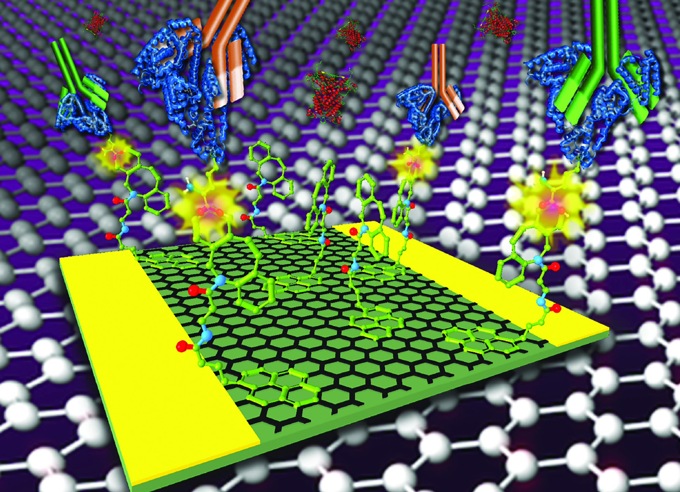
Read her Emerging Investigator Series article “Graphene-based biosensor for on-chip detection of Bio-orthogonally Labeled Proteins to Identify the Circulating Biomarkers of Aging during Heterochronic Parabiosis” and find out more about her in the interview below:


Your recent Emerging Investigator Series paper focuses on digital detection of tagged proteins in vivo to identify the circulating biomarkers in aging. How has your research evolved from your first article to this most recent article?

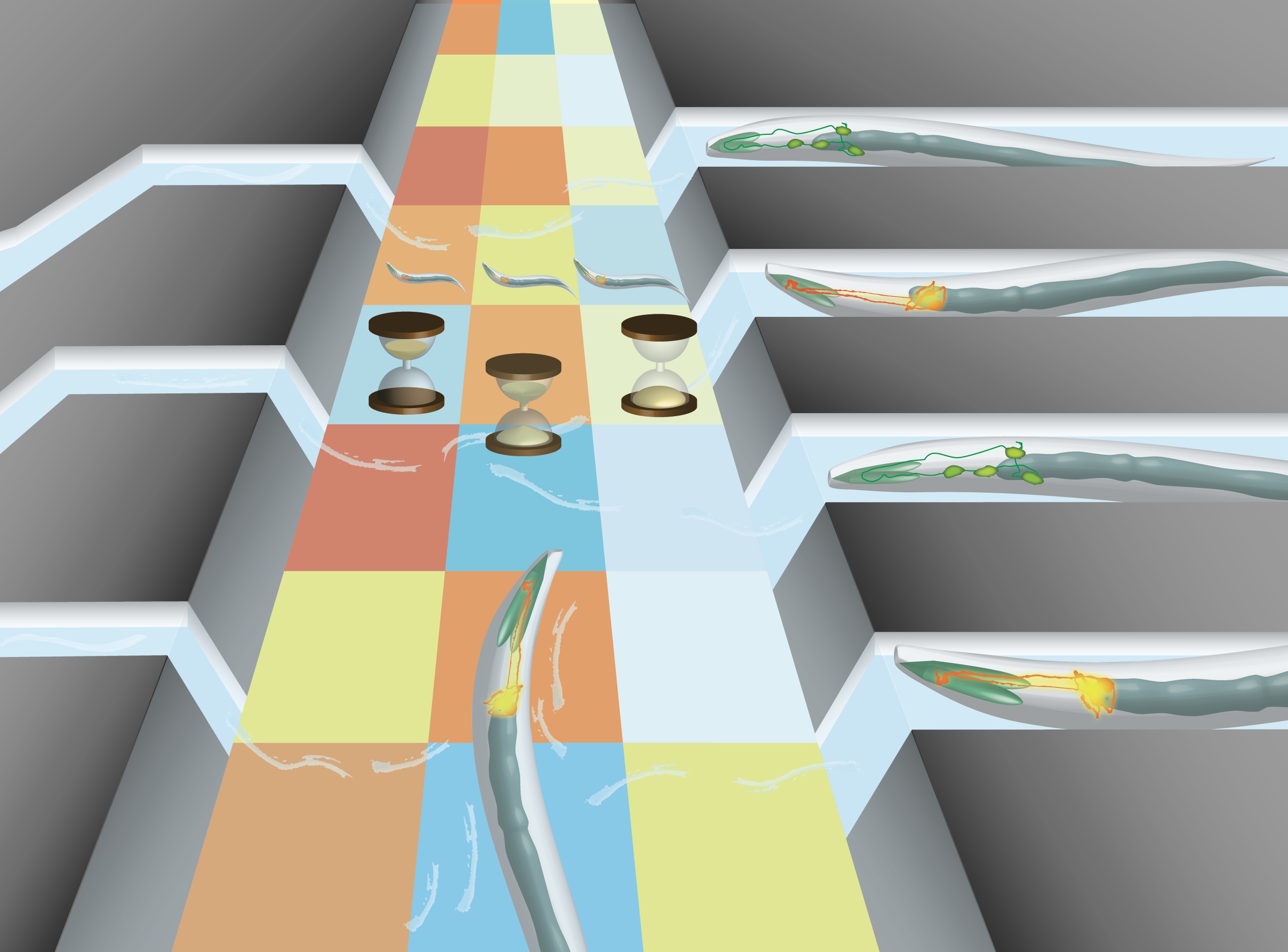
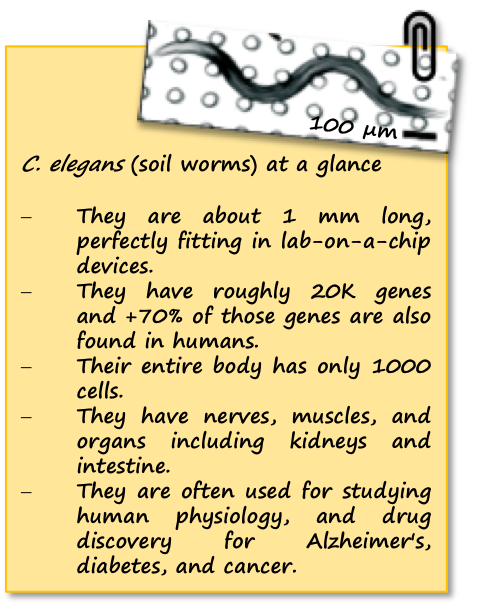
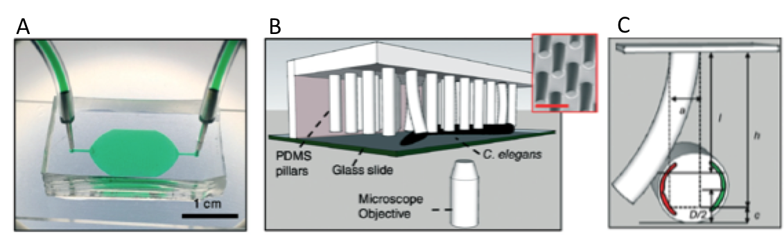
I was a recipient of an NIH T32 award during my last years of postdoc training at UC Berkeley (2015-2017) which provided me with the opportunity to work with Dr. Irina Conboy, a pioneer in utilizing bio-orthogonally labeled proteins for research on aging. This approach enabled the detection of several rejuvenating protein candidates from the young parabiont, which were transferred to the old mammalian tissue through their shared circulation. Although this method was very powerful, there were a lot of challenges with the detection of these tagged proteins including very time consuming, complex and expensive assays, large sample requirement and false positive. I started working on this project as a side project along with other projects I had with Dr. Conboy just to make the process more facile so we can detect these protein faster and more continuously. This work is a great example of utilizing a lab on a chip technology for a real application. I have also started collaboration with nanomedical diagnostics, a startup company in San Diego, to enable mass production of our chips with high reproducibility. With my academic and industrial collaborators, we are planning to utilize this technology for at least two different applications in the near future to better understand the biology of aging.

What aspect of your work are you most excited about at the moment?

I am very excited to see my technologies go beyond publications. Two of my inventions have been licensed toward developing medical devices for drug delivery and medical diagnostics and the joy from that have been the biggest drive for me to work harder.

In your opinion, what is the future of portable devices for biosensing?

Even though still in its emerging stage, digital diagnostics and 2D materials will shape the future of biosensors.

What do you find most challenging about your research?

As a junior faculty, it is difficult to recruit highly motivated postdoctoral researchers. Unfortunately, the majority of postdoctoral candidates often look for a well-established laboratory headed by a leading scientist. Students and postdoc should know that working in a new lab can be challenging but can provide researcher with opportunities to strengthen their scientific and management skills.

In which upcoming conferences or events may our readers meet you?

I will be presenting some of my biosensor work in the upcoming IEEE EMBS MNMC Conference

Above: Dr Aran supports a community in Africa by building a school

How do you spend your spare time? 

I love what I do so I can not define my spare time from my work time. I have recently co-founded a startup in digital diagnostics so that takes my weekend but I love it. I also paint when I feel like painting.  But I do run, swim and work out routinely after work and enjoy a variety of sports.  I have also started traveling to small villages around the world with my friends where we help in building schools.

Which profession would you choose if you were not a scientist?

I love painting and would have explored a career in art.

Can you share one piece of career-related advice or wisdom with other early career scientists?

Have a vision. Sometimes we get lost in small career goals such as publishing but if you have a vision of what you are trying to accomplish you can define your career path, you will start working with the right people who support your vision, you will attend the right conferences and workshops for your career development and you will definitely be successful.

Lab on a Chip Issue 21

Digg This
Reddit This
Stumble Now!
Share on Facebook
Bookmark this on Delicious
Share on LinkedIn
Bookmark this on Technorati
Post on Twitter
Google Buzz (aka. Google Reader)

Emerging Investigator Series: Adriana San Miguel

Adriana San Miguel is an Assistant Professor in the Department of Chemical & Biomolecular Engineering at NC State University. She is part of the Synthetic and Systems Biology Chancellor´s Faculty Excellence Program. Her work combines engineering and biology and focuses on developing tools to perform high-throughput automated experiments with the model organism C. elegans. Her team uses these tools and this organism to better understand aging, stress, and the nervous system.

Adriana is originally from San Luis Potosi, Mexico. After receiving a BS in Chemical Engineering at the Monterrey Institute of Technology (ITESM) in 2005, she worked in the water treatment and cement industries for 2 years. She obtained a Ph.D. in Chemical Engineering from the Georgia Institute of Technology in 2011. She then took on a Postdoctoral Research position at Georgia Tech, and a second one at the Dana-Farber Cancer Institute in Boston. In 2013, Adriana was awarded the NIH K99 Pathway to Independence Award to study the mechanisms regulating synaptic plasticity and aging in the nematode C. elegans.

Read her Emerging Investigator Series article “A microfluidic platform for lifelong high-resolution and high throughput imaging of subtle aging phenotypes in C. elegansand find out more about her in the interview below:

Your recent Emerging Investigator Series paper focuses on a microfluidic platform for high-resolution and high throughput imaging of aging phenotypes in C. elegans. How has your research evolved from your first article to this most recent article? 

We have previously been interested in developing systems for high-throughput imaging and analysis of subcellular features in C. elegans, and gained interest in quantitatively assessing how these change during aging. Aging studies pose several technical challenges that could not be addressed with previous microfluidic devices, thus our interest in developing a system capable of performing high-throughput analysis of aging phenotypes.

What aspect of your work are you most excited about at the moment?

I am excited about the possibility of doing systems biology enabled by microfluidics. Lab on chip approaches provide an excellent level of precision in experimental conditions which are unfeasible with traditional C. elegans techniques. Microfluidics also allows high-throughput studies that result in large data sets, and as a consequence, a need to perform unsupervised quantitative data analysis. When combined, these powerful approaches enable better understanding of biological processes from a systems perspective.

In your opinion, what are the challenges with using microfluidics for whole organism behaviour?

Behavioral studies are challenging, particularly because there is a large degree of variability within a population. Microfluidics can help by facilitating animal handling and ensuring the stimuli used is precise (in time, concentration, and localization). Although it is difficult to determine how representative a microfluidic environment (where animals navigate in 2D) resembles their natural 3D environment, microfluidic chips enable single animal tracking, thus increasing the confidence of noisy behavioral readouts.

What do you find most challenging about your research?

Our research requires the integration of several different fields. We develop microfluidic tools, and use them to answer fundamental biological questions. In addition, we use automation, image processing, machine learning, and analysis tools for the large data sets we acquire. Integrating all of these can certainly be challenging, but achieving fully integrated systems is what we find very motivating.

In which upcoming conferences or events may our readers meet you?

I typically attend the American Institute of Chemical Engineers Annual Meetings, as well as the International C. elegans meeting, among others.

How do you spend your spare time?

I enjoy working out every day, it is a necessary part of my routine. I also enjoy baseball, live music, reading, and spending time with family and friends.

Which profession would you choose if you were not a scientist?

I’d probably still be an Engineer, or maybe an artist of some sort.

Can you share one piece of career-related advice or wisdom with other early career scientists?

Something I could suggest is to not be afraid of testing new ideas and methods or venturing into different fields. Although it can be challenging to develop something that is completely new, if it sparks your interest, give it a try. It is very rewarding and motivating to get something new working.


San Miguel Journal Front Cover

Digg This
Reddit This
Stumble Now!
Share on Facebook
Bookmark this on Delicious
Share on LinkedIn
Bookmark this on Technorati
Post on Twitter
Google Buzz (aka. Google Reader)

How soil-worms allow for realistic human physiology studies

Mice, fruit fly, zebra fish easily come to mind when thinking of animal models for human physiology studies – but one animal is often forgotten, although it is as functional as the others. Have you guessed already? We are talking about soil-dwelling worms, aka C. elegans! These animals combine the simplicity of single-cell systems with the complexity of animal models, therefore they can provide significant insights into human disorders. Please take a moment to look at our note below summarizing the key features of C. elegans.

Muscular strength is a good example of human physiology studies  and it relies on calcium-initiated muscle contraction, sarcomere composition and organization, and translocation of actin and myosin molecules. Analysis of such parameters can reveal the formation of muscular dystrophy, a muscle degenerative disorder. However, the measurement of these parameters has been a challenge due to the dependence on random animal-behavior that yields irreproducible results. Recently, researchers from Texas Tech University collaborated with Rutgers University and University of Nottingham to study muscular strength in C. elegans. They achieved to obtain results independent of animal behavior and gait in a miniature system consisting of an elastic micropillar forest (Figure 1a and 1b).

The microfluidic system is made of PDMS, and contains bendable micropillars hanging from the microchannel ceiling. The pillars are bent upon the action of the body muscles when C. elegans crawls through the pillar array. Individual pillar bending events can be quantified using a microscope-camera system and image analysis (Figure 1c). The pillar density is designed to create high mechanical resistance to locomotion, therefore maximum exertable force can be measured independent of animal behavior. Here, maximum exertable force corresponds to the peak force exerted by human quadriceps muscle in a standardized knee extension resistance test.

Figure 1. (a) Image of the microfluidic device with the pillar forest and the ports. (b) Schematic demonstration of the C. elegans strength measurement apparatus. Inset shows a scanning electron microscope picture of the pillars. (c) A sketch of interaction with a pillar by the worm body. The pillar is bent due to the action of the body muscles (shown in red and green). Image from Rahman et al.

The authors of this study explain that animals produce strong forces in highly resistive areas and demonstrate different locomotion regimes based on the body size relative to gap between pillars. Besides the body size, body configuration and behavioral characteristics can be the sources to the magnitude of the force exerted on the pillars. Thanks to the probabilistic nature of the parameters sourcing of the force exerted, a reproducible algorithm can be defined for quantifying muscle strength. Using this strategy, researchers showed for the first time that locomotion between microfluidic pillars comprises of three regimes: non-resistive (worm contacts with 1-2 pillars and doesn’t adjust body posture), moderately resistive (worm contacts with >2 pillars and minimally adjust body posture), and highly resistive (worm contacts with multiple pillars and body posture adjustment is disabled). When operated at highly-resistive regime, the microfluidic system suppresses the animal behavior. This system allows for (1) discriminating between the muscle strength or weakness levels of individual worms of different ages, (2) determining body length decrease and muscular contraction levels led by levamisole treatment, (3) comparing the muscular strength in the wild and mutant C. elegans types. According to the researchers, the future studies can help us to obtain deeper understanding in molecular and cellular circuits of neuromuscular function as well as dissection of degenerative processes in disuse, aging, and disease.

To download the full article for free* click the link below:

NemaFlex: a microfluidics-based technology for standardized measurement of muscular strength of C. elegans
Mizanur Rahman, Jennifer E. Hewitt, Frank Van-Bussel, Hunter Edwards, Jerzy Blawzdziewicz, Nathaniel J. Szewczyk, Monica Driscolld, and Siva A. Vanapalli
Lab Chip, 2018, Lab on a Chip Recent Hot Articles
DOI: 10.1039/c8lc00103k


About the Webwriter

Burcu Gumuscu is a postdoctoral fellow in Herr Lab at UC Berkeley in the United States. Her research interests include development of microfluidic devices for quantitative analysis of proteins from single-cells, next generation sequencing, compartmentalized organ-on-chip studies, and desalination of water on the microscale.

Digg This
Reddit This
Stumble Now!
Share on Facebook
Bookmark this on Delicious
Share on LinkedIn
Bookmark this on Technorati
Post on Twitter
Google Buzz (aka. Google Reader)