Archive for June, 2018

Emerging Investigator Series – Rebecca Pompano

We are delighted to introduce our latest Lab on a Chip Emerging Investigator, Rebecca Pompano.

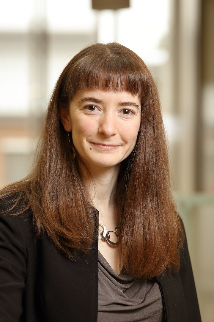
Dr. Rebecca Pompano is an Assistant Professor in the Departments of Chemistry and Biomedical Engineering at the University of Virginia, and a member of the Beirne B. Carter Center for Immunology Research.  She completed a BS in Chemistry at the University of Richmond in 2005, and a PhD in 2011 at the University of Chicago, working in the laboratory of Dr. Rustem Ismagilov.  She completed a postdoc in the University of Chicago Department of Surgery, leading a collaboration between Dr. Joel Collier, a tissue engineer, and Dr. Anita Chong, an immunologist.  Since 2014, she has been a faculty member at UVA, where her research interests center on developing microfluidic and chemical assays to unravel the complexity of the immune response.  She received an Individual Biomedical Research Award from The Hartwell Foundation and the national 2016 Starter Grant Award from the Society of Analytical Chemists of Pittsburgh.  Recently, her lab was awarded an NIH R01 to develop hybrids of microfluidics and lymph node tissue to study inflammation.  In addition to her research, she is active in advocating for continued funding for education and biomedical research on Capitol Hill.

Read her Emerging Investigator Series article “User-defined local stimulation of live tissue through a movable microfluidic port” and find out more about her in the interview below:

Your recent Emerging Investigator Series paper focuses on stimulation of live tissue through a movable microfluidic port. How has your research evolved from your first article to this most recent article?

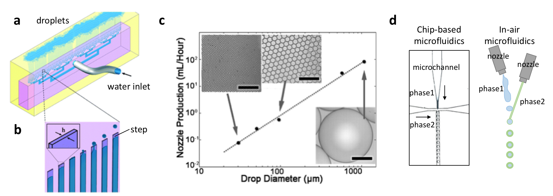
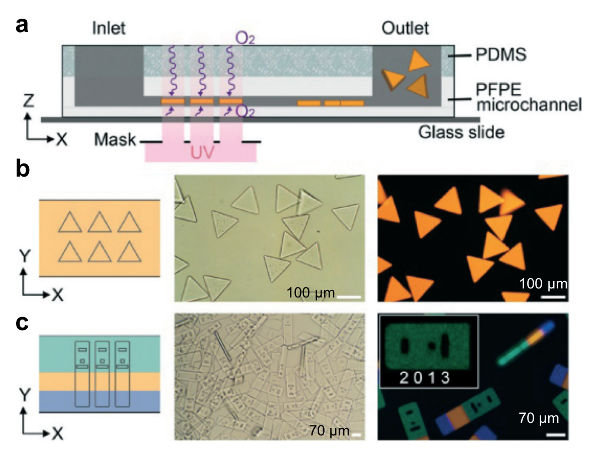
My current research combines some seemingly disparate themes from my prior work.  My first article in graduate school used droplet microfluidics to study blood clotting, and I became fascinated with how spatial organization affects the function of complex biological systems. Later, I also worked on the physics of fluid flow in a reconfigurable SlipChip device… and both of these ideas make a comeback in this current paper!  Then in my postdoc, I had the fabulous opportunity to work in both a bioengineering lab and an immunology lab, studying the mechanism of action of a new non-inflammatory vaccine. The research in my lab now is really at the intersection of bioanalytical chemistry, bioengineering, and immunology.  We develop new tools to study the immune system and how it is organized. This particular paper offers a new technology to pick and choose where to deliver a drug or stimulant to a piece of live tissue, and we demonstrated it for lymph nodes, our favorite immune organ.

What aspect of your work are you most excited about at the moment?

I am very excited about the ideas we are pursuing, specifically that our tools to control and detect how tissue is organized might prove useful for other researchers.  As a chemist by training, I’m thrilled to collaborate with creative bioengineers and immunologists like Jennifer Munson (Virginia Tech) and Melanie Rutkowski (U Virginia) to work on inflammatory diseases and tumor immunology.  Seeing our chips at work in their labs is very rewarding.

In your opinion, what is the biggest advantage of using local stimulation over global stimulation for measuring tissue responses?

Local stimulation, by which I mean delivering fluid or a drug to one region of tissue, rather than bathing the entire sample in media, gives you the chance to ask unique questions about spatial organization.  For example, I envision using this microfluidic technique to determine whether a drug is more effective when delivered to one area of tissue than another, and then developing a nanoparticle that targets just the right region.  It can also be used to mimic local biological events, like diffusion of signals from a blood vessel, to determine how inflammation initiates and propagates through live tissue.

What do you find most challenging about your research?

Studying the immune system – its complexity is what I love about it, but it is challenging when the cells and tissue do exactly the opposite of what you expected!  This happens over and over when we ask a real biological question. I suppose it shows how much there is still to learn, and why new tools are so desperately needed to predict and control immunity.

In which upcoming conferences or events may our readers meet you?

I’m looking forward to MicroTAS in Taiwan this year. I also bounce around between Pittcon (analytical chemistry), the Society for Biomaterials annual meeting, and the annual AAI Immunology conference.  This fall I’ll be attending the BMES annual meeting (Biomedical Engineering Society) for the first time!  There is not yet a focused conference for immunoanalysis and immunoengineering, but I’m hoping one will form soon.

How do you spend your spare time?

A few years ago I would have said knitting… I had a great group of friends in graduate school who would get together to knit every week.  I still wear those socks and sweaters!  Now though, my husband Drew and I spend most of our free time playing together with our 2-year old, Jasper.  Sometimes I also go to Drew’s gigs to be a rock star’s spouse instead of a chemistry professor for a while.  He’s a bassist in several great bands in Charlottesville (check a few of them out – Pale Blue Dot and 7th Grade Girl Fight).

Which profession would you choose if you were not a scientist?

I almost went into science policy instead of academia. I seriously entertained the idea of working at USAID helping promote vaccines internationally, or working in a think tank to help guide health-related policies.  I’m still very passionate about the need for scientists to inform the public and our elected officials about the science underlying issues like health, education, and care of the environment.

Can you share one piece of career-related advice or wisdom with other early career scientists?

A former mentor recommended me the book, Ask For It, by Linda Babcock and Sara Laschever, and it completely changed how I operate.  I think many early career scientists could benefit from this book, which is about overcoming self-doubt to ask for what you really need. Although ostensibly written for women, in science I see so many men and women who could achieve something great with just a little confidence booster.

Digg This
Reddit This
Stumble Now!
Share on Facebook
Bookmark this on Delicious
Share on LinkedIn
Bookmark this on Technorati
Post on Twitter
Google Buzz (aka. Google Reader)

Microparticles: Good things come in small packages

Microparticles were first described in 1967 by Peter Wolf, a physician, as ‘minute particulate materials’ when he investigated the platelet activity in human plasma. They were initially used as drug delivery agents because their size is as small as pollens, which can easily go into the human body. Not long after the great promise of microparticles has been realized, and today we use microparticles in numerous applications including pharmaceuticals, biomedicine, bioengineering, cosmetics, printing, and food science. The widespread use is not a coincidence, they can be synthesized from a multitude of materials, i.e. metal, polymer, gel etc. Especially, polymer microparticles, conferring a great versatility in size, shape, and chemistry, gained more attention in industry. Just like their usage areas, fabrication techniques of microparticles vary a lot. Polymer microparticle production is typically done in two ways, first microfluidics-assisted techniques including droplet-based fabrication, flow-lithography-based fabrication, and microjetting; second other techniques including centrifugation, electrohydrodynamics, and molding. With a focus on microfluidics-assisted techniques, we bring a few remarkable and commercialized studies on high throughput production of spherical-shaped and irregular-shaped microparticles to your attention.

Spherical microparticles

Particle monodispersity has to be compromised for high-througput production when using coaxial microfluidic devices, and both features are highly desired in medical applications and industry. Luckily, as a droplet-based fabrication technique, high-throughput step emulsification of microparticles addresses this fundamental problem. David Weitz’s research group at Harvard University has recently reported a droplet generator microchip with 135 step-emulsifier nozzles that produce monodisperse emulsions of polymers at an exceptional throughput of 10K mL/h (Figure 1a).1 This means, monodisperse microparticle production with this device is thousands times higher than a typical droplet generator microchip with one droplet maker and a throughput of 10 mL/h. The chip is made of PDMS, which is a flexible and inexpensive material. Monodispersity  at high flow rates is maintained using microchannels connected through an array of parallelized nozzles (Figure 1b). Microparticles are formed at the step between each nozzle and the continuous-phase channel. The formation can be explained by the Laplace pressure difference developing between the nozzle and the symmetric polymer bulb, resulting in suction of the dispersed phase into the bulb. The growing polymer bulb increases the pressure gradient and a neck forms between the nozzle and the bulb due to depletion of dispersed phase, resulting in release of a droplet. This geometry can produce spherical-shaped microparticles. The production efficiency scales linearly with droplet diameter (Figure 1c). Weitz demonstrated the production of oil microcapsules in water with the envision of standardizing the process by converting the emulsifier into a pipettor tip. Such a technology can replace the existing pipettor technology tools including multi-well and robots, and this replacement can serve for parallelizing and automation of the encapsulation chemi- and bio-assays. This technology has recently been introduced to the market by a Switzerland-based startup company called Microcaps.

As an alternative concept, in-air microfluidics is based on the idea of producing droplets at higher flow rates without using microfluidic channels. In the research groups of Detlef Lohse and Marcel Karperien at University of Twente, microparticles were generated using two nozzles, and one of the nozzles is mounted on a vibrating piezoelectric element (Figure 1d). The breakup of the liquid jet ejected from the first nozzle leads to formation of monodisperse droplets, which hit onto a continuous liquid jet ejected from the second nozzle. After passing ‘the meeting point’, both liquids react with each other to form physically-encapsulated microparticles. This technique provides with hundreds to thousands times faster microparticle production when compared to coaxial microchip setups. Such constructs can be especially beneficial in tissue engineering, where rapid fabrication of multi-scale materials with multiple cell types is an ongoing challenge. This technology has recently been introduced to the market by a Dutch startup company called IamFluidics.

Figure 1. Up-scaled step-emulsification device producing monodisperse droplets. (a) A schematic of the entire microfluidic chip actively producing oil-in-water droplets. (b) The emulsification process. (c) Maximum production rates per nozzle plotted against drop diameter, scale bars are 400 µm.The image is modified from Stolovicki et al. (see the references below). (d) Chip-based microfluidics comparison with in-air microfluidics.

Irregular-shaped microparticles

Another microfluidics-assisted fabrication concept is stop-flow lithography, introduced by Patrick Doyle’s research group at Massachusetts Institute of Technology.2 In this concept, while two (or more) streams of monomers flow side by side through a microchannel made of PFPE coated PDMS, the streams are exposed to intermittent illumination of ultraviolet light through a photomask, which blocks the light selectively. Due to the chemical reaction initiated by ultraviolet light, the liquid solidifies, and forms an individual microparticle (Figure 2a). Upon polymerization, gel particles do not stick to the PFPE microchannel walls, allowing for the production of free-floating particles by the virtue of oxygen lubrication layers. As the ultraviolet light is projected onto the stream through the photomask, each particle takes on the shape of the mask, making the microparticles customizable (Figure 2b). Microparticles composed of multiple monomers can be fabricated by combining multiple monomer streams. The single-step production is advantageous to reduce the production costs, however the particle shape is limited by the photomask and the microchannel geometry – not allowing for generation of spherical-shaped particles. For a proof-of-concept demonstration, upconverting nanocrystal laden-microparticles were synthesized and emitted homogenous visible spectrum of light. The technique allows for synthesis of striped microparticles without losing their homogeneous emission property. The microparticles were also encoded with multiple dot-patterns (Figure 2c), each specific to a target molecule (such as DNA) reacting with the other ingredients in the particle. Such a reaction leads to the formation of a fluorescent color in the microparticle, so the reaction can be traced by microscopy. This technology has been introduced to the market by Firefly Bioworks (acquired by Abcam in 2015), and Motif Micro (acquired by YPB Systems in 2018) startup companies.

microparticles

Figure 2. Stop Flow Lithography concept. (a) A schematic demonstration the coaxial microchip. (b) Bright-field and fluorescent images show triangle-shaped particles (c) A mask with an array of barcode particle shapes was aligned on three phase laminar flows in the microchip. Bright-field and fluorescent images show the barcoded particles with three distinct compartments with a region coding “2013”. The image is modified from Bong et al. (see the references below).

To download the full articles click the links below:

1Throughput enhancement of parallel step emulsifier devices by shear-free and efficient nozzle clearance
Elad Stolovicki, Roy Ziblat, and David A. Weitz
Lab Chip, 2018.
DOI: 10.1039/C7LC01037K

2Stop flow lithography in perfluoropolyether (PFPE) microfluidic channels
W. Bong, J. Lee, and P. S. Doyle
Lab Chip, 2014.
DOI: 10.1039/C4LC00877D

About the Webwriter

Burcu Gumuscu is a postdoctoral fellow in Herr Lab at UC Berkeley in the United States. Her research interests include development of microfluidic devices for single-cell analysis, next generation sequencing, compartmentalized organ-on-chip studies, and desalination of water on the microscale.

Digg This
Reddit This
Stumble Now!
Share on Facebook
Bookmark this on Delicious
Share on LinkedIn
Bookmark this on Technorati
Post on Twitter
Google Buzz (aka. Google Reader)

Roll-to-roll PDMS-chips for the masses in molecular diagnostics

PDMS microfluidic devices for molecular diagnostics are now produced at scale using roll-to-roll manufacturing

If there is one material that has enabled microfluidic research in academia, poly(dimethylsiloxane) (PDMS) is surely it. PDMS is cheap and easy to prototype with, and its elastomeric properties have led to complicated structures (e.g. valving) in microfluidic channels. Although it is great for rapid prototyping, there is often a disconnect between the prototype and high throughput manufacturing due to a lack of scalable production methods. Researchers at VTT-Technical Research Centre of Finland and the University of California Berkeley have recently reported a roll-to-roll method for fabricating PDMS microfluidic chips.

In roll-to-roll (R2R) processing—common to the paper industry—long sheets of materials are continuously processed, feeding through rollers and modules with different functionalities. To form R2R microfluidic devices, PDMS was applied to an aluminized paper substrate and then embossed by a heated nickel imprinting cylinder which also cured the PDMS. The devices had good reproducibility and channel depths around 100 µm were achieved. Replication from the nickel master was automated and performed at high throughput of 1.5 m/min. Olli-Heikki Huttunen, one of the authors on the paper, said that “although the process required a lot of fine tuning, it was surprisingly simple.” Like other high-throughput manufacturing techniques (e.g. injection moulding), the nickel tool is quite expensive, but these costs can be overcome by the volume of production.

As a proof-of-principle application, the authors demonstrated nucleic acid detection by loop-mediated isothermal amplification (LAMP). Reagents were spotted and dried in the microchannels using a roll-to-roll compatible dispensing machine, and PDMS lids with vias for fluidic and vacuum connections were formed by a roll-to-roll process (though vias were manually punched) and then bonded manually. Huttunen said that the next steps are to figure out how to manufacture the entire device roll-to-roll, but that it should not be too challenging.

Using aluminized paper as the base substrate for the devices offered a couple advantages. One is that the aluminium dramatically reduced the paper’s autofluorescence. Another advantage was the aluminum reflected back both excitation and emission light, resulting in stronger signals. Results from the test could be read within 20 minutes, suggesting that these devices would be useful for low-cost point-of-care testing.

The challenge for the future, says corresponding author Luke Lee, will be “to learn what the new rules of thinking and design are for roll-to-roll microfluidics in order to solve the problem of mass production in integrated molecular diagnostics for all.” This is an exciting new prospect for both PDMS and the microfluidics community.

To read the full paper for free*, click the link below:

PDMS microfluidic devices for molecular diagnostics are now produced at scale using roll-to-roll manufacturing

*article free to read from 06/06/2018 – 06/07/2018

About the Webwriters

Darius Rackus (Right) is a postdoctoral researcher at the University of Toronto working in the Wheeler Lab. His research interests are in combining sensors with digital microfluidics for healthcare applications.

Digg This
Reddit This
Stumble Now!
Share on Facebook
Bookmark this on Delicious
Share on LinkedIn
Bookmark this on Technorati
Post on Twitter
Google Buzz (aka. Google Reader)

2018 Joint Ontario-on-a-Chip and Training Program in Organ-on-a-Chip Engineering & Entrepreneurship (TOeP) Symposium, Toronto, Ontario, Canada

This May, the University of Toronto hosted the 13th annual Ontario-on-a-Chip (OOAC) symposium in conjunction with the Training Program in Organ-on-a-Chip Engineering & Entrepreneurship (TOeP) annual research day. This two-day event has a tradition of bringing together the local microfluidics community as well as an exceptional programme of keynote and invited speakers. One highlight of this year’s program included the keynote lecture from Howard Stone (Princeton) at the start of the event. Dr. Stone gave a fascinating talk describing his group’s work trying to understand bacterial motility in flow environments as well as the use of diffusiophoresis—generating electric fields through liquid junction potentials—to separate particles in flow, and this generated a lot of discussion over the two days. Two great overviews of emerging topics were also given: Sabeth Verpoorte (U. Groningen) provided an engaging perspective on the journey from cells in microchannels to organ-on-a-chip technology, and Dan Huh (U. Penn.) spoke on his lab’s efforts to develop various complex organs-on-a-chip, including a blinking eye. In the same vein, Ravi Selvaganapathy (McMaster U.) shared his work on developing tools and materials for low-cost bioprinting.

Lab-on-a-Chip first place poster award presented to Jae Bem You (left) by Edmond Young (right)


In addition to a great program of keynote and invited speakers, student presentations and posters are at the core of the symposium. This year, Jae Bem You (Sinton Lab, U. Toronto) won the Lab on a Chip sponsored Top Poster Prize for his poster on isolation and immobilization of single sperm cells for motility and genetic analysis. The symposium was organized by Edmond Young (U. Toronto), Scott Tsai (Ryerson) and Milica Radisic (U. Toronto). The organizers are grateful to Lab on a Chip for their support, and look forward to bringing the microfluidics community together again next year!

About the Webwriters

Darius Rackus (Right) is a postdoctoral researcher at the University of Toronto working in the Wheeler Lab. His research interests are in combining sensors with digital microfluidics for healthcare applications.

Digg This
Reddit This
Stumble Now!
Share on Facebook
Bookmark this on Delicious
Share on LinkedIn
Bookmark this on Technorati
Post on Twitter
Google Buzz (aka. Google Reader)