This year’s 21st International Conference on Miniaturized Systems for Chemistry and Life Sciences (more commonly known as MicroTAS) was held in Savannah, Georgia. As with previous MicroTAS conferences, the event brought together the international microfluidics and lab-on-a-chip community for an outstanding week of talks and posters. Rather than starting with a talk, MicroTAS 2017 opened with a conversation with George Whitesides moderated by Thomas Laurell, who asked questions pre-selected from the conference attendees. Whitesides, one of the pioneers of microfluidics, provided salient and humorous takes on the past, present, and future of our field. Two of Whitesides’ memorable takeaways were: (1) keep things simple, and (2) make sure you can answer the question “who cares?” The spirit of open discussion that started at the beginning of the conference continued through the many oral presentations and poster sessions at the conference. While the talks and posters represented the usual gamut of microfluidic technologies, 3D printing made a big splash this year. There was a 3D printing session, with a great keynote from Greg Nordin, as well as posters and companies featuring 3D-printed microfluidics. The sense of community was also palpable during many ferry rides to the Savannah International Trade & Convention Centre, the student trivia night, female faculty mixer event, and the conference ending banquet (and unofficial after party!).
In addition to all the great talks, we’ve highlighted some of our favourite “Late News” posters for our readers:
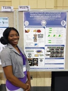
Rapid Extraction and Concentration of Magnetic Particles from Whole Blood with Microfluidic Magnetic Ratcheting,
Oladunni Adeyiga, Coleman Murray, Dino Di Carlo
Magnetic particles are a useful tool for extracting and concentrating target analytes. Conventionally, pipetting, centrifuge tubes, and magnetic racks are used, but this approach is prone to loss of particles and errors from pipetting. Dunni presented a new microfluidic device that concentrates magnetic particles using magnetic ratcheting. A rotating permanent magnet was used to induce magnetic fields in permalloy micropillars embedded within the devices, which could then move magnetic particles along like on a conveyor belt. At the end of the process, the particles are concentrated into a drop within an immiscible fluid. While demonstrated as a standalone device, this tool could also function as a pre-concentrator in an integrated microfluidic device.
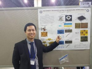
High Density, Reversible 3D Printed Microfluidic Interconnects,
Hua Gong, Adam T. Woolley, Greg P. Nordin
Earlier this year, Lab on a Chip published a paper describing the first 3D printed microchannels that truly had microscale dimensions. You can read the blog post summarizing the work here. It was nice to see this poster from Hua demonstrating the capabilities of their custom 3D printer, including printing complex valves. Hua explained how their 3D printer has a very small print area, and so one of the challenges to overcome was how to fit all the interconnects needed to control the valves into such a small space. This was achieved by printing a world-to-chip interface that connects tubing to the smaller, more densely packed inlets and outlets on the microfluidic chip.
3D-printing, Ink Casting and Lamination (3-D PICL): A Rapid, Robust, and Cost Effective Process Technology Toward The Fabrication of Microfluidic and Biological Devices,
Tariq Ausaf, Avra Kundu, and Swaminathan Rajaraman
Tariq, Avra, and Swami presented an application of 3D-printing aimed at developing microelectrode arrays, microneedles, and microfluidic chips without cleanroom facilities. This work was motivated by a desire for disposable microelectrode arrays that can be developed from concept to functional prototype in < 24 hours. The authors integrated stereolithographic 3D-printing to form the device body, selective ink casting to define conductive traces, lamination of an insulating layer, and micromachining of electrodes and connecting layers. Combining multiple benchtop fabrication techniques could increase the functionality of the developed microdevices and could speed up the transition from prototype to final product in a cost effective manner.
An Automated Modular Microsystem For Enzymatic Digestion With Gut-On-A-Chip Applications
Pim de Haan, Margaryta A. Ianovska, Klaus Mathwig, Hans Bouwmeester and Elisabeth Verpoorte.
What is the primary function of the gastrointestinal tract? Digestion. However, most human gut-on-a-chip models tend to focus on cultured cells in one region of the gut (typically the small intestine) and do not model the initial digestive processes that take place in the mouth, stomach, and small intestine. Pim’s work is focused on developing bioreactors to study the conversion of food into chyme, as it moves from the mouth, through the stomach, and into the small intestine. This requires accurate modelling and realization of the vast pH differences that take place from one region of the gut to the next and verifying enzyme activity within his gut-on-a-chip model. Future work will focus on integration of this upstream digestion model with the downstream study of absorption of nutrients across an intestinal cell layer.
__________
Darius Rackus (Right) is a postdoctoral researcher at the University of Toronto working in the Wheeler Lab. His research interests are in combining sensors with digital microfluidics for healthcare applications.
Ayok unle Olanrewaju (Left) is an industrial postdoctoral fellow at McGill University working in the Juncker lab (dj.lab.mcgill.ca). He is excited about projects that use engineering design to effect real world change, especially in healthcare. Currently, he builds portable and self-powered microchips that rapidly detect bacteria in urine.


