Archive for September, 2017

Zenith in “artery”

When cutting a finger, thrombocytes and fibrin in the blood make up the blood-clotting mechanism, aka. haemostasis, to stop the blood loss. Another way to trigger this mechanism is having an artery damaged by atherosclerosis, which is often caused by several genetic or acquired factors. In the latter case, thrombosis develops within a vein or artery, obstructing or stopping the blood flow to major organs like the heart and eventually causing heart attack. Considering every year over 14 million lives worldwide are lost to heart attacks, more investigation on this topic is needed without any doubt.

Recently, a research team led by Andries van der Meer published a research article in Lab on a Chip on mimicking arterial thrombosis in 3D vascular structures, representing a major step forward in the development of accurate and faster methods of studying arterial thrombosis without using animals. The authors highlighted the inconvenience of using animal models to predict arterial thrombosis in humans. This is mainly due to fundamental differences between human and animal physiology, the researchers explain. For instance, rodent platelet biology, coagulation dynamics, and shear stress in mice arteries significantly vary between humans and mice.

thrombosis on chip

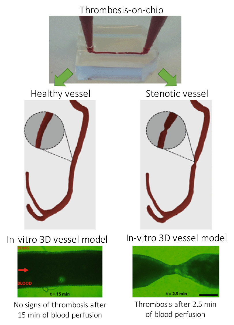
Figure 1. Three-dimensional models of a healthy and stenotic vessels and thrombosis formation upon blood perfusion through the channels.

The paper uses miniaturized vascular structures mimicking 3D architectures found in both healthy and stenotic blood vessels in-vitro (Figure 1). They combined stereolithography and 3D printing of computed tomography angiography data to construct 3D-printed templates of vessels in PDMS microchips. The 3D printed vessels are then coated with human umbilical vein endothelial cells, forming a monolayer fully covering the surface. In the next step, the artificial vessels are perfused with blood at normal arterial shear rates, allowing a blood clot to form as it would happen in the human body. The 3D printed vessel is clinically more relevant when compared to 2D vessel models, since the realistic flow profiles of blood and even distribution of shear stress across the vessel are of great importance when researching arterial thrombosis. Hugo Albers, the co-first author of the paper explains what led the team to try 3D models: “Other groups have worked on thrombosis-on-a-chip before, but we wanted to incorporate flow profiles that are similar to what one would find in-vivo. So we opt for a round and thus 3D shape. Since the stenotic geometry is an important part of this work, we wanted to find a technique that allowed us to make almost any shape we could come up with. Thus 3D-printing seemed to be the way to go.”

When it comes to defining the challenges in 3D organ-on-chip modeling and fabrication, “we needed to replicate the cellular environment using human endothelial cells and human whole blood to fully mimic the nature of vasculature” says Albers. “Incorporating the shape of vasculature to recreate the flow profiles found in-vivo and recreating the shape of vasculature on a small scale was quite challenging, since the resolution of 3D-printing quickly started to be the limiting factor. Furthermore, we ran into problems related to working with whole blood. We had to figure out how to perfuse small channels with blood without instigating thrombosis outside of the microfluidic channel.” The researchers successfully overcame the challenges mentioned by Albers and mimicked the formation of thrombosis in a stenotic vessel model as seen in Figure 1 (bottom).

The researchers note that the next step involves co-culturing arterial endothelial cells and smooth muscle cells with human umbilical vein endothelial cells or moving to different cell lines such as differentiated human induced pluripotent stem cells. “I think we can also apply the 3D-printing technique to create thrombosis-on-a-chip devices with different geometries, e.g. aneurysms or bifurcated geometries”, says Albers.

To download the full article for free* click the link below:

Mimicking arterial thrombosis in a 3D-printed microfluidic in vitro vascular model based on computed tomography angiography data

Pedro F. Costa, Hugo J. Albers, John E. A. Linssen, Heleen H. T. Middelkamp, Linda van der Hout, Robert Passier, Albert van den Berg, Jos Malda and Andries D. van der Meer

Lab Chip, 2017, Paper

DOI: 10.1039/C7LC00202E

This paper is included in our Organ-, Body- and Disease-on-a-Chip Thematic Collection. To read other articles in the collection, visit – rsc.li/organonachip

About the Webwriter

Burcu Gumuscu is a postdoctoral fellow in Herr Lab at UC Berkeley in the United States. Her research interests include development of microfluidic devices for quantitative analysis of proteins from single-cells, next generation sequencing, compartmentalized organ-on-chip studies, and desalination of water on the microscale.

Digg This
Reddit This
Stumble Now!
Share on Facebook
Bookmark this on Delicious
Share on LinkedIn
Bookmark this on Technorati
Post on Twitter
Google Buzz (aka. Google Reader)

Emerging Investigators series – Ian Wong

We are delighted to introduce our latest Lab on a Chip Emerging Investigator – Ian Wong!

Ian Y. Wong is currently an Assistant Professor of Engineering and of Medical Science at Brown University. He completed an A.B. in Applied Mathematics from Harvard University in 2003, Ph.D. in Materials Science and Engineering with Nick Melosh at Stanford University in 2010, and postdoctoral training at Massachusetts General Hospital with Mehmet Toner and Daniel Irimia in 2013. He has been recognized with an NSF Graduate Research Fellowship, a Damon Runyon Cancer Research Fellowship, the Brown University Pierrepont Prize for Outstanding Advising, as well as a Biomaterials Science Emerging Investigator. His research interests include the development of miniaturized technologies to investigate cancer cell invasion, phenotypic plasticity and drug resistance. Moreover, his group engineers unconventional fabrication techniques for printing and patterning nano/bio materials.

Read his Emerging Investigators paper “Stereolithographic printing of ionically-crosslinked alginate hydrogels for degradable biomaterials and microfluidics“, watch the associated video and find out more about his research in the interview below:

Your recent Emerging Investigator Series paper focuses on stereolithographic printing of ionically-crosslinked alginate hydrogels. How has your research evolved from your first article to this most recent article?

I give complete credit to my graduate student, Tom Valentin, who came up with this approach to light-based 3D printing via ionic crosslinking – and then actually got it to work. In retrospect, my Ph.D. thesis focused on biomolecular self-assembly based on ionic interactions, so it’s serendipitous that my current research has circled back to some of these concepts.

 What aspect of your work are you most excited about at the moment?

My lab integrates biomaterials, microfluidics, and computer vision to investigate cancer cell migration and drug resistance. Our first few papers set down the foundations for these different technologies, but now we’re starting to put these pieces together to gain some fascinating insights into cancer biology.

 In your opinion, what is the biggest advantage of stereolithographic printing of hydrogels over other printing techniques?

 Conventional light-based 3D printing of soft materials is based on covalent crosslinking, which results in strong but irreversible bonds. Our demonstration of light-based patterning using reversible ionic crosslinks should enable smart and “biomimetic” properties such as self-healing and stimuli-responsiveness. These properties have been previously demonstrated in bulk hydrogels, but remain relatively nascent for 3D printed structures.

What do you find most challenging about your research?

I work at the interface of engineering and cancer biology, and I find that it takes a lot of effort to bridge between these two communities and become fluent in both disciplines. Moreover, there are twice as many things that can go wrong with the experiments! Nevertheless, it has been extremely worthwhile to see how our technologies could potentially make an immediate and highly meaningful impact.

In which upcoming conferences or events may our readers meet you?

I will be attending BMES this October in Phoenix, AZ.

How do you spend your spare time?

Whenever possible, I enjoy dining out with my wife. I also enjoy cycling, which helps to burn off all those calories

Which profession would you choose if you were not a scientist?

I’ve always been interested in entrepreneurship, and this is something I will likely revisit once my lab and technologies become more established.

Can you share one piece of career-related advice or wisdom with other early career scientists?

Early career scientists are constantly pulled in many directions and have limited time to commit to anything. Nevertheless, I try my best to spend a lot of time with my students and postdocs early on. Such mentoring helps trainees transition towards independence and can also catch problems before they become serious, so it is incredibly worthwhile in the long run.

Digg This
Reddit This
Stumble Now!
Share on Facebook
Bookmark this on Delicious
Share on LinkedIn
Bookmark this on Technorati
Post on Twitter
Google Buzz (aka. Google Reader)

New YouTube Videos

Fluorescence Activated Cell Sorting via a Focused Traveling Surface Acoustic Beam

 
Microfluidic Bead Trap as a Visual Bar for Quantitative Detection of Oligonucleotides


Configurable Microfluidic Platform for Investigating Therapeutic Delivery from Biomedical Device Coatings

Digg This
Reddit This
Stumble Now!
Share on Facebook
Bookmark this on Delicious
Share on LinkedIn
Bookmark this on Technorati
Post on Twitter
Google Buzz (aka. Google Reader)

Emerging Investigators series – Steve Shih

We are delighted to introduce our latest Lab on a Chip Emerging Investigator – Steve Shih!

Steve Shih completed his BASc in Electrical Engineering from Toronto and then went to University of Ottawa to complete his Master’s degree in Chemistry. He then returned to Toronto to complete his Ph.D in Biomedical Engineering in Aaron Wheeler’s laboratory.  He then spent two years at UC Berkeley and at the Joint BioEnergy Institute (JBEI) as a postdoctoral researcher and worked closely with collaborators Jay Keasling and Nathan Hillson.  He learned pathway engineering of microbes for biofuel production using synthetic biology tools and published four papers related to this research.  As of January 2016, he became an Assistant Professor at Concordia University in the Department of Electrical and Computer Engineering with appointments in the Department of Biology and the Center for Applied Synthetic Biology.  His current research entails combining new microfluidic platforms with synthetic biological tools to solve challenges in the health, energy, and medical fields.

Read his Emerging Investigators paper “Image-based Feedback and Analysis System for Digital Microfluidics” and find out more about his research in the interview below:

Your recent Emerging Investigator Series paper focuses on an image-based feedback system for digital microfluidics How has your research evolved from your first article to this most recent article?

Wow it has evolved immensely! I started off as a naïve graduate student dabbling in the field of NMR and using that technique to determine structures of membrane proteins. My first paper described how we used computational and experimental techniques to optimize the determination of membrane protein structures. I learned so much in the field of chemistry and molecular biology, especially coming from an engineering background.

Now my research is in microfluidics and I am using this technique to solve some challenging biological problems.  Although the topics are completely different – the techniques that I learned previously has helped to find interesting solutions to engineering problems.  I am always excited to dabble in new and exciting fields and integrating traditional fields with the new.

What aspect of your work are you most excited about at the moment?

I just recently started my lab and there are so many exciting new projects. My lab is currently working on integrating microfluidics and automating processes related to synthetic biology. Synthetic biology has evolved towards engineering new organisms to produce vast quantities of valuable products, such as biorenewable fuels. This promise (and among many others) has been inspired by biologists that believe genetic engineering of biological cells can be more like the engineering of any hardware. However, challenges loom at multiple steps in the process and our lab is using microfluidics that will overcome these (or least some) challenges.

In your opinion, what is the biggest advantage of this technology and how will this impact digital microfluidics?

I am very excited about this paper because it is the first time that imaging techniques for feedback has been applied to digital microfluidics. One of the biggest challenges with digital microfluidics is the reliability of droplet movement – i.e. an application of a potential does not always equate to a droplet movement. This problem is exacerbated when we are multiplexing droplet movement – more droplets will fail during operation. We have developed a method in which we can individually detect all the droplets on the device using image-based techniques. This is a huge advantage since we only require applying a feedback mechanism to only those droplets that failed in movement while it does not delay the movement of other droplets that translated successfully on the device. This optimizes the time a droplet rests on an electrode and can minimize other effects that prevent droplet movement (e.g., biofouling).

What do you find most challenging about your research?

Everything, but this is why I love my inter-disciplinary research field since it involves so many different aspects. Some examples are trying to understand the underlying mechanisms of breast cancer to resolving issues of integrating synthetic biology techniques at the microscale.

In which upcoming conferences or events may our readers meet you?

I will be attending MicroTAS this year in Savannah and two of my students will be presenting posters. I will also be attending the 4BIO gene editing and synthetic biology conference at London, UK in December, where I will be giving a talk to describe some our microfluidic work with synthetic biology.

How do you spend your spare time?

Spare time is so rare among new professors. But I spend most of my time chasing my kids and trying to excite them for what is to come…

Which profession would you choose if you were not a scientist?

This is a tough question. I really love my job as an educator and everything else that comes with it. But if I had to choose something else, I think I would be a sports broadcaster on ESPN. I love sports and I am an avid fan of tennis and basketball. It would be my dream to commentate a Roger Federer game or to call a Toronto Raptors game.

Can you share one piece of career-related advice or wisdom with other early career scientists?

Failure is inevitable. I failed so many times during my career and it is all part of the learning process. Young scientists should embrace failure and learn as much as they can from it – don’t be afraid of it! They do not realize that failure is where new innovation and ideas come from. I definitely would not be where I am today if it was not for failure.

Digg This
Reddit This
Stumble Now!
Share on Facebook
Bookmark this on Delicious
Share on LinkedIn
Bookmark this on Technorati
Post on Twitter
Google Buzz (aka. Google Reader)