Archive for August, 2015

A closer look at erythrocytes in motion

Blood analysis is usually the first step involved in the diagnosis of various diseases, such as typhoid and malaria. The biochemical and cellular equilibrium of blood is very sensitive to physiological variations occurring in the body at various disease stages. Thus, a fast and accurate examination of blood properties is essential. The morphological and biochemical changes in erythrocytes are used as  the pathological signatures of various diseases.

Flow cytometry is used  to examine blood cells, which requires hydrodynamic sheath flow alignment and fluorescence antibody labelling, making it time-consuming and expensive. Advanced light scattering techniques (such as digital holography) are often seen as suitable alternatives, as they provide fast and label-free measurements.

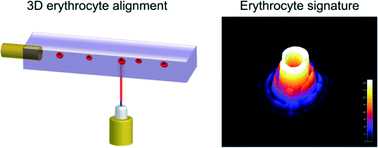
In a recent Lab On A Chip articleNetti et al. from the Italian Institute of Technology, in collaboration with scientists from Germany and Russia, presented a camera-based light scattering approach, coupled with a viscoelasticity -induced cell migration technique. This new system is used to characterise the morphological properties of erythrocytes in microfluidic flows.

They obtained light scattering profiles (LSPs) of individual living cells in microfluidic flows over a wide angular range and matched them with scattering simulations to characterise their morphological properties. A healthy erythrocyte diameter lies between 6 and 9 µm. The diameter values obtained from the experiment lie between 7 and 8.3 µm, which is in good agreement with the existing literature.

‘The results demonstrate the ability of a rapid and cost effective way to measure the average dimensions of an erythrocyte population which can be easily related to the health of a patient,’ concludes Netti.


To gain deeper insight into LSP acquisition and simulation, you can read the full article for free* by following the link below.
Optical signature of erythrocytes by light scattering in microfluidic flows
D. Dannhauser, D. Rossi, F. Causa, P. Memmolo, A. Finizio, T. Wriedt, J. Hellmers, Y. Eremin, P. Ferraro and   P. A. Netti
Lab Chip, 2015,15, 3278-3285
DOI: 10.1039/C5LC00525F

*Access is free until 27/09/2015 through a registered RSC account.
Digg This
Reddit This
Stumble Now!
Share on Facebook
Bookmark this on Delicious
Share on LinkedIn
Bookmark this on Technorati
Post on Twitter
Google Buzz (aka. Google Reader)

New YouTube Videos

Centrifugal microfluidic platform for single-cell level cardiomyocyte-based drug profiling and screening 


Size Reduction of Cosolvent-Infused Microbubbles to Form Acoustically Responsive Monodisperse Perfluorocarbon Nanodroplets 


A programmable microfluidic static droplet array for droplet generation, transportation, fusion, storage, and retrieval 

Digg This
Reddit This
Stumble Now!
Share on Facebook
Bookmark this on Delicious
Share on LinkedIn
Bookmark this on Technorati
Post on Twitter
Google Buzz (aka. Google Reader)

New YouTube Videos

One-Touch-Activated Blood Multidiagnostic System using a Minimally Invasive Hollow Microneedle Integrated with a Paper-Based Sensor 


Vortex Chain Formation in Regions of Ion Concentration Polarization 


Bi-content micro-collagen chip provides contractility-based biomechanical readout for phenotypic drug screening with expanded and profiled targets 

Digg This
Reddit This
Stumble Now!
Share on Facebook
Bookmark this on Delicious
Share on LinkedIn
Bookmark this on Technorati
Post on Twitter
Google Buzz (aka. Google Reader)

Cell pinballs in microfluidic channels

Researchers from the Kaneko Higashimori Lab at Osaka University and the Arai Lab at Nagoya University have observed an interesting phenomenon when studying red blood cells in microfluidic channels. Instead of flowing along the channel in a smooth motion as expected, some cells bounce back and forth between the channel walls in a pinball-like motion at much slower speed. In addition to these ‘cell pinballs’, there are also cells that move at a similar reduced speed, but don’t hit the channel walls.

This altered behaviour could have detrimental effects on microfluidic devices, caused by non-uniform movement of the cells in the channels. In order to prevent these potential problems, the authors have investigated the cause of this behaviour. They noted that cell pinballs only occur when the saline medium is hypotonic, as this causes the cells to inflate due to intake of water. By attaching microbeads to the cells and using a high speed camera, the motion of the cells were studied in more detail. The pinball cells rotated clockwise as they moved to the left of the channel and anticlockwise as they moved to the right of the channel (relative to the direction of the flow).

This observation, combined with the knowledge that the cells were inflated in the hypotonic solution, led the authors to believe that the pinball-motion was occurring due to both the shape of the red blood cell and contact with the channel walls. 3D images obtained using confocal microscopy showed that the upper and lower surfaces of the cells were flattened, confirming that the cells were in contact with the walls.

By studying the different possible deformations of the inflated red blood cells when subjected to flow, the authors found that the contact line (between the cell and wall) and the centre line of the cell were not the same. This explains both types of unexpected cell motion – if the contact line is downstream of the centre line, the cell is unstable to rotational motion and this causes it to move at an angle to the flow, leading to the pinball cells, whereas if the contact line is upstream of the centre line the cell is stable to rotational motion and no displacement occurs, leading to the slow moving non-pinball cells.

From these studies, the authors were able to propose mechanisms that successfully explained the two types of altered red blood cell behaviour in hypotonic solutions, and hopefully in the future this should allow microfluidic systems to be used which will avoid this pinball-motion occurring.


To download the full article for free* click the link below:
Cell pinball: phenomenon and mechanism of inertia-like cell motion in a microfluidic channel
Ryo Murakami, Chia-Hung Dylan Tsai, Makoto Kaneko, Shinya Sakuma and Fumihito Arai
Lab Chip, 2015, 15, 3307-3313
DOI: 10.1039/ c5lc00535c

—————-

About the webwriter

Claire Weston is a PhD student in the Fuchter Group, at Imperial College London. Her work is focused on developing novel photoswitches and photoswitchable inhibitors.

—————-

*Access is free until 06/09/2015  through a registered RSC account.

Digg This
Reddit This
Stumble Now!
Share on Facebook
Bookmark this on Delicious
Share on LinkedIn
Bookmark this on Technorati
Post on Twitter
Google Buzz (aka. Google Reader)

µ-Med A 2015 workshop

µ-Med A 2015, an international workshop on microsystems technologies for African health. This interesting workshop will be held during 16-19 September 2015 at Protea hotel, Stellenbosch, South Africa.

The event will bring together researchers, technologists, entrepreneurs, non-governmental organizations and funding bodies to interact on the latest developments and future trends in the multidisciplinary field of microsystems technology.

The workshop will focus on the following themes:

  • Burden of disease in Africa
  • Microfluidic diagnostic technologies
  • Point of care diagnostics
  • Paper based diagnostics

Feel free to read more about the success of the first workshop held in 2011.


Register now and contribute to the efforts to improve health in Africa!


For additional information, please visit µ-Med A website.

Digg This
Reddit This
Stumble Now!
Share on Facebook
Bookmark this on Delicious
Share on LinkedIn
Bookmark this on Technorati
Post on Twitter
Google Buzz (aka. Google Reader)