Archive for March, 2015

When microfluidics meets inkjet printing

Since its inception over 50 years ago, inkjet printing has become the most widely adopted method for the reproduction of images and text on paper. Inherently a microfluidic technology, inkjet printing is a process in which individual ink droplets, between 10 to 100 µm diameters, are placed at software-configurable locations on a substrate. The resulting dots can combine to create photo-quality images with resolutions of up to several thousand dots per inch. Such exquisite precision and flexibility make inkjet printing particularly attractive for the scalable fabrication of fine features. Indeed, in addition to coloured inks, this technology has also been adapted to deposit a wide range of materials for manufacturing, including metals, ceramics, polymers, and even biological cells.1

Not surprisingly, the lab on a chip community is also beginning to leverage printing technologies for the fabrication of microfluidic components.2 However, this is not straightforward because the quality of the printed features often depends on the interaction of the ink droplets with the substrate material. Since microfluidic devices are predominantly prototyped using polydimethylsiloxane (PDMS),3 recent research has focused on the development of PDMS-compatible methods for inkjet printing.

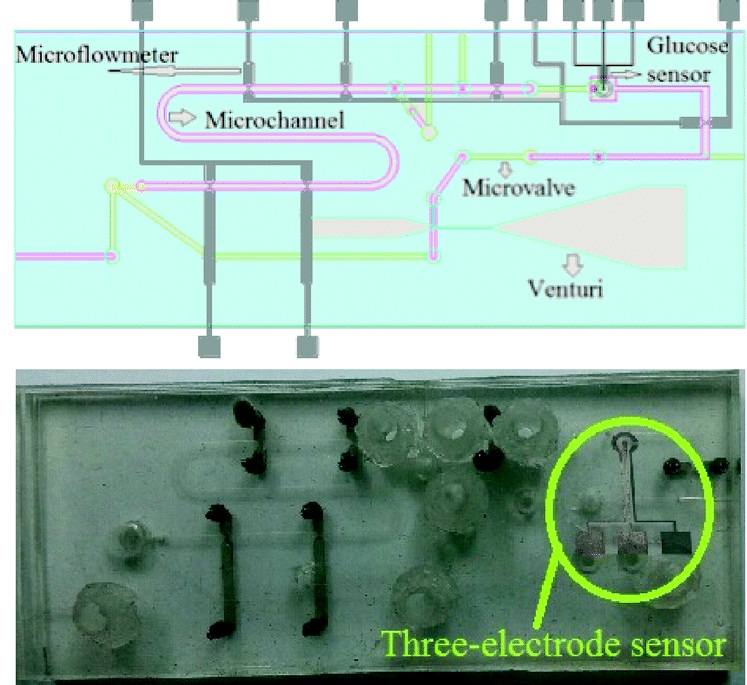
In one example, Profs Dachao Li at the Tianjin University, China and Robert C. Roberts at the University of Hong Kong used inkjet printing to form robust silver microelectrodes on PDMS-based microfluidics for glucose sensing.4 In forming these electrodes, the PDMS material posed two challenges: 1) poor adhesion of silver to PDMS inhibited robust electrode formation and, 2) the inherent hydrophobicity of PDMS promoted coalescence of neighboring ink droplets, which impaired the precision of printed features.

To overcome these challenges, the researchers modified the substrate using (3-Mercaptopropyl)trimethoxysilane, a coupling reagent that presents thiol groups on the PDMS surface. The thiol groups not only served as covalent anchoring sites for the inkjet-printed silver but also increased the wettability of the ink droplet, which improved the precision of the printed features.

Remarkably, the printed electrodes formed on these substrates tolerated very harsh stress tests, including immersion in water, exposure to high pressure air stream, and even ultrasonication. This new fabrication strategy enabled the team to create an all-in-one PDMS system with fluid handling and a three-electrode electrochemical cell for transdermal detection of glucose (Picture 1).

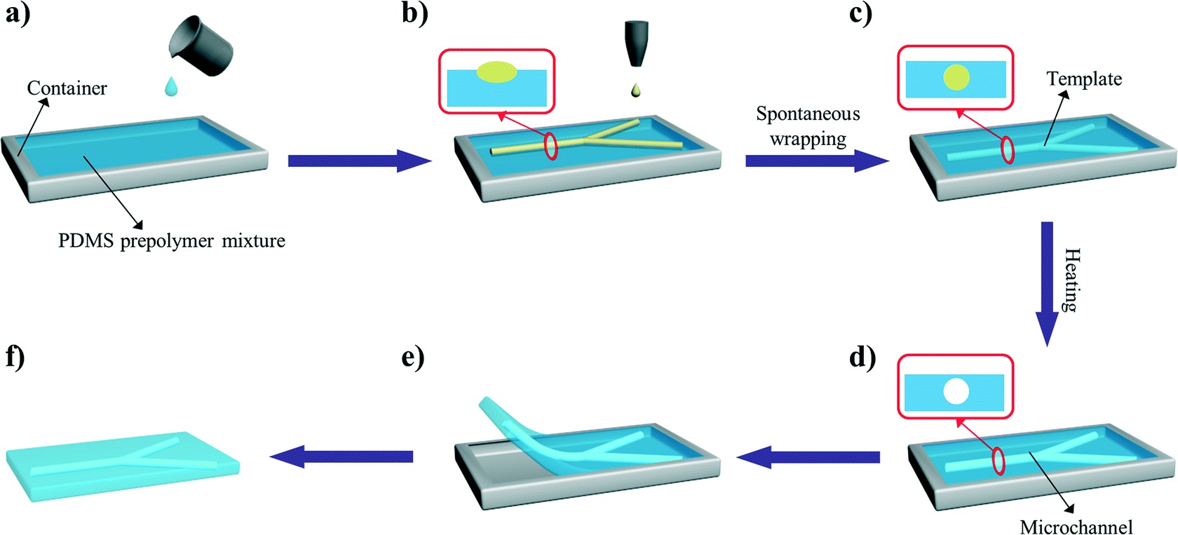
In another example, Profs Yanlin Song and Fengyu Li at the Chinese Academy of Sciences, China developed an elegant inkjet-printed method 5 for the fabrication of enclosed PDMS microchannels. This process conventionally requires at least 4 steps: 1) create a solid template via photolithography, 2) cast PDMS prepolymer mixture in the template, 3) cure and separate the PDMS from the template, and 4) bond the molded PDMS to a substrate.

To simplify this procedure, the researchers implemented a liquid template formed by inkjet-printing (Picture 2). Here, an immiscible liquid pattern is printed directly on a pool of PDMS prepolymer solution. Within seconds, the liquid spontaneously submerges below the prepolymer solution and acts as a template. Upon heating, the polymer solidifies while the liquid template evaporates, leaving behind an enclosed PDMS microchannel device.

In the development of this method, one major challenge was maintaining the stability of the liquid template in the prepolymer solution. Due to surface tension, printed liquid templates tend to break up and relax into discontinuous spherical droplets.  Although this instability can be inhibited by increasing the liquid viscosity, high viscosities are not compatible with inkjet printing. To address this challenge, the researchers used an ink whose viscosity is tunable by temperature. At room temperature, the ink has relatively low viscosity (10 centipoise), which is compatible with inkjet printing. When the ink is printed on a cooled (3-4oC) pool of prepolymer solution, the viscosity of the liquid increases dramatically (>40 centipoise), which enabled the formation of a stable liquid template with no breakages.

This inkjet-printed approach enabled the formation of remarkably versatile PDMS microchannels. The diameters of the fabricated channels can range from 100 to 900 µm just by changing the template design. In addition, the interior surface of the channel can be modified by including vinyl-terminated functional molecules in the ink composition, which covalently incorporates in the PDMS matrix during thermal curing. Applying this technique with vinyl-terminated poly(ethylene glycol)methacrylate molecules, the researchers effortlessly imbue their devices with protein fouling resistance.

In summary, the lab on a chip community is beginning to leverage the benefits of inkjet printing in the fabrication of microfluidic components. By engineering the interaction of the ink droplets with the substrate material, researchers are devising innovative ways to fabricate robust electrodes and versatile microchannels without the need for cleanroom facilities and complicated procedures.

1.    I. M. Hutchings and G. D. Martin, Inkjet technology for digital fabrication, John Wiley & Sons, 2012.
2.    P. Tseng, C. Murray, D. Kim and D. Di Carlo, Lab on a Chip, 2014, 14, 1491-1495.
3.    E. Berthier, E. W. K. Young and D. Beebe, Lab on a Chip, 2012, 12, 1224-1237.
4.    J. Wu, R. Wang, H. Yu, G. Li, K. Xu, N. C. Tien, R. C. Roberts and D. Li, Lab on a Chip, 2015, 15, 690-695.
5.    Y. Guo, L. Li, F. Li, H. Zhou and Y. Song, Lab on a Chip, 2015, 15, 1759-1764.

Digg This
Reddit This
Stumble Now!
Share on Facebook
Bookmark this on Delicious
Share on LinkedIn
Bookmark this on Technorati
Post on Twitter
Google Buzz (aka. Google Reader)

Free access to February’s HOT Articles

These HOT articles published in February 2015 were recommended by our referees and are free* to access for 4 weeks


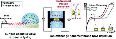
On-chip surface acoustic wave lysis and ion-exchange nanomembrane detection of exosomal RNA for pancreatic cancer study and diagnosis
Daniel Taller, Katherine Richards, Zdenek Slouka, Satyajyoti Senapati, Reginald Hill, David B. Go and Hsueh-Chia Chang
Lab Chip, 2015,15, 1656-1666
DOI: 10.1039/C5LC00036J, Paper


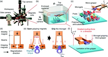
Three-dimensional heterogeneous assembly of coded microgels using an untethered mobile microgripper
Su Eun Chung, Xiaoguang Dong and Metin Sitti
Lab Chip, 2015,15, 1667-1676
DOI: 10.1039/C5LC00009B, Paper

A flexible lab-on-a-chip for the synthesis and magnetic separation of magnetite decorated with gold nanoparticles
Flávio C. Cabrera, Antonio F. A. A. Melo, João C. P. de Souza, Aldo E. Job and Frank N. Crespilho
Lab Chip, 2015, Advance Article
DOI: 10.1039/C4LC01483A, Paper

Take a look at our Lab on a Chip 2015 HOT Articles Collection!

*Access is free until 30.04.15 through a publishing personal account. It’s quick, easy and free to register!


Digg This
Reddit This
Stumble Now!
Share on Facebook
Bookmark this on Delicious
Share on LinkedIn
Bookmark this on Technorati
Post on Twitter
Google Buzz (aka. Google Reader)

New YouTube videos

Woven Electrochemical Fabric-based Test Sensors (WEFTS): A new class of multiplexed electrochemical sensors 


Dynamic formation of a microchannel array enables kinesin-driven microtubule transport between separate compartments on a chip 


Electromechanical cell lysis using a portable audio device: enabling challenging sample preparation at the point-of-care 

Digg This
Reddit This
Stumble Now!
Share on Facebook
Bookmark this on Delicious
Share on LinkedIn
Bookmark this on Technorati
Post on Twitter
Google Buzz (aka. Google Reader)

New YouTube Videos

3D-Printed Microfluidic Automation 


Microfluidics-based single cell analysis reveals drug-dependent motility changes in trypanosomes 


A Fast and Switchable Microfluidic Mixer Based on Ultrasound-Induced Vaporization of Perfluorocarbon 

Digg This
Reddit This
Stumble Now!
Share on Facebook
Bookmark this on Delicious
Share on LinkedIn
Bookmark this on Technorati
Post on Twitter
Google Buzz (aka. Google Reader)

Scientists can quickly detect waterborne pathogens using a smartphone!

More than half a billion people have to survive with unimproved water, as providing safe drinking water is still a problem in many parts of the developing world. Of the waterborne pathogens, Giardia lambia (G. lambia) is one of the most common intestinal parasites that are difficult to remove using traditional water purification methods. Current methods for their detection take up to two days and require analysis laboratories with trained specialists and expensive equipment. Because of this there is an ongoing effort to design low-cost and field-portable methods that can rapidly analyse large volumes of water.

Ozcan and co-workers at UCLA have developed a method for the detection of G. lambia cysts in water using a light weight attachment to a smartphone. The attachment consists of a fluorescence microscope, aligned to the smartphone camera, and a disposable water sample cassette that can hold 20 mL of water. The whole test can be carried out in just 1 hour, from taking the sample from the source, to receiving the total number of cysts detected in the sample.

The process is relatively simple, with the test sample first being fluorescently labelled and then filtered through a membrane that traps the G. lambia cysts. A fluorescence image is taken and wirelessly transmitted to servers using an app designed by the group. Digital analysis is carried out using a machine learning algorithm that can specifically recognise the cysts over other fluorescent micro-objects. The results of this analysis are then transmitted back to the phone and displayed on the app.

The group were able to achieve an impressive limit of detection of 12 cysts per 10 mL of sample, citing several factors that led to this limit. They have put forward a number of suggestions for how they hope to further improve their system, so it will be interesting to hear more from this group.

To download the full article for free* click the link below:

Rapid imaging, detection and quantification of Giardia lamblia cysts using mobile-phone based fluorescent microscopy and machine learning
Hatice Ceylan Koydemir, Zoltan Gorocs, Derek Tseng, Bingen Cortazar, Steve Feng, Raymond Yan Lok Chan, Jordi Burbano, Euan McLeod and Aydogan Ozcan
DOI: 10.1039/C4LC01358A

*Access is free until 31st March 2015 through a publishing personal account. It’s quick, easy and free to register.

About the web writer

Claire Weston is currently studying for a PhD at Imperial College London, focussing on developing novel photoswitches and photoswitchable inhibitors.

Digg This
Reddit This
Stumble Now!
Share on Facebook
Bookmark this on Delicious
Share on LinkedIn
Bookmark this on Technorati
Post on Twitter
Google Buzz (aka. Google Reader)

New You Tube videos

Three-dimensional heterogeneous assembly of coded microgels using an untethered mobile microgripper 


Cell culture on microfabricated one-dimensional polymeric structures for bio-actuator and bio-bot applications 


A droplet microchip with substance exchange capability for the developmental study of C. elegans  

Digg This
Reddit This
Stumble Now!
Share on Facebook
Bookmark this on Delicious
Share on LinkedIn
Bookmark this on Technorati
Post on Twitter
Google Buzz (aka. Google Reader)