Archive for October, 2013

Paper device spots antibiotic-resistant bacteria

Scientists in Canada have developed a paper-based device that checks if bacteria are resistant to certain antibiotics. The simple system could help users in remote areas pick the most appropriate treatment for bacterial infections.

Testing bacteria to see which antibiotics will be effective against them is vital for properly medicating patients. Methods exist to assess bacteria but the equipment needed is often expensive and requires highly trained operators and laboratory conditions. Now, Ratmir Derda and colleagues at the University of Alberta have designed a portable device made from paper and other low-cost materials. The team sought the help of high school students to make and test the devices to show how easy they are to construct and use.

Derda’s device consists of a paper support with a clear plastic window over an area of nutritious media permeated into a sheet of thick blotting paper. This culture media has a uniform pattern of hydrophobic spots to ensure samples disperse evenly across it. Two zones of antibiotics are added onto the media before the device is sterilised in an autoclave. After autoclaving, a cell viability dye is dropped on the culture media and the bacterial sample is then added on top of the dye. Finally, the device is sealed and incubated overnight.

In areas with no bacterial growth the dye remains blue and in areas where bacteria do grown the dye turns pink. The colours are easy to see through the device’s plastic window – there is no need to open the device. A blue area around the antibiotic zones indicates that the bacteria are susceptible to the antibiotics. The hydrophobic spots on the culture media aid quantification of the blue area to give an idea of how sensitive the bacteria are to the antibiotics.

Preparing and using the device is simple. (M: culture media; A: antibiotic zone; C: culture zone)

‘This work shows that living microorganisms can be grown and screened for antibiotic resistance using paper devices that are small and light enough to store in a person’s pocket,’ says Marya Lieberman, an expert in paper-based sensors at the University of Notre Dame in Indiana, US.

The team also found that the device could easily be stored long-term. After assembling to the point that it contained the culture media and antibiotics, it could be left for up to 70 days in a sealed bag. This storage ability along with the device’s cheap components and ease of use make it very promising for use in remote areas.

Visit Chemistry World to read this article and others by clicking here.

Or read the full paper: Antimicrobial susceptibility assays in paper-based portable culture devices, DOI: 10.1039/C3LC50887K

Digg This
Reddit This
Stumble Now!
Share on Facebook
Bookmark this on Delicious
Share on LinkedIn
Bookmark this on Technorati
Post on Twitter
Google Buzz (aka. Google Reader)

Top ten most accessed LOC articles in Q2 2013

This month sees the following articles in Lab on a Chip that are in the top ten most accessed April – July:-

Microfluidic approaches for cancer cell detection, characterization, and separation
Jian Chen, Jason Li and Yu Sun
Lab Chip, 2012,12, 1753-1767
DOI: 10.1039/C2LC21273K

Label-free biodetection using a smartphone
Dustin Gallegos, Kenneth D. Long, Hojeong Yu, Peter P. Clark, Yixiao Lin, Sherine George, Pabitra Nath and Brian T. Cunningham
Lab Chip, 2013,13, 2124-2132
DOI: 10.1039/C3LC40991K

Paper-based microfluidic point-of-care diagnostic devices
Ali Kemal Yetisen, Muhammad Safwan Akram and Christopher R. Lowe
Lab Chip, 2013,13, 2210-2251
DOI: 10.1039/C3LC50169H

Enrichment, detection and clinical significance of circulating tumor cells
Sunil K. Arya, Bing Lim and Abdur Rub Abdur Rahman
Lab Chip, 2013,13, 1995-2027
DOI: 10.1039/C3LC00009E

Nanotechnology for membranes, filters and sieves A series of mini-reviews covering new trends in fundamental and applied research, and potential applications of miniaturised technologies
Jan C. T. Eijkel and Albert van den Berg
Lab Chip, 2006,6, 19-23
DOI: 10.1039/B516903H

Microfluidic probes for use in life sciences and medicine
Mohammad A. Qasaimeh, Sébastien G. Ricoult and David Juncker
Lab Chip, 2013,13, 40-50
DOI: 10.1039/C2LC40898H

Droplet microfluidics
Shia-Yen Teh, Robert Lin, Lung-Hsin Hung and Abraham P. Lee
Lab Chip, 2008,8, 198-220
DOI: 10.1039/B715524G

Lab-on-a-chip technologies for single-molecule studies
Yanhui Zhao, Danqi Chen, Hongjun Yue, Jarrod B. French, Joseph Rufo, Stephen J. Benkovic and Tony Jun Huang
Lab Chip, 2013,13, 2183-2198
DOI: 10.1039/C3LC90042H

Rapid electrochemical detection on a mobile phone
Peter B. Lillehoj, Ming-Chun Huang, Newton Truong and Chih-Ming Ho
Lab Chip, 2013,13, 2950-2955
DOI: 10.1039/C3LC50306B

External cavity laser biosensor
Chun Ge, Meng Lu, Sherine George, Timothy A. Flood, Clark Wagner, Jie Zheng, Anusha Pokhriyal, J. Gary Eden, Paul J. Hergenrother and Brian T. Cunningham
Lab Chip, 2013,13, 1247-1256
DOI: 10.1039/C3LC41330F

Why not take a look at the articles today and blog your thoughts and comments below.

Fancy submitting an article to Lab on a Chip? Then why not submit to us today or alternatively email us your suggestions.

Digg This
Reddit This
Stumble Now!
Share on Facebook
Bookmark this on Delicious
Share on LinkedIn
Bookmark this on Technorati
Post on Twitter
Google Buzz (aka. Google Reader)

Cells in a pinch

Figure

Researchers at the University of California, Los Angeles have developed a method to rapidly quantify the mechanical properties of single cells at high resolution using hydrodynamic and inertial fluid focusing. 

Cells undergo structural changes during disease states, particularly in certain cancer cell lines. For example, tumor cells with increased invasive potential exhibit increased deformability, which aids them during their migration into surrounding tissues1. Measuring the mechanical properties of single cells as a biomarker of cell health using conventional Atomic Force Microscopy (AFM) or micropipette aspiration is time-consuming and requires extensive sample prep, limiting the analysis to a few cells. Dino Di Carlo’s group and collaborators developed a way to apply fluids to stretch single cells along two axes using hydrodynamic flow and perpendicular cross-flows without waiting for cells to stop within the channel. This method, termed ‘hydropipetting’, is compatible with inline microfluidic rinsing, capable of processing 65000 cells/sec, and utilizes automatic image processing to rapidly derive the mechanical phenotype of cell populations. 

Hydropipetting starts with inertial fluid focusing of cells to position them precisely in a flow channel by balancing lift forces and secondary flows by fine control over the Reynolds number of the fluid. Cell-free liquid is then excluded and cells are deformed both parallel and perpendicular to the main channel flow. Automated image data analysis then extracts metrics of cell strain, viscosity, diameter and deformability from high speed observation during cell deformation. 

Di Carlo and his group demonstrated the hydropipetting technique on two cancer cell lines (HeLa and Jurkat cells) and observed increases in cell deformability upon drug treatment to increase invasiveness, metastatic potential, and when disrupting structural cellular filaments. 

References:
[1] S. E. Cross, Y. Jin, J. Rao and J. K. Gimzewski, Nature Nanotechnology, 2007, 2, 780-783. 

Pinched-flow hydrodynamic stretching of single-cells
Jaideep S. Dudani, Daniel R. Gossett, Henry T. K. Tse and Dino Di Carlo. Lab Chip, 2013, 13, 3728-3734.
DOI: 10.1039/C3LC50649E

Digg This
Reddit This
Stumble Now!
Share on Facebook
Bookmark this on Delicious
Share on LinkedIn
Bookmark this on Technorati
Post on Twitter
Google Buzz (aka. Google Reader)

Free to access HOT articles!

These HOT articles were recommended by our referees and are free to access for 4 weeks*

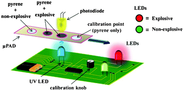
A portable explosive detector based on fluorescence quenching of pyrene deposited on coloured wax-printed μPADs
Regina Verena Taudte, Alison Beavis, Linzi Wilson-Wilde, Claude Roux, Philip Doble and Lucas Blanes  
DOI: 10.1039/C3LC50609F


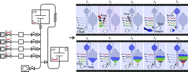
Capillarics: pre-programmed, self-powered microfluidic circuits built from capillary elements
Roozbeh Safavieha and David Juncker  
DOI: 10.1039/C3LC50691F

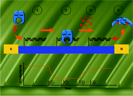
Detection of real-time dynamics of drug–target interactions by ultralong nanowalls
Andreas Menzel, Raphael J. Gübeli, Firat Güder, Wilfried Weber and Margit Zachariasa  
DOI: 10.1039/C3LC50694K

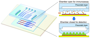
Ultrasensitive microfluidic solid-phase ELISA using an actuatable microwell-patterned PDMS chip
Tanyu Wang, Mohan Zhang, Dakota D. Dreher and Yong Zeng  
DOI: 10.1039/C3LC50783A

Protein–DNA force assay in a microfluidic format
Marcus Otten, Philip Wolf  and Hermann E. Gaub  
DOI: 10.1039/C3LC50830G

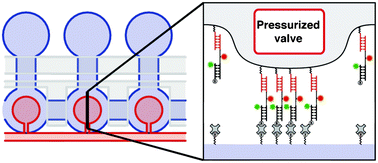
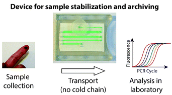
A microfluidic device for dry sample preservation in remote settings
Stefano Begolo, Feng Shen and Rustem F. Ismagilov  
DOI: 10.1039/C3LC50747E

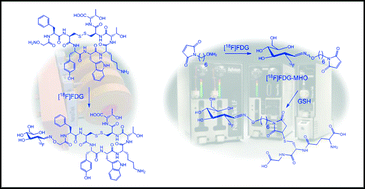
Application of [18F]FDG in radiolabeling reactions using microfluidic technology
Vincent R. Bouveta and Frank Wuest  
DOI: 10.1039/C3LC50797A

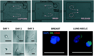
Size-selective collection of circulating tumor cells using Vortex technology
Elodie Sollier, Derek E. Go, James Che, Daniel R. Gossett, Sean O’Byrne, Westbrook M. Weaver, Nicolas Kummer,Matthew Rettig, Jonathan Goldman, Nicholas Nickols, Susan McCloskey, Rajan P. Kulkarni and Dino Di Carlo 
DOI: 10.1039/C3LC50689D

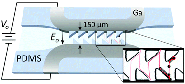
Ratcheted electrophoresis for rapid particle transport
Aaron M. Drews, Hee-Young Lee and Kyle J. M. Bishop  
DOI: 10.1039/C3LC50849H

Parallel measurements of reaction kinetics using ultralow-volumes
Etienne Fradet, Paul Abbyad, Marten H. Vos and Charles N. Baroud  
DOI: 10.1039/C3LC50768H

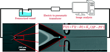
Controlling thread formation during tipstreaming through an active feedback control loop
Todd M. Moyle, Lynn M. Walker and Shelley L. Anna  
DOI: 10.1039/C3LC50946J

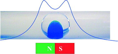
Micro magnetofluidics: droplet manipulation of double emulsions based on paramagnetic ionic liquids
Viktor Misuk, Andreas Mai, Konstantinos Giannopoulos, Falah Alobaid, Bernd Epple and Holger Loewe  
DOI: 10.1039/C3LC50897H

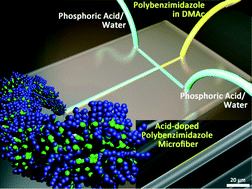
A microfluidic approach to synthesizing high-performance microfibers with tunable anhydrous proton conductivity Mohammad Mahdi Hasani-Sadrabadi,  Jules J. VanDersarl, Erfan Dashtimoghadam, Ghasem Bahlakeh, Fatemeh Sadat Majedi, Nassir Mokarram, Arnaud Bertsch, Karl I. Jacob and Philippe Renaud  
DOI: 10.1039/C3LC50862E

One-step microfluidic generation of pre-hatching embryo-like core–shell microcapsules for miniaturized 3D culture of pluripotent stem cells
Pranay Agarwal, Shuting Zhao, Peter Bielecki, Wei Rao, Jung Kyu Choi, Yi Zhao, Jianhua Yu, Wujie Zhang and Xiaoming He  
DOI: 10.1039/C3LC50678A

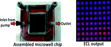
High-throughput metabolic genotoxicity screening with a fluidic microwell chip and electrochemiluminescence
Dhanuka P. Wasalathanthri, Spundana Malla, Itti Bist, Chi K. Tang, Ronaldo C. Fariaa and James F. Rusling  
DOI: 10.1039/C3LC50698C

*Free access to individuals is provided through an RSC Publishing personal account. It’s quick, simple and more importantly – free – to register!

Digg This
Reddit This
Stumble Now!
Share on Facebook
Bookmark this on Delicious
Share on LinkedIn
Bookmark this on Technorati
Post on Twitter
Google Buzz (aka. Google Reader)

Simplifying Microfluidic Flow Control

New research shows that it is possible to carry out flow switching in microfluidic devices via a single active hardware element: a tunable frequency periodic pressure source.

A common hindrance to the design of microfluidic systems is the large amount of unwieldy and expensive hardware (valves, actuators, etc.) required to control fluid flow rates in different parts of the chip. A team of engineers and chemists from UC Santa Barbara, the University of Virginia, and the University of Southampton has devised a method to address this problem. In the new method, a single variable-frequency pressure source communicates with the microfluidic chip via tubes connected to deformable films (“capacitors”) on the chip. The length of each tube is chosen such that the tubes will have well-separated resonance frequencies. Then, the frequency of the actuator is tuned to drive fluid flow in the desired channel only.

As demonstrated in their recent paper, the authors produced three separate devices with clearly differentiated excitation frequencies (see figure below). In addition, they demonstrated a single device with two separate flow channels that could be switched between by modulation of the driving frequency.

Fluid flow in the chip (left, from Figure 1E) is controlled by a periodic pressure source connected to a deformable film by a tube. By using tubes of different lengths, one can selectively drive fluid flow in a channel by tuning the pressure source to that channel’s resonant frequency (right, from Figure 2A).

Fluid flow in the chip (left, from Figure 1E) is controlled by a periodic pressure source connected to a deformable film by a tube. By using tubes of different lengths, one can selectively drive fluid flow in a channel by tuning the pressure source to that channel’s resonant frequency (right, from Figure 2A).

The authors state that by utilizing a large range of excitation frequencies it should be possible to independently control up to 10 flow channels on a single chip using their technique. They also project that it should be possible to control multiple channels simultaneously by employing an excitation signal incorporating multiple frequencies. Thus, this new flow control technique has the potential to be an elegant and low-cost solution for many types of diagnostic applications.

Read this article in Lab on a Chip today:

Flow switching in microfluidic networks using passive features and frequency tuning
Rachel R. Collino, Neil Reilly-Shapiro, Bryant Foresman, Kerui Xu, Marcel Utz, James P. Landers, and Matthew R. Begley
DOI: 10.1039/c3lc50481f

Digg This
Reddit This
Stumble Now!
Share on Facebook
Bookmark this on Delicious
Share on LinkedIn
Bookmark this on Technorati
Post on Twitter
Google Buzz (aka. Google Reader)

Insect-powered tweezers

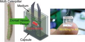
Scientists in Japan have developed the first biohybrid microdevice that can function in air. The microtweezers powered by insect muscle tissue could be used to handle cells and other fragile objects as part of a microelectromechanical system (MEMS).

cyborg jellyfish driven by rat heart cells is part of a recent flurry of research into devices that use mammalian muscle cells to provide motor power. Higher energy efficiency and the ability to self-repair are just some of the benefits of using cells in tiny devices. However, biohybrid devices don’t typically work outside cell culture medium. Now, a team lead by Keisuke Morishima at Osaka University has used insect muscle tissue to fabricate a microtweezer device that can operate in the open air.

Morishima‘s team assembled the microtweezers from polydimethylsiloxane and muscle cells taken from moth larvae and packaged them into a capsule containing a small amount of cell medium. The capsule’s clever design exploits a surface tension effect to stop medium from spilling even if the device is flipped upside down.

‘These results indicate that it will be possible to utilise insect muscle tissue and cells as microactuators both in a wet environment, such as in a microchannel, and on a dry substrate, such as on a silicon MEMS structure,’ says Morishima. When the team layered the medium surface with paraffin to slow evaporation from the opening of the device the microtweezers were functional for up to five days.


Shuichi Takayama a microfluidics and nanotechnology specialist at the University of Michigan in the US says that ‘while a variety of muscle-based actuators have been reported, this device is interesting as it is capable of operating in harsh environments. It is a great idea to combine robust muscle tissue from insects with clever packaging.’

The group now hopes to integrate muscle tissue that functions in both wet and dry environments into devices to expand their work into the nanorobotics field.

This article was taken from Chemistry World. To read this one, and many more, click here!

Digg This
Reddit This
Stumble Now!
Share on Facebook
Bookmark this on Delicious
Share on LinkedIn
Bookmark this on Technorati
Post on Twitter
Google Buzz (aka. Google Reader)