Archive for 2011

New filters for fluorescence detection – UK special issue paper

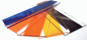
A simple technique for making non-emissive colour filters for microfluidics applications has been developed by John deMello and colleagues at Imperial College, London. Their approach, described recently in Lab on a Chip, employs a solely dye-based method to create high performance filters avoiding the expense and variability inherent in interference filters. The authors highlight the advantages of lower cost and lower auto-fluorescence which could be good news for the analytical chemistry community.

Read the full article here and why not take a sneak preview of the special issue in which it will be included, the UK 10th Anniversary issue, by reading the introductory Editorial by Andrew deMello and Hywel Morgan.

Digg This
Reddit This
Stumble Now!
Share on Facebook
Bookmark this on Delicious
Share on LinkedIn
Bookmark this on Technorati
Post on Twitter
Google Buzz (aka. Google Reader)

Guess the baby competition – the answers!

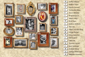
Congratulations to Professor Brian Kirby from Cornell who won the ‘guess the baby’ competition held during the microTAS meeting in Groningen and NanoBioTech conference in Montreux last year. Our lucky winner received a state of the art digital photo frame.

The competition, organized by Lab on a Chip Editor Harp Minhas, challenged participants to correctly match the name of a leading member of the microfluidics community with the childhood photo of that person.  Why not take a look yourself and see how many you can correctly identify!

Find out more about what happened at microTAS by viewing the abstracts for free here and read about the Pioneer Prize winner, Steve Quake. Take a look at issue 6 of Lab on a Chip which includes an article covering the 2010 Art in Science Award given at mTAS 2010 and featured on the cover of this issue.

Digg This
Reddit This
Stumble Now!
Share on Facebook
Bookmark this on Delicious
Share on LinkedIn
Bookmark this on Technorati
Post on Twitter
Google Buzz (aka. Google Reader)

HOT article: rapid identification of ‘bird-flu’

Reverse transcriptase PCR (RT-PCR) has been introduced as the most sensitive method for detecting and pathotyping the avian influenza virus (AIV) or ‘bird flu’, however the number of targets that can be amplified in a single run is limited.

Now chemists at the Technical University of Denmark have developed a device for the rapid and unambiguous detection of AIV by integrating DNA microarray-based solid-phase PCR on to a microfluidic chip. This combines the advantages of microfluidic devices, the high-throughput capabilities of microarrays and the superior specificity of solid-phase PCR. The whole process takes under an hour and uses a sample volume 10 times less than anything previously, meaning that this device can be widely employed by veterinarians for rapid on-site screening of AIV in wild and domestic poultries.

Find out more by reading this HOT article, which is free to access for the next 4 weeks!

A lab-on-a-chip device for rapid identification of avian influenza viral RNA by solid-phase PCR
Yi Sun, Raghuram Dhumpa, Dang Duong Bang, Jonas Høgberg, Kurt Handberg and Anders Wolff
Lab Chip, 2011, Advance Article
DOI: 10.1039/C0LC00528B

Digg This
Reddit This
Stumble Now!
Share on Facebook
Bookmark this on Delicious
Share on LinkedIn
Bookmark this on Technorati
Post on Twitter
Google Buzz (aka. Google Reader)

HOT article: precise, high speed droplet formation

Isolating biological or biochemical content in aqueous droplets within an immiscible oil medium on a microfluidic device allows samples to be transported without cross-contamination or dispersion.  But generating droplets at a suitably high speed with precise volume control has been a challenge.

Now Pei-Yu Chiou and Sung-Yong Park from UCLA have developed a pulse laser-driven droplet mechanism that allows droplet formation of up to 10000 droplets per second with controllable volumes between 1-150 pL and >1% volume variation.

Their device (shown below) consists of two microfluidic channels connected by a nozzle-like opening. A highly focused intense laser pulse induces a rapidly expanding cavitation bubble to push the nearby water into the oil channel for droplet formation.

This HOT article is free to access until the end of March – so download it today and see how they did it!

High-speed droplet generation on demand driven by pulse laser-induced cavitation
Sung-Yong Park, Ting-Hsiang Wu, Yue Chen, Michael A. Teitell and Pei-Yu Chiou
Lab Chip, 2011, 11, 1010-1012
DOI: 10.1039/C0LC00555J

Digg This
Reddit This
Stumble Now!
Share on Facebook
Bookmark this on Delicious
Share on LinkedIn
Bookmark this on Technorati
Post on Twitter
Google Buzz (aka. Google Reader)

ISMM 2011 in conjunction with the KBCS Spring Meeting

Lab on a Chip will be attending the 2011 International Symposium on Microchemistry and Microsystems, hosted at the KBCS Spring Meeting in Seoul, Korea from 2-4 June 2011.

ISMM is the international forum on Micro Total Analysis Systems (μTAS) in Asia region. Following Kanazawa (2009) and Hong Kong (2010), the main topic in the year 2011 will be the “Future of Miniaturized Systems”.  ISMM 2011 in Seoul will be jointly held at the same place in the conjunction with an Annual Spring Meeting of the Korean BioChip Society. We anticipate about 400+ scientists and professionals engaged in research of micro and nanosystems for chemistry and life science.

Chaired by SangHoon Lee (Korea University) the program includes many excellent speakers, including:

Luke P. Lee (University of California at Berkeley, USA)
Marc Madou (University of California at Irvine, USA)
Andrew de Mello (Imperial College London, UK)
Minoru Seki (Chiba University, Japan)
Bingcheng Lin (Chinese Academy of Sciences, China)
DongPyo Kim (Chungnam National University, Korea)

The deadline for abstract submissions has just been extended to March 14th 2011, so hurry and submit yours today! Submission information can be found online here.

Digg This
Reddit This
Stumble Now!
Share on Facebook
Bookmark this on Delicious
Share on LinkedIn
Bookmark this on Technorati
Post on Twitter
Google Buzz (aka. Google Reader)

Lab on a Chip Issue 6 now online

In this issue Michael Gaitan and Laurie Locascio introduce the 3rd annual μTAS Art in Science award, sponsored by Lab on a Chip and presented in October 2010 to Nicholas Gunn from the University of California, Irvine.

Nicholas Gunn’s winning image, entitled Cell Block 9, can be seen on the front cover of Issue 6.

The issue also features a highly recommended Critical Review from David Erickson at Cornell University on nanoscale manipulation techniques using near field photonics technology, a Communication on high speed droplet formation in microfluidic channels from Sung-Yong Park and Pei-Yu Chiou at UCLA.

HOT papers in this issue:

Nanomanipulation using near field photonics
David Erickson, Xavier Serey, Yih-Fan Chen and Sudeep Mandal
Lab Chip, 2011, 11, 995-1009
DOI: 10.1039/C0LC00482K, Critical Review

High-speed droplet generation on demand driven by pulse laser-induced cavitation
Sung-Yong Park, Ting-Hsiang Wu, Yue Chen, Michael A. Teitell and Pei-Yu Chiou
Lab Chip, 2011, 11, 1010-1012
DOI: 10.1039/C0LC00555J, Communication

Towards a fast, high specific and reliable discrimination of bacteria on strain level by means of SERS in a microfluidic device
Angela Walter, Anne März, Wilm Schumacher, Petra Rösch and Jürgen Popp
Lab Chip, 2011, 11, 1013-1021
DOI: 10.1039/C0LC00536C

A ‘microfluidic pinball’ for on-chip generation of Layer-by-Layer polyelectrolyte microcapsules
Chaitanya Kantak, Sebastian Beyer, Levent Yobas, Tushar Bansal and Dieter Trau
Lab Chip, 2011, 11, 1030-1035
DOI: 10.1039/C0LC00381F

A polyacrylamide microbead-integrated chip for the large-scale manufacture of ready-to-use esiRNA
Huang Huang, Qing Chang, Changhong Sun, Shenyi Yin, Juan Li and Jianzhong Jeff Xi
Lab Chip, 2011, 11, 1036-1040
DOI: 10.1039/C0LC00564A

Integrated DNA purification, PCR, sample cleanup, and capillary electrophoresis microchip for forensic human identification
Peng Liu, Xiujun Li, Susan A. Greenspoon, James R. Scherer and Richard A. Mathies
Lab Chip, 2011, 11, 1041-1048
DOI: 10.1039/C0LC00533A

Digg This
Reddit This
Stumble Now!
Share on Facebook
Bookmark this on Delicious
Share on LinkedIn
Bookmark this on Technorati
Post on Twitter
Google Buzz (aka. Google Reader)

Unravelling chromosomes

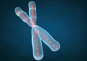
Chromosomes were extracted by washing cell extracts with a protein digester in a microfluidic trap

Danish scientists have used a micro device to isolate centimetre-long portions of human DNA to help study the genetic make-up of diseased cells.

Rodolphe Marie at the Technical University of Denmark, Kongens Lyngby, and colleagues made the device to isolate chromosomes from cell extract samples and manipulate them in such a way that the strands remain intact.

Being able to sequence DNA accurately is a challenge, but Marie’s technique allows the DNA to remain intact so that the gene sequence can be read in one go. The device is made from silicon and consists of an isolation zone in which cell samples are trapped and washed to obtain chromosomes.

Read Catherine Bacon’s Chemistry World article online here or go straight to the HOT Lab on a Chip paper:

A device for extraction, manipulation and stretching of DNA from single human chromosomes
Kristian H. Rasmussen, Rodolphe Marie, Jacob M. Lange, Winnie E. Svendsen, Anders Kristensen and Kalim U. Mir
Lab Chip, 2011, Advance Article
DOI: 10.1039/c0lc00603c

Digg This
Reddit This
Stumble Now!
Share on Facebook
Bookmark this on Delicious
Share on LinkedIn
Bookmark this on Technorati
Post on Twitter
Google Buzz (aka. Google Reader)

Diagnosing diseases with CDs

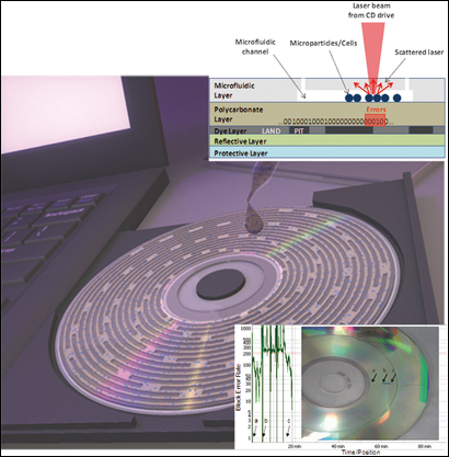
Cells in the microfluidic channels interfere with the laser beam in a standard CD drive and cause errors in reading and decoding the digital data previously burned on the CD

A digital compact disc integrated with a microfluidic device to analyse cells has been developed by scientists in the US. The disc can be inserted into a standard computer disc drive for analysis and could be used to diagnose diseases.

Gang Logan Liu and coworkers at the University of Illinois at Urbana-Champaign built a layer onto a CD that converts biological information into digital information to detect the presence of cells and their size and number. Blood cell counting and sizing are often standard medical practices in the diagnosis of diseases such as leukemia, anemia and Aids.

The team made the device by burning an audio file consisting of a repeating sequence of binary numbers onto a CD’s surface. On top of this, they added a microfluidic layer with a channel. They injected small fluid samples of cells into the channel and focused the laser beam from a CD drive onto the disc’s data layer, which reads the binary code. Any particles on the layer interfere with the laser beam, interrupting the code and altering the data readout in graphs of error against time. The size of the response correlates with the cells’ shape, concentration and optical density.

Read the full Chemistry World article online here or go straight to the Lab on a Chip paper:

Microparticle and cell counting with digital microfluidic compact disc using standard CD drive
Syed M. Imaad, Nathan Lord, Gulsim Kulsharova and Gang Logan Liu
Lab Chip, 2011, Advance Article
DOI: 10.1039/c0lc00451k

Digg This
Reddit This
Stumble Now!
Share on Facebook
Bookmark this on Delicious
Share on LinkedIn
Bookmark this on Technorati
Post on Twitter
Google Buzz (aka. Google Reader)

microTAS 2010 abstracts now available for free online!

Thanks to the Chemical and Biological Miniaturisation Society (CBMS) Lab on a Chip is pleased to offer free access to the microTAS  abstracts from the 2010 conference in Groningen, The Netherlands.

The microTAS meeting abstracts from 2003-2010 can be found on our website here as click-through pdf files.

Digg This
Reddit This
Stumble Now!
Share on Facebook
Bookmark this on Delicious
Share on LinkedIn
Bookmark this on Technorati
Post on Twitter
Google Buzz (aka. Google Reader)

Mapping brain networks

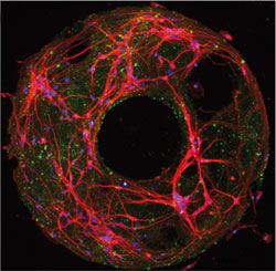
The ring structure of the neuron networks was prepared by stamping proteins onto a glass substrate and growing neurons on the proteins

US scientists have created a model of the ring-shaped networks of neurons in the brain, which could help researchers to understand small changes within diseased brain cells.

Henry Zeringue, and colleagues at the University of Pittsburgh, developed the model to enable the study of brain circuitry in an in vitro system instead of using brain slices. In neurological diseases, such as Alzheimer’s disease, subtle changes in this activity take years, or even decades, to become clinically detectable. Most studies only explore the electrical properties of a single neuron or the connection between two neurons. Until now, there hasn’t been a good model to study network activity in vitro.

Zeringue and his team are considering  using their platform to characterise network-level changes based on small experimental perturbations, like changes in network geometry or expression of mutant proteins in 10 per cent of the neurons. ‘Deciphering the relationship between the connectivity of the neurons and the activity in the ring culture will be challenging and unravelling the connections between the neurons may still prove to be a daunting task,’ he concludes.

Read Carl Saxton’s Chemistry World article online here or go straight to the Lab on a Chip paper:

Ring-shaped neuronal networks: a platform to study persistent activity
Ashwin Vishwanathan, Guo-Qiang Bi and Henry C. Zeringue,
Lab Chip, 2011, 11, 1081
DOI: 10.1039/c0lc00450b

Digg This
Reddit This
Stumble Now!
Share on Facebook
Bookmark this on Delicious
Share on LinkedIn
Bookmark this on Technorati
Post on Twitter
Google Buzz (aka. Google Reader)