Chips & Tips Moderator – Glenn Walker

Glenn Walker

Glenn M. Walker received B.S. and M.S. degrees in biomedical engineering from Louisiana Tech University in 1996 and 1998 respectively, and his Ph.D. degree in biomedical engineering from the University of Wisconsin-Madison in 2002. From 2002 to 2004 he was a Postdoctoral Fellow in the Department of Molecular Physiology & Biophysics at Vanderbilt University Medical Centre. From 2004-2018 he was with the Joint Department of Biomedical Engineering at the University of North Carolina and North Carolina State University. In 2018 he joined the Biomedical Engineering Program at the University of Mississippi, where he is an Associate Professor. His current research interests are in the areas of paper-based fluidics and tumor-on-a-chip devices.

Faculty webpage

Lab webpage

Digg This
Reddit This
Stumble Now!
Share on Facebook
Bookmark this on Delicious
Share on LinkedIn
Bookmark this on Technorati
Post on Twitter
Google Buzz (aka. Google Reader)

Efficient cleaning of a microfluidic chip

David Fernandez Rivas

Mesoscale Chemical Systems group, University of Twente, The Netherlands


Why is this useful?

Microfluidic chips and lab-on-a-chip devices are used at small scales where contamination has a significant impact on operation, or on the outcome of the measurement. Therefore, several cleaning steps are normally required during the production stage of the device, as well as before its actual use, to ensure that chips and connectors are properly clean. After precise connections and experimental preparation, the last thing a microfluidics user wants to observe on its microscope is a clogged channel with debris that could have been avoided. If this happens, the options to remove contamination inside microchannels and connections are unfortunately limited.

Typical chip cleaning protocols involve consecutive use of detergents, alcohol, acetone and demineralized water. Each of these liquids is used in glass or plastic beakers and placed in an ultrasonic bath for several minutes. These actions include handling steps that make the process tedious and time consuming. For example, the containers used need to be cleaned afterwards or in between cleaning steps, in order to avoid recontamination from previous jobs. Also, they significantly block the ultrasound pressure waves and therefore reduce the cleaning potential of the ultrasonic bath.

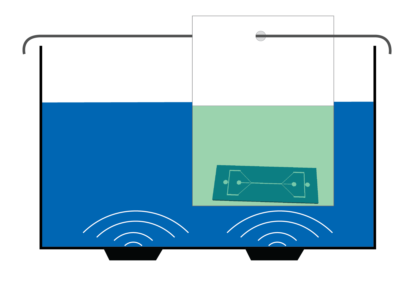
An alternative is to use disposable plastic bags, such as those you can find in regular shops and supermarkets, to replace the use of beakers [1-3]. Using bags can reduce the time and the complexity of cleaning microfluidic chips and other laboratory tools (connectors, tweezers, glass slides prior to plasma bonding to PDMS). Additionally, we have reduced the amount of liquids used routinely.

Figure 1. Sketch of the use of a bag for indirect cleaning of a chip inside an ultrasonic bath.


What do I need?

  • Ultrasonic bath, filled with only water up to the indicated filling height
  • Cleaning liquids
  • Blunt tweezers
  • Plastic bags from a nearby shop or commercially available bags (e.g., BuBble bags)
  • Rod (e.g. 2 mm diameter stainless steel; length larger than one side of the ultrasonic bath)

What do I do?

  1. Place your chip or connectors inside the bag.
  2. Add the cleaning liquid to the bag. Volume: about 10-50 mL, depending on the size of the bag.
  3. Use the rod to hang the bag inside the ultrasonic bath, in a hot spot (above one of the transducers).
  4. Start the ultrasonic bath. We expect that it will take less time than what you would have used in the past.
  5. After ultrasonic cleaning, use the tweezers to retrieve the chip from the bag.
  6. Dispose of the liquid in the usual way, and throw away the bag (plastic recycling). Don’t reuse the bag, since it now contains contamination from the cleaning process, and this should not end up on the next chip.

What are the advantages of doing this?

Using ultrasound for cleaning is a common practice in almost all laboratories around the world. Ultrasonic baths are used specially when mechanical brushing or other cleaning procedures are not possible; e.g. fragile structures or small dimensions in a microchannel which are difficult to reach.

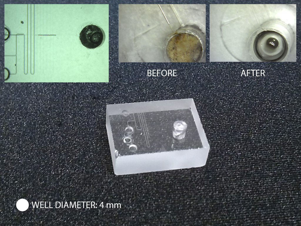
Figure 2. A glass microfluidic chip containing a large reaction/analysis well and several inlets and channels. Such a chip is difficult and not cheap to make; however the chip becomes unusable when the well is clogged with by-product of the reaction. Using ultrasonic cleaning inside a bag, the well can be emptied and the device reused.

The basic principle is based on the creation of small bubbles in the liquid. When these bubbles collapse, they emit powerful shockwaves and liquid jets, among other interesting effects. Combined with liquid motion induced by acoustic streaming, the collapse of bubbles contributes to remove debris from surfaces, even when the bubbles are not in direct contact with the surface[4]. That is the reason why, even when a closed microfluidic channel is placed in an operating ultrasonic bath, it is possible to clean the interior of the device. The more bubbles that exist in a liquid being sonicated, the more cleaning effect takes place.

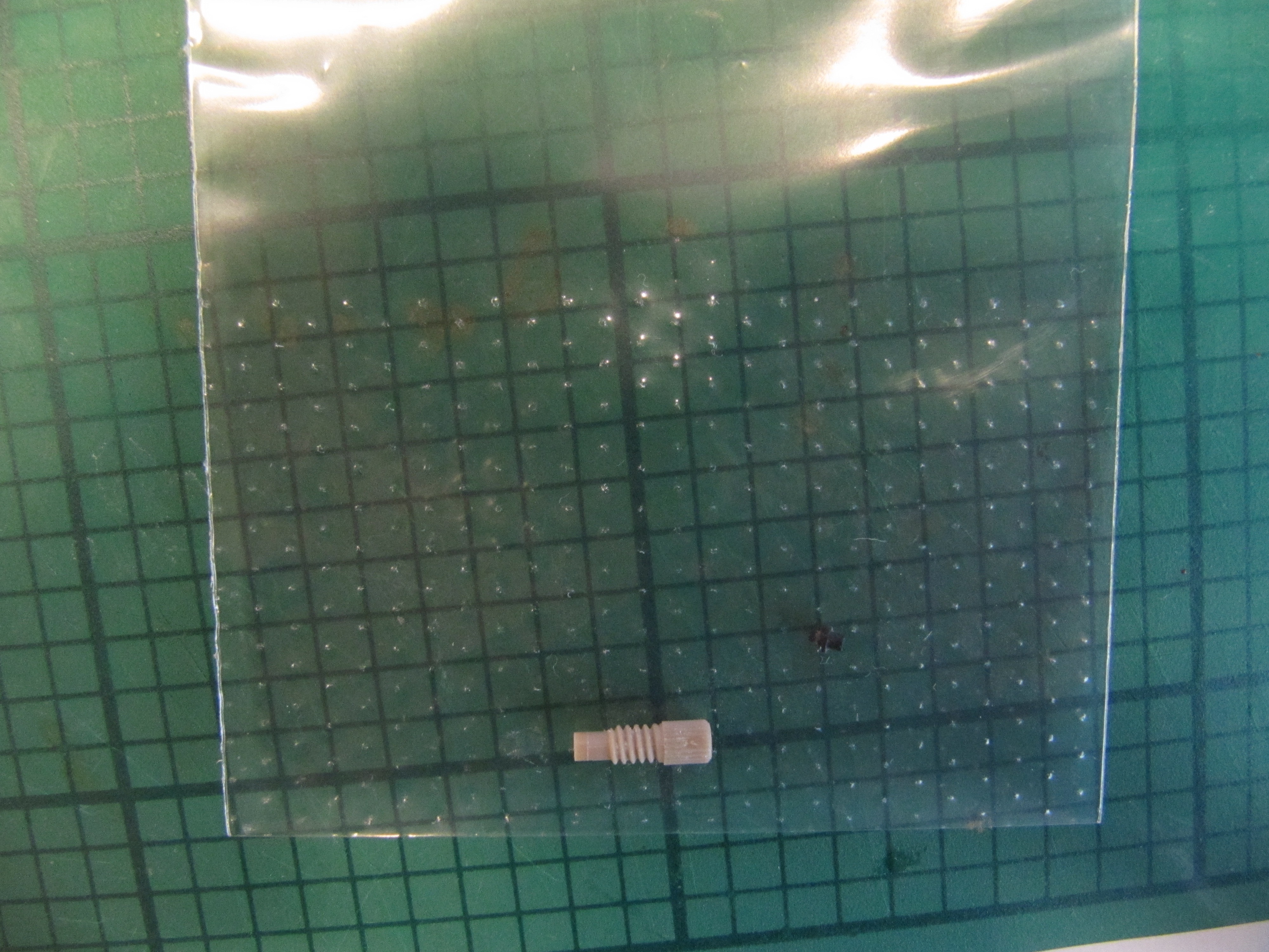
Figure 3. An Upchurch connector can become contaminated and is difficult to clean due to the small grooves. Ultrasonic cleaning ensures rapid cleaning of the connectors.

In plastic bags with thin walls, ultrasound is better transmitted than in glass or plastic beakers, leading to more bubbles and more efficient cleaning. Since the bags only need 10-50 mL instead of ~250 mL for a beaker, you save on chemicals and the volume of used solvents to be disposed. There is also an advantage in using less detergent for washing the glassware or other container used for cleaning, whereas plastic bags can be recycled for a milder ecological impact. Last but not least, when always using new bags, the risk of cross-contamination is drastically reduced.

Figure 4. An Upchurch ferrule is too small to clean manually, yet it may become clogged with particles or deposits. The panels on the right show a through-view of the ferrule, before use (note the particles already present), after clogging and after ultrasonic cleaning in a bag.


What else should I know?

A few tips & tricks:

  • The water in the ultrasonic bath doesn’t need to be refreshed frequently, since all contamination remains inside the bags.
  • Use blunt tweezers to avoid puncturing the bags.
  • Fill the bag with the correct amount of liquid using a dosing bottle or fluid dispenser.
  • Most plastic bags (PE or PP plastics) are compatible with alcohols, acetone and water-based cleaning liquids, but possibly not compatible with acidic or basic cleaning liquids. Making a simple test on chemical resistance is a fun thing to do; please keep us posted of what you find!
  • Bags specifically designed to enhance ultrasonic cleaning by increasing microbubble production are commercially available (e.g., BuBble bags[5-8])

We would like to know if you find it useful or have suggestions to improve cleaning!

Figure 5. Image showing the connector and ferrule inside a bag for ultrasonic cleaning.

[1] http://www.practicalmachinist.com/vb/general-archive/tips-wanted-using-ultrasonic-cleaner-home-shop-96298/

[2] http://www.practicalmachinist.com/vb/general-archive/solvent-ultrasonic-cleaner-89507/

[3] http://www.ccrexplorers.com/showthread.php?t=16892

[4] Fernandez Rivas D, Verhaagen B, Seddon JRT, Zijlstra AG, Jiang L-M, Van der Sluis LWM, Versluis M, Lohse D, Gardeniers JGE. ‘Localized removal of layers of metal, polymer or biomaterial by ultrasound cavitation bubbles’, Biomicrofluidics 6, 034114 (2012).

[5] Verhaagen, B. and Fernandez Rivas, David (2015)  Measuring cavitation and its cleaning effect.  Ultrasonics sonochemistry . 619 – 628. ISSN 1350-4177

[6] Fernandez Rivas D, Verhaagen B, Galdamez Perez A, Castro-Hernandez E, van Zwieten R,  Schroen K. ‘A novel ultrasonic cavitation enhancer’, Journal of Physics: Conference Series 656, 1 1742-6596  (2015).

[7] www.bubclean.nl/bubble-bags-2

[8] Verhaagen, B. , Y. Liu, A. Galdames Pérez, E. Castro-Hernandez, D. Fernandez Rivas, ‘Scaled-up sonochemical microreactor with increased efficiency and reproducibility,’ Chemistry Select, 1(2), 136-139 (2016).

*Conflict of Interest Statment

David Rivas is a co-founder of BuBclean and is the CTO of the company. He does not receive financial compensation from BuBclean.

Digg This
Reddit This
Stumble Now!
Share on Facebook
Bookmark this on Delicious
Share on LinkedIn
Bookmark this on Technorati
Post on Twitter
Google Buzz (aka. Google Reader)

Use of gelatin as intermediate thin passivating layer in PDMS soft lithography technology

Gabriele Pitingolo1,3, Raffaele Vecchione1,3 and Paolo A. Netti1,2,3

1 Center for Advanced Biomaterials for Healthcare, Istituto Italiano di tecnologia (IIT@CRIB), Largo Barsanti e Matteucci, 53, 80125, Naples, Italy.

2 Dipartimento di Ingegneria Chimica, dei Materiali e della Produzione Industriale D.I.C.MA.P.I, Università di  Napoli  Federico II, Naples 80125, Italy.

3 Centro di Ricerca Interdipartimentale sui Biomateriali (CRIB), Università di Napoli Federico II, p.le Tecchio 80, Naples, 80125, Italy

gabriele.pitingolo@iit.it


Why is this useful?

Microfluidic channels, and microstructures in general, are made by various techniques including photolithography coupled with wet etching, reactive ion etching, stamp-based techniques, such as soft lithography, hot embossing and injection molding, as well as ablation technologies like conventional machining, laser ablation and finally direct 3D printing.1 Among these techniques, PDMS soft lithography is commonly and used to replicate polymer microstructures, and particularly microchannels.2, 3

Conversely, casting a PDMS replica from a PDMS mold is challenging as both PDMS layers significantly adhere to each other and demoulding is, if at all, only possible after a careful manual cutting and peeling. A less fiddly but more elaborate approach is the passivation of the first PDMS copy by silanisation in order to reduce adhesion. Particularly, in order to prevent adhesion of the PDMS replica on the master, in a conventional process the master is treated with oxygen plasma to activate the surface and immersed for about 2 min into a silane solution (i.e., a mixture of 94% v/v isopropanol (Sigma Aldrich), 1% v/v acetic acid (Sigma Aldrich), 1% v/v Fluorolink S10 (Solvay), and 4% v/v deionized water) and then placed in an oven at 80 °C for 1 h, thus allowing a complete reaction of the master surface with the fluorinated polymer. This long and expensive process uses materials that are toxic if not removed thoroughly from the master. Recently Gitlin et al. proposed an alternative method utilising hydroxypropylmethylcellulose (HPMC) to passivate a PDMS mold4. Wilson and colleagues presented an “incubation” procedure using a 1% gelatin solution to passivate the PDMS mold5, but this method lacks the ability to control the gelatin layer thickness. Our tip shows a precise method for preparing a thin gelatin layer by spin coating technology which helps preserve the geometry of microstructures on the PDMS mold. In addition, the use of the spin coater makes controlling the gelatin thickness easier.

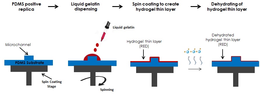
Here we propose the use of a thin hydrogel layer created with spin coating technology or other thin layer depositing techniques as a passivating material which is easy to use and less toxic than other passivating materials. In addition, this process yields hydrogel coated microstructures since gelatin remains on the replicated structures unless it is removed by peel off.

General scheme of the process


What do I need?

  • Poly(methyl metacrilate) (PMMA) sheet
  • Poly(dimethylsiloxane) PDMS pre-polymer
  • Porcine gelatin type A
  • Micromachining machine or similar
  • Spin coater
  • Oxygen plasma machine (optional)
  • Tweezers (optional)

What do I do?

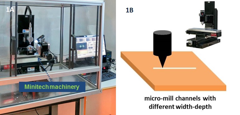
1. Mill microstructures and related inlet/outlet holes (in the case of microchannels) using a micromilling machine (Minitech CNC Mini-Mill) (fig. 1A-1B). To design a draft of the microstructures we created a layout with Draftsight (Cad Software). During micromilling, spindle speed, feed speed and plunge rate per pass were set to 12 000 rpm, 15 mm/s, and 20, respectively.

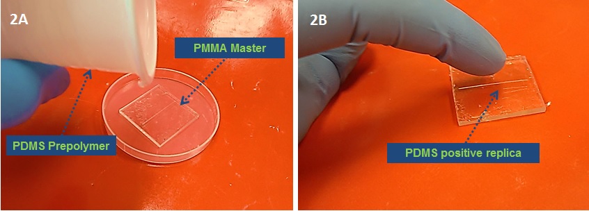
2. After micromilling, the PMMA master is ready to use. Pour liquid PDMS prepolymer (10:1) onto the master to fabricate a PDMS positive replica and cure at 80 °C for 2 h (Fig  2A-2B). The PDMS precursor is previously exposed to vacuum to eliminate air bubbles for at least 30 min.

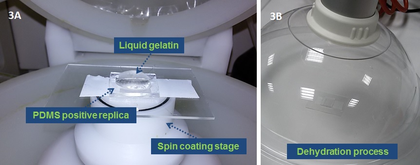
3. Place the PDMS positive replica onto a spin coating stage,  deposit a small (~1 ml) droplet of liquid 10% w/v gelatin, previously degassed with nitrogen for 10 min at the center of the substrate and then spin at high speed (2000 rpm for 20 sec) (Fig. 3A). Afterwards put the system into the fridge for 20 min at 4°C, to finalize the gelling process. Dehydrate the hydrogel layer at room temperature for 5 hours under hood aspiration. (Fig. 3B) Alternatively, prepare the gelatin coating via spray deposition.

4. The Hydrogel-PDMS positive replica (HPPR) is completely dehydrated and ready to cast a new PDMS replica. IMPORTANT: only use a curing temperature below 37° C when making replicas.

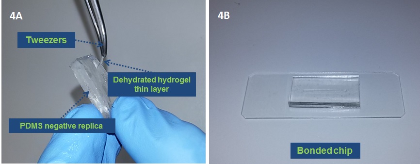
5. (Optional) After PDMS curing remove the dehydrated hydrogel layer with a tweezers from the PDMS negative replica (Fig 4A).

6. (Optional) Treat the PDMS replica with O2 plasma and bond  the chip to make it ready to use (Fig 4B).

CONCLUSIONS: In this tip a double replica of PDMS was obtained by the use of an intermediate layer of gelatin. Spin coating or other thin layer deposition techniques ensure the manufacture of a very thin hydrogel layer which preserves the initial geometry of the microstructure by changing the gelatin concentration. Furthermore the dehydrated hydrogel layer ensures a biocompatible coating of the microstructures.

References

1. H. Becker and C. Gartner, Electrophoresis, 2000, 21, 12-26.

2. Y. N. Xia and G. M. Whitesides, Angewandte Chemie-International Edition, 1998, 37, 550-575.

3. S. Brittain, K. Paul, X. M. Zhao and G. Whitesides, Physics World, 1998, 11, 31-36.

4. L. Gitlin, P. Schulze and D. Belder, Lab on a Chip, 2009, 9, 3000-3002.

5. M. E. Wilson, N. Kota, Y. Kim, Y. D. Wang, D. B. Stolz, P. R. LeDuc and O. B. Ozdoganlar, Lab on a Chip, 2011, 11, 1550-1555.

Digg This
Reddit This
Stumble Now!
Share on Facebook
Bookmark this on Delicious
Share on LinkedIn
Bookmark this on Technorati
Post on Twitter
Google Buzz (aka. Google Reader)

How to remove plasma-bonded PDMS from glass?

Wojciech Adamiak, Martin Jönsson-Niedziolka

Institute of Physical Chemistry, Polish Academy of Sciences, Kasprzaka 44/52, 01-224 Warsaw, Poland

wadamiak@ichf.edu.pl


Why is this tip useful?

It allows to remove and bind different PDMS structures many times to one and the same glass surface. It is especially useful for electrochemical measurements where the glass surface is coated with metal electrodes [1,2]. It allows to test different channel geometries on one and the same electrodes which saves money and time required for depositing new electrodes.

What problem does it solve?

If the channel is clogged, damaged or simply we want to do electrochemistry with different channel geometry, we do not have to make new electrodes on glass. We can remove the unwanted PDMS structure and re-use the glass plate with the new channels.


What do I need?

Concentrated H2SO4, glass pipette, glass beaker, tweezers, gloves and lab glasses to work with concentrated H2SO4.


What do I do?

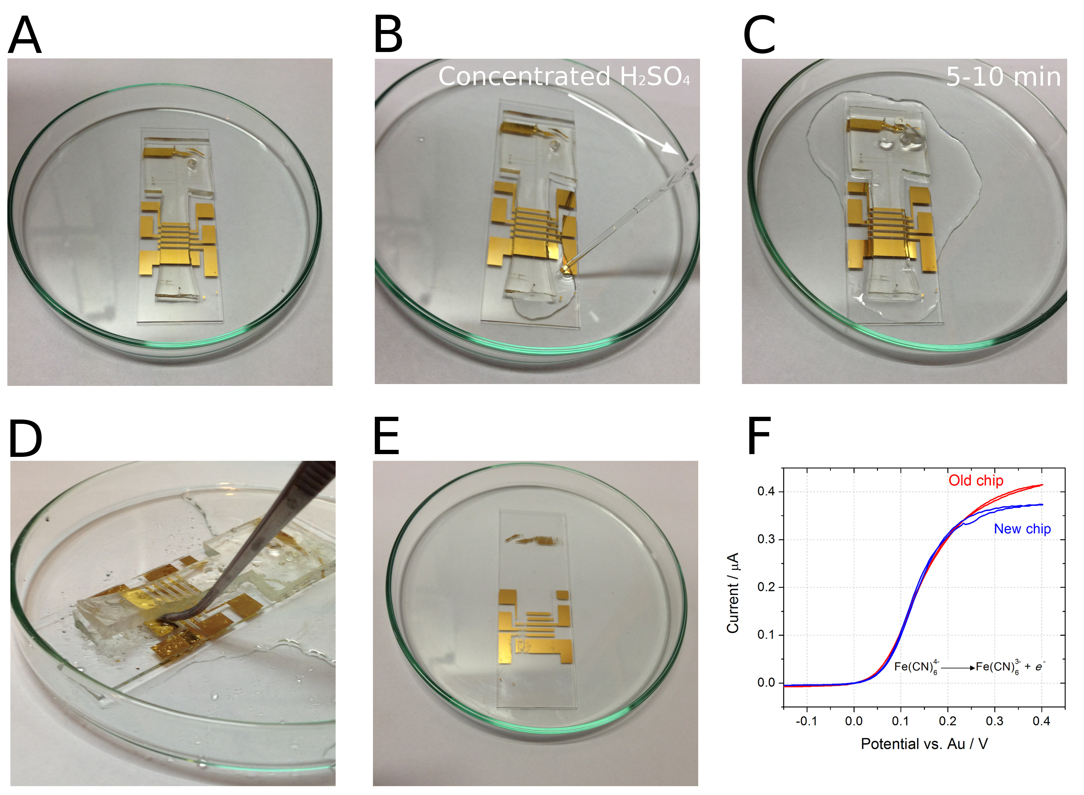
  1. Put the PDMS chip in a Petri dish (Figure A).
  2. With a glass pipette, add concentrated H2SO4 along the edges of PDMS (Figure B).
  3. Leave the chip in contact with H2SO4 for a few minutes.
  4. Gently peel off PDMS from the glass using tweezers (Figure D).
  5. Wash the glass plate with water.
  6. The glass plate is ready for plasma-binding to a new PDMS structure.

References

[1] D. Kaluza, W. Adamiak, T. Kalwarczyk, K. Sozanski, M. Opallo, M. Jönsson-Niedziolka, Langmuir 2013, 29, 16034-16039.

[2] D. Kaluza, W. Adamiak, M. Opallo, M. Jönsson-Niedziolka, Electrochim. Acta 2014, 132, 158-164.

Digg This
Reddit This
Stumble Now!
Share on Facebook
Bookmark this on Delicious
Share on LinkedIn
Bookmark this on Technorati
Post on Twitter
Google Buzz (aka. Google Reader)

Precise Microchannel Fabrication and Alignment with Rapid Prototyped Apparatus

Sukru U Senveli, Rajapaksha WRL Gajasinghe, and Onur Tigli

Department of Electrical and Computer Engineering, University of Miami, Coral Gables, FL, USA and
Biomedical Nanotechnology Institute at University of Miami (BioNIUM), Miami, FL, USA


Why is this useful?

PDMS (Polydimethylsiloxane) is a widely used material for fabricating microchannels through molding processes. In some studies, the exact position or dimensions of the PDMS microchannel on an active substrate is not very important, especially if no active manipulation or sensing is desired with microfluidics. However, there are many studies that can benefit from precise aligning of microchannels to structures on a substrate for optimum system performance. In such cases, coarse alignment using only tweezers may not be an option as overlay accuracy is limited to around millimeter scale. Plasma activated PDMS is a good example of this case where irreversible bonding requires bonding with one try in a short amount of time. Furthermore, there are cases in which the outer dimensions of the PDMS slab become important where cutting the PDMS using razors alone is not precise enough. Possible reasons for needing well-defined outlines include housing requirements and necessity of electrical access.

This study deals with methods to

  • fabricate microchannels with well-defined shapes using a plastic template on a handle substrate and
  • align and bond these microchannels onto an active substrate with high precision.

The error in outline dimensions of the microchannels was seen to be approximately 100 µm but it is ultimately limited by the 3D-printer’s accuracy. On the other hand, the overlay error was measured to be as small as <10 µm in our experiments.

We demonstrate the method on bonding of microchannels to surface acoustic wave devices on a lithium niobate substrate. The microchannel and the sidewalls need to be in the center of the region between the two devices facing each other whereas the electrical pads should be accessible and not covered with PDMS.


What do I need?

  • 3D-printer with ABS filament
  • 3D-printed microchannel template
  • 3D-printed alignment apparatus
  • Probe station with microscope
  • Silicon wafer with SU-8 patterns for molding
  • Tri-chloro-silane (#175552 Sigma Aldrich)
  • Vacuum desiccator (Bel-Art)
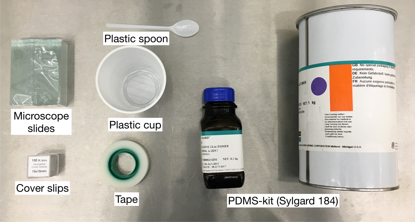
  • PDMS kit (Sylgard 184)
  • Microscope slides
  • Razors
  • Large binder clips
  • 3M Scotch tape
  • Tweezers with blunt tips
  • Biopsy punch
  • Neodymium magnets
  • Pipettes

What do I do?

Microchannel Fabrication with 3D-Printed Template

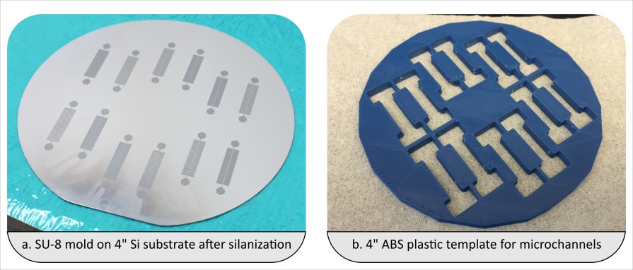
1. It is assumed that SU-8 patterns are already formed on a silicon wafer as shown in Fig. 1(a). A template mask is designed with a CAD program and fabricated using a 3D-printer with ABS type plastic to define the outline of the microchannels. A template thickness of at least 3 mm is recommended for durable microchannels and less than 6 mm is recommended for ease of punching. Fig. 1(b) shows the template structure made of ABS.

2. Under a chemical fume hood, one drop of tri-chloro-silane is placed on a microscope slide, which is in turn placed in a small vacuum chamber. The silicon wafer with SU-8 microchannel patterns is placed inside alongside the microscope slide. The chamber is closed and pumped down. After pumping down for 5 minutes to -0.8 atm of relative vacuum, the chamber is left for 2 hours for silane deposition on the silicon surface. Silane formation is visually checked after the prescribed duration as shown in Fig. 1(a).

Fig. 1 (a) Silanized mold with SU-8 features of the microchannels. (b) The 3D-printed template for microchannel outlines.

3. The 3D-printed template is carefully aligned to the substrate and held in place using paperclips. PDMS (Sylgard) mixed at a ratio of 10:1 is poured onto the holes in the filter. The amount of PDMS leaking between the filter and the substrate should be kept to a minimum.

4. A razor or another flat and sharp object is used to sweep off the excess PDMS from the top. This makes it easier in the eventual alignment step using the microscope. The assembly with the plastic template in place on the substrate and filled with PDMS is shown in Fig. 2(a). Binder clips are used to hold them close together.

5. PDMS is left to cure overnight.

6. The template is separated from the substrate by first cutting through the interface with a razor from the sides and then wedging in with tweezers from four different locations around the substrate. A removed template is shown in Fig. 2(b).

7. Once the template is removed, the backsides of the microchannels are immediately covered entirely with Scotch tape to avoid any dust particles on the bonding surface.

8. Individual microchannels are popped out from the filter by applying uniform pressure from the top side.

9. Inlet and outlet holes are formed using biopsy punches with appropriate gauges. An example of individual channels obtained like this is shown in Fig. 2(c).

Fig. 2 (a) Template after aligning with the substrate is held in place using binder clips. (b) Template after separation from the substrate. (c) A microchannel popped out and punched for inlet and outlet.

Microchannel Alignment with 3D-Printed Alignment Apparatus on a Probe Station

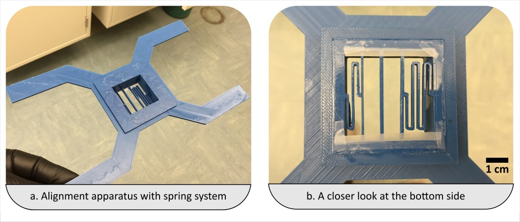
1. The microchannels are placed in the alignment apparatus shown in Fig. 3(a) between the appropriate spring and the stationary beam which are displayed in Fig. 3(b). The choice of spring depends on the spring constant and the size of the microchannel. The microchannel should be sitting flat between the spring and the beam. This can be checked by looking from the side.

Fig. 3 (a) Alignment apparatus as prototyped. (b) Bottom side of the apparatus showing the stationary beam in the middle and two springs on either side for clamping the microchannel for alignment.

2. After correct placement of the microchannel, the scotch tape can be removed slowly by shearing it in order not to disturb the PDMS piece.

3. The alignment apparatus is slowly placed on the platen of the probe station. Neodymium magnets are placed on its four legs to balance and secure the alignment piece in place as shown in Fig. 4(a). A PDMS piece loaded against the spring with a lower spring constant and the stationary beam is shown in Fig. 4(b).

Fig. 4 (a) Alignment apparatus is secured using neodymium magnets on the probe station platen. (b) A closer look at a PDMS piece loaded onto the apparatus. (c) Side view of alignment.

4. Alignment is carried out while observing the overlay using the microscope of the probe station. The overlay is controlled by the stage which moves in plane with the substrate.

5. When the alignment is made, the microchannel is lowered carefully and slowly onto the substrate to bring them into contact as shown in Fig. 4(c).

6. Blunt tipped tweezers are used to apply pressure on the top of the PDMS piece to hold it in place and for a more uniform bond.

7. The spring is released with another set of tweezers. The alignment apparatus is raised by hoisting the platen without touching the sample again. The microchannel has been bonded at this step as displayed in Fig. 5 (a).

8. Microchannel alignment and bonding is double checked using the microscope as shown in Fig. 5(b-c).

Fig. 5 (a) Photograph of after successfully completed alignment and bonding. (b) Evaluation of the alignment of the microchannel to a substrate containing a set of SAW devices. The PDMS is designed in such a way as to bond on areas excluding the interdigitated electrodes of the devices marked as “Devices of interest”. The microchannel was aligned down to an alignment error of <10 µm. (c) The PDMS slab outline does not cover the pads which are available for electrical access using probes.


What else should I know?

  • Materials other than ABS might also be convenient for forming templates too but ABS was preferred due to its higher temperature resilience (in case a high temperature curing of PDMS is desired).
  • If there is a considerable amount of leakage between the template and the handle substrate, it becomes harder to separate the two. This is where the silanization helps. Also, the microchannel outlines might need to be traced using a razor in case there is a substantial amount of residue.
  • Overnight curing is optional. However, lower temperature/longer duration curing was seen to perform better than higher temperature/shorter duration curing step as it results in somewhat softer PDMS.
  • The PDMS piece sitting flat on the alignment apparatus is of utmost importance. The bottom of a 4mm tall PDMS piece should be peeking about 2 mm from the bottom of the alignment apparatus.
  • The Scotch tape can also be removed from PDMS before placement onto the apparatus but this can increase the chances of it collecting dust if not in a cleanroom environment.
  • Activation of PDMS piece and/or substrate is optional. In our studies, we did not use an oxygen plasma and the microchannel was able to withstand microfluidic operations with estimated pressure levels about -200 kPa.
  • Scotch tape was applied to the bottom side of the alignment apparatus to keep the springs from buckling down with the added mass from the PDMS piece.

Acknowledgment

Support from the National Science Foundation (NSF) under grant No. ECCS-1349245 is gratefully acknowledged by the authors.

Digg This
Reddit This
Stumble Now!
Share on Facebook
Bookmark this on Delicious
Share on LinkedIn
Bookmark this on Technorati
Post on Twitter
Google Buzz (aka. Google Reader)

Highly precise alignment for the rapid fabrication of Plexiglas® microfluidic devices

G. Simone

University of Naples Federico II, Piazzale Tecchio 80, 80125 Napoli, Italy.


Why is this useful?

Most of microfluidic devices use channels with rectangular cross-sections. The microfabrication of rectangular shaped channels is straightforward with standard tools such as photolithography.

Fluid dynamics, rheology, soft matter and, recently, biology-based investigations need circular cross-section microchannels; indeed, pre-fabricated capillaries are normally used to carry out the studies. However, capillaries are impractical for some investigations requiring complicated designs.

For Plexiglas® (or other plastic) devices, microfabrication by micromilling is a low cost procedure, which, in the last decades, has gained popularity in the field of microfluidic applications.1-2 The fabrication and the sealing of Plexiglas® microchannels with circular cross-section can be challenging.

Here, a method of fabrication of plastic microfluidic devices with circular cross-section is presented. The method is low cost and it can be performed by minimally trained users. The alignment step builds on a procedure first introduced by Lu et al. that used circular magnets to align layers of polydimethylsiloxane (PDMS).3 The protocol is validated for circular cross-section channels, but it can be used for fabricating rectangular channels or special inlets as well.


What do I need?

  • Plexiglas® sheets (thickness 1mm) Rohm Italy
  • CNC MicroMill (Minitech, US)
  • Ball nose end-mills (0.001 inch, PMT Endmill)
  • Magnets 4 mm × 4 mm× 4 mm, DX
  • Microscope Bresser 58-02520
  • Clamps RS Italy
  • Ethanol
  • Microscope (or stereomicroscope)
  • Glass slides for microscopy (50 mm × 75 mm)


What do I do?

1. Computer aided design of the device (CAD).

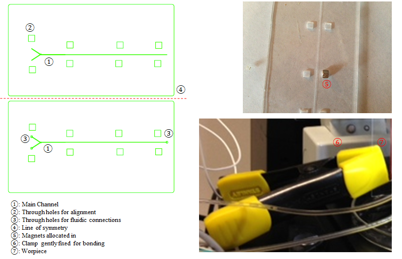
  • Design the microfluidic channels with CAD software. Fig. 1a displays the top and bottom layer of the main channel and, in particular, aligned square through-holes in each layer.
  • Translate the CAD file to Computer Numerical Control (CNC) code for micromilling.

2. Micromilling

  • The endmill should be aligned to set the point z=0 by using a microscope.
  • Then, the microchannels (top and bottom) must be milled. Channel with depends on the endmill diameter.4 It is worth emphasizing that setting the correct work parameters (such as tool speed and depth) is crucial. Indeed, if the latter are not appropriate, the plastic workpiece develops internal stresses that during the bonding result in cracks and chip breakdown.
  • Finally, the through holes and the frame have to be milled. The number of through holes for locating the magnets depends on the dimension of the microfluidic device. It is good practice for the rectangular through-holes to have a pitch of roughly 10 mm (this dimension depends on the size of the microchannels and of the magnets).

3. Alignment

  • Align the top and bottom layers with a stereomicroscope (Fig. 1b). The bottom layer should be set on a solid surface and the magnets inserted into the through-holes. Then, the second layer should be placed on the first and a second set of magnets inserted in the square through-holes of the second layer.
  • The magnets located in the first and second layer naturally provide a good “first alignment” of the microchannels, while leaving freedom to slightly adjust the layers (i.e., this is a reversible sealing). Once the magnets are fixed, the quality of the alignment should be checked with a microscope.

    Fig. 1. The Fabrication. a. CAD design of a microfluidic channel with circular cross section. The length of the microfluidic device is 40mm, the width is 15mm. The depth of grooves is 50μm, width 25μm. b. Magnets employed for the bonding. C. Clamps used for sealing the microchannel.

4. Bonding

  • Clamp the Plexiglas® layers together and submerge assembly in ethanol for 15 minutes (Fig. 1c). For optimal bonding, the clamping should be done with clamps positioned at the edges of the Plexiglas®.5
  • After 15 minutes, the sealing of the microfluidic channels should be checked. If the Plexiglas® is sufficiently bonded, the magnets can be removed and glass slides placed on either side of the Plexiglas® layers. Clamp the glass slides and allow the assembly to rest for another 5-15 minutes.
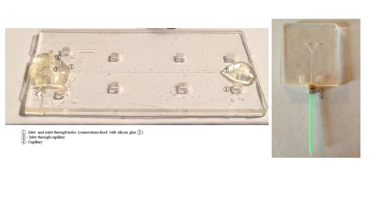
  • The device is ready to be used for different applications as shown in Fig. 2a. Capillary tubing can be easily connected to the channel for modular design (Fig. 2b).

    Fig. 2. Examples of microfluidic devices. a. Perfusion of the samples through hole at the inlet. b. Perfusion through capillaries.

In conclusion, analyzing the protocol, the following advantages can be emphasized:

  1. The process is designed for different materials, but it fits perfectly with Plexiglas®
  2. The equipment necessary for the fabrication and assembly includes simply a micromilling machine and a (stereo)microscope
  3. The use of square magnets (instead of circular ones) allows for more precise alignment due to further restriction to the sliding of the top and bottom layers.


References

  1. G. Simone, G. Perozziello, J. Nanosc. Nanotech., 2010, 11, 2057.
  2. G. Simone, RSC Advances, 2015, 5, 56848.
  3. J-C Lu, W-H Liao, Y-C Tung, J. Micromech. Microeng. 2012, 22, 075006-075014.
  4. G. Perozziello, G. Simone, P. Candeloro, F. Gentile, et al. Micro and Nanosystems, 2010, 2, 227-238.
  5. G. Medoro, G. Perozziello, A. Calanca, G. Simone, N. Manaresi, 2010, US Patent App. 13/257,545.

Digg This
Reddit This
Stumble Now!
Share on Facebook
Bookmark this on Delicious
Share on LinkedIn
Bookmark this on Technorati
Post on Twitter
Google Buzz (aka. Google Reader)

Direct Delivery of Reagents from a Pipette Tip to a PDMS Microfluidic Device

Hoon Suk Rho1, Yoonsun Yang2, Henk-Willem Veltkamp1, and Han Gardeniers1

1Mesoscale Chemical Systems Group, MESA+ Institute for Nanotechnology, University of Twente, The Netherlands.

2Medical Cell BioPhysics Group, MIRA Institute for Biomedical Technology and Technical Medicine, University of Twente, The Netherlands.


Why is this useful?

Most common way to handle and transport reagents in chemical or biological labs is by using a pipette. However, tubing connection is generally used for the delivery of reagents into a microfluidic device. Even though the connection with commercial tubing and connectors allows various choices on the sizes and materials of the tubing and easy connection, difficult sampling from stock solutions, dead volume in tubing and connectors, and extra sterilization on tubing and connectors for biomaterials, still remain challenges. Here we demonstrate a direct connection of pipette tips to a PDMS device and loading reagents by pressure driven flow.


What do I need?

  • Stainless still puncher (Syneo LLC)
  • Precision stainless steel tip (23 gauge, #7018302, Nordson Corporation)
  • Tygon tubing (0.020″ x 0.060″OD, #EW-06418-02, Cole-Parmer Instrument Company)
  • 3D printed plug
  • Pliers

What do I do?

Punching inlet and outlet on a PDMS device

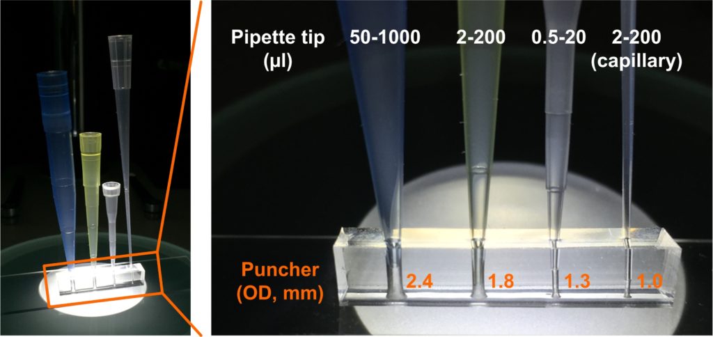
1. Select the size of a puncher based on the size of a pipette tip will be connected. We achieved tight connections of pipette tips on a PDMS substrate when we punched holes by using punchers with outer diameter of 2.4 mm, 1.8 mm, 1.3 mm, and 1.0 mm for 50 – 1000 µl, 2 – 200 µl, 0.5 – 20 µl, and 2 – 200 µl (capillary) pipette tips, respectively.

Fig. 1 Direct connection of pipette tips on a PDMS device.

Plug connection preparation

1. Separate a pin from a precision stainless steel tip. The pin can be easily removed by twisting the plastic part while the pin is held by pliers.

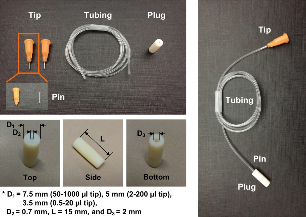
2. 3D-print a plug. The outer diameter of the plug depends on the size of the pipette tip that will be connected. The detailed dimensions of the plug are shown in Fig. 2.

3. Connect a precision stainless steel tip, tubing, a pin, and a plug. Because the plastic part of the tip is luer tapped, it can be connected to male luer connectors and commercial plastic syringes.

Fig. 2 Tubing connection with a 3D-printed plug.

Solution loading

1. Pipette the sample, connect the pipette tip into the inlet of a PDMS device, and take off the pipette. When an empty pipette tip is connected into the outlet of the device, the sample from the outlet can be collected in the tip.

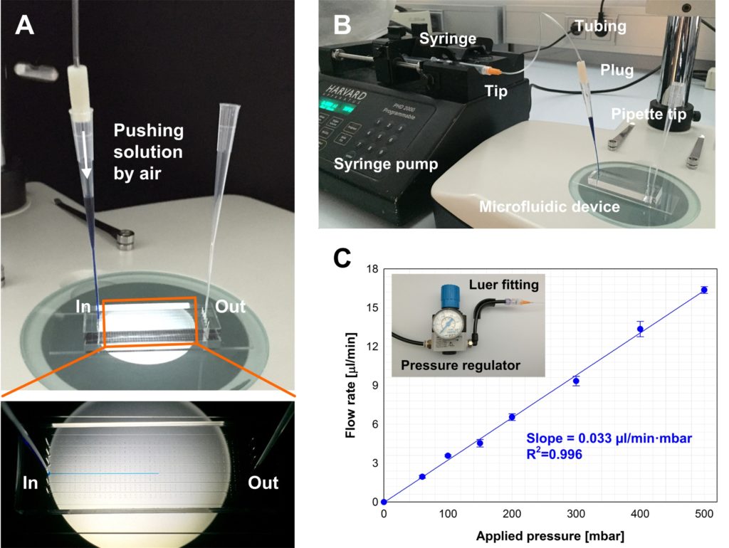
2. Insert the plug into the tip and connect the tip to a pressure source. When pressure is applied into the pipette tip, the solution in the tip is pushed into a microchannel (Fig. 3A). The luer tip can be connected to a syringe and a syringe pump can be used as a pressure source (Fig. 3B). The flow rate of the sample solution can be controlled by the syringe pump. The luer tip also can be connected to a pressure regulator with a luer fitting. Fig. 3C shows the flow rate of sample loading at various applied pressures controlled by a pressure regulator. In this case an external gas source is required. However, this system is cheaper than commercial microfluidic flow control systems. Also a digital pressure regulator can be used for accurate flow rate control at the low flow rate regime less than 1 µl/min.

Fig. 3 A. Loading blue food dye solution into a microchannel, B. Solution loading by a syringe pump, and C. Solution loading by using a pressure regulator.


What else should I know?

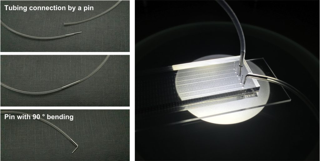
No leakage of the solution was observed in the connection of a pipette tip onto a 1mm thick PDMS substrate. However at least a thickness of 3 mm is highly recommended to achieve tight fitting and stable support for the pipette tip. The pin obtained from a precision stainless tip is very useful for tubing connections. For example two separated tubes can be connected by the pin and also the pin can be inserted into an inlet or outlet of a PDMS device punched by a puncher with an outer diameter of 1mm. Also the pin can be easily bent by using pliers for compact connection to a PDMS device.

Fig. 4 Tubing connection by using the pin from a precision stainless tip.

Digg This
Reddit This
Stumble Now!
Share on Facebook
Bookmark this on Delicious
Share on LinkedIn
Bookmark this on Technorati
Post on Twitter
Google Buzz (aka. Google Reader)

Syringe funnels for facile loading of precious samples

Alexander Price, Wesley Cochrane and Brian Paegel
Department of Chemistry, The Scripps Research Institute, Jupiter FL 33458


Why is this useful?

Syringe pumps are the most popular tool for transporting fluids within microfluidic devices. In the process of loading sample into a syringe, air bubbles (derived from the syringe dead volume) frequently migrate into the barrel and require removal to achieve consistent flow. Ideally, a researcher would have a large excess of sample so that the barrel can be filled and evacuated multiple times. During loading, syringes are held vertically with the sample directly below the tip, necessitating forceful evacuation to dislodge rising bubbles, however this not feasible for low/intermediate-volume “precious” samples (50-500 µL). Here, we present a simple funnel to aid bubble removal during syringe loading.


What do I need?

Plastic transfer pipets (FisherBrand 13-711-7M)

Razor blade

Pipette and pipette tips

Syringe (we use Hamilton Gastight 1700 series w/ TLL tips)

What do I do?

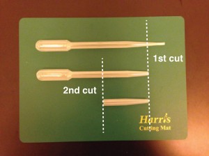
1.  Using the razor blade, carefully cut the end off of the transfer pipet so that it fits snugly over the tip of your syringe (Fig. 1). It might take a couple tries, but you can use this as a template once you have found the right location to cut.

2.  Cut the transfer pipette again, roughly 2 inches up from the previous cut (Fig. 1). Your funnel is complete.

3.  Attach the funnel onto the tip of the syringe. Holding the syringe vertically (funnel up), load your sample into the bottom of the funnel (Fig. 2).

4.  Fill and evacuate the syringe barrel as needed to eliminate any air bubbles (Fig. 3).

5.  Dispose of the funnel.

Figure 1. Construction of a syringe funnel from a transfer pipet.

Figure 2. The funnel is attached to the syringe (left), and sample is loaded into the bottom of the funnel (middle and right).

Figure 3. Air bubbles in the barrel are expelled into the funnel and syringe is filled.

Digg This
Reddit This
Stumble Now!
Share on Facebook
Bookmark this on Delicious
Share on LinkedIn
Bookmark this on Technorati
Post on Twitter
Google Buzz (aka. Google Reader)

Rapid technique for UV-curable adhesive bonding of glass coverslips to polystyrene microdevices

David J. Guckenberger,*a Jake Kanack,*a Loren Stallcop,b David J. Beebea

aDepartment of Biomedical Engineering, Wisconsin Institutes for Medical Research, University of Wisconsin-Madison, Madison, WI, USA
bDepartment of Materials Science and Engineering, University of Wisconsin
Madison, Madison, WI, USA
* Authors contributed equally


Why is this useful?


With several microfabrication techniques now available, including: 3D-printing,1 micromilling,2 and hot embossing,3 in-house fabrication of thermoplastic microdevices has become cheaper, faster, and easier. However, for many applications – such as cell culture and microscopy – these devices must be bonded to optically-transparent substrates such as glass. While bonding similar materials, such as Polystyrene (PS) to PS, is relatively simple, bonding dissimilar materials, such as PS to glass, presents a particular challenge. Current methods to circumvent these challenges include spin coating adhesives, such as polydimethylsiloxane (PDMS), onto sacrificial substrates4 and injecting adhesive directly into the bond interface.5 However, equipment requirements, associate long cure times, heterogeneity in glue uniformity, and complexity limit acceptance of these techniques.

Here we present simple technique for applying uniform layers of adhesive to enable rapid – less than a minute – bonding of PS to glass. Using UV-curable adhesives, readily accessible materials, and a simple techniques, we demonstrate how to apply thin uniform layers of adhesive to a microchannel. We provide design suggestions that will improve bonding repeatability, and additional information that may help apply this technique to materials beyond PS and glass.


What do I need?


  • Polystyrene                                            (1.2 mm, #ST313120, Goodfellow)
  • Glass coverslip                                      (#260450, Ted Pella, Inc.)
  • UV curable adhesive                           (Ultra Light-Weld 3025, Dymax)
  • Silicone foam (Textured)                   (#31943970, MSC Industrial Supply Co.)
  • Isopropyl alcohol                                 (#190764, Sigma-Aldrich)
  • Low particulate wipers                       (#TX609, Texwipe)
  • Wooden flat stick                                 (#704, Brightwood)
  • UV lamp                                                 (OmniCure S1000, Exfo)
  • Adhesives are often material-specific. Consult the manufacturer to determine the best adhesive for your application.

Tip: Some adhesives may require post-treatment / aging to reach a full cure.

  • We have tested this protocol with Ultra Light-Weld 3025 (Dymax) and Norland Optical Adhesive 68 (Thor Labs, Inc.). These adhesives had similar performance, however the protocol may need to be tailored for other adhesives.
  • If the adhesive is too viscous or does not adequately wick around the rib, heat may be applied to achieve thinner adhesive layers, or to improve the wicking of the adhesive.
  • This protocol is amenable to wide variety of materials, including: cyclic olefin copolymer (COC), glass, metal, PS, and various rapid-prototyping materials.
  • Creating the rib and allowing the adhesive to wick eliminates excess adhesive and prevents adhesive from squeezing into the microchannel.


What do I do?

Fig.1 Channel border design

Step 1: Fabricate the microdevice. To improve bonding repeatability and adhesive distribution we recommend fabricating a groove (thickness > 0.5mm) around the channel – leaving a rib (0.5 mm < thickness < 1.5 mm) around the perimeter of the channel.

Tip: Rib thickness may need to be tuned for individual adhesives
Tip: Extra caution while applying the adhesive may be necessary for channels shallower than 0.1 mm

Step 2: Thoroughly clean the surface of the microdevice, silicone foam sheet, and coverslip using isopropyl alcohol and low-particulate wipers. Remaining particulates can be blown off with compressed air. Ensure PS and glass surfaces remain clean throughout the bonding process.

Step 3: Apply a dollop of UV curable adhesive to the foam sheet.

Step 4: Use a tongue depressor to spread the adhesive into a uniformly thin layer across the foam. The area of the adhesive should be larger than the microdevice – add more adhesive if necessary.

Step 5: Position the device onto the adhesive bonding surface down. Press down gently; avoid sliding the microdevice to prevent build-up of adhesive within the channels. Pick up the device and repeat this step two or three times to ensure the bonding surface is completely covered with adhesive.

Tip: Take care to ensure no adhesive is transferred from gloves to surfaces of the device not intended to be bonded.
Tip: Minimize the delay between step 4 and step 5 to help ensure a uniform thickness of adhesive

Step 6: Position the microdevice above the coverslip, and gently lower it until it makes contact. Once contact is made, release the device, taking extra caution to avoid sliding the microdevice.
Tip: The adhesive may have a yellow color after bonding. If necessary, allow 24 hours for adhesive to clear

Step 7: Allow a few seconds for the adhesive to wick along the ribs, then cure device for 20 seconds with ~350 nm UV light at [Intensity]

Fig. 2 Process workflow


What else should I know?

Fig. 3 Cross sectional image of a PS microchannel bonded to a glass coverslip. Scale bar represent 0.5 mm

•          Adhesives are often material-specific. Consult the manufacturer to determine the best adhesive for your application.

Tip: Some adhesives may require post-treatment / aging to reach a full cure.

•          We have tested this protocol with Ultra Light-Weld 3025 (Dymax) and Norland Optical Adhesive 68 (Thor Labs, Inc.). These adhesives had similar performance, however the protocol may need to be tailored for    other  adhesives.

•          If the adhesive is too viscous or does not adequately wick around the rib, heat may be applied to achieve thinner adhesive layers, or to improve the wicking of the adhesive.

•          This protocol is amenable to wide variety of materials, including: cyclic olefin copolymer (COC), glass, metal, PS, and various rapid-prototyping materials.

•          Creating the rib and allowing the adhesive to wick eliminates excess adhesive and prevents adhesive from squeezing into the microchannel.


References


1. Au, A. K., Lee, W., Folch, A., Lab Chip, 2014, 14(7), 1294-1301.
2. Guckenberger, D. J., de Groot, T., Wan, A. M.-D., Beebe, D., & Young, E., Lab Chip, 2015, 15(11), 2364–2378.
3. Young, E. W. K., Berthier, E., Guckenberger, D. J., Sackmann, E., Lamers, C., Meyvantsson, I., Beebe, D. J., Analytical Chemistry, 2011, 83(4), 1408–1417.
4. Gu, P., Liu, K., Chen, H., Nishida, T., Fan, Z. H., Anal. Chem., 2011, 83(1), 446-452
5. Lu, C., Lee, L. J., & Juang, Y. J., Electrophoresis, 2008, 29(7), 1407–1414.


Digg This
Reddit This
Stumble Now!
Share on Facebook
Bookmark this on Delicious
Share on LinkedIn
Bookmark this on Technorati
Post on Twitter
Google Buzz (aka. Google Reader)

DIY peristaltic pump

Shannon Faley, Bradly Baer, Matthew Richardson, Taylor Larsen, and Leon M. Bellan*

Vanderbilt University, Department of Mechanical Engineering, Nashville TN, 37235, USA

*leon.bellan@vanderbilt.edu

Why is this useful?

Figure 1: Fully assembled peristaltic pump


The majority of microfluidic applications require an external pumping mechanism.  Multi-channel, individually addressable pumps are expensive, often large, and prone to failure when operated inside cell culture incubators at 95% humidity.  The number of experiments that can be run at a given time is limited by the availability and expense of pumps.  Perfusing artificial tissue scaffolds containing engineered vasculature requires long-term (days to weeks) continuous flow at low rates.  We designed an inexpensive (~$100 for 2 pumps, ~$70 for each additional set of 2 pumps) peristaltic pumping system using an Arduino- controlled stepper motor fitted with a custom 3D-printed pump head and laser-cut mounting bracket. Each pump has a footprint roughly that of the NEMA 17 stepper motor and is easily controlled individually using open source software.  Up to 64 motor shields can be stacked for a given Arduino Uno R3, each capable of supporting two stepper motors, and thus has the expansion potential to control 128 pumps in parallel.  We have successfully implemented two stacked motor shields driving four independent stepper motors. Flow rate is dependent upon both tubing diameter and step rate.  We found flow rates to range between ~50-250 μl/min for 1/16” tubing and ~500-1500 μl/min for 1/4″ tubing.  We anticipate that this pump design will likely prove more resilient to incubator humidity compared to standard peristaltic pump powered by DC motors.  Since implementation, these pumps have functioned without fail for 3 months (intermittent) under humid conditions. In the event of failure, however, cost of motor replacement is an economical $14.

Figure 2

What do I need?


Materials:

  • Nema 17 stepper motor ($14, spec, vendor)
  • Arduino Uno R3 Controller ($25, spec, vendor)
  • Arduino Motor Shield ($20, spec, vendor)
  • M3 machine screws (4) & hex bolts (4) ($1, McMaster-Carr)
  • DB9 Male & Female Solder Connectors ($9, StarTech)
  • 18AWG 4C speaker cable ($10, Monoprice)
  • Spring steel
  • ABS Filament
  • 6-32 machine screws & square nuts (3) ($1, McMaster-Carr)

Equipment:

  • 3D printer
  • Laser/Metal cutter
  • Soldering iron & solder
  • Butane torch

What do I do?


Pump head fabrication:

  1. Using ABS filament, 3D print pump head from file pumphead.crt.9
  2. Cut three 15 mm (length) sections from rigid ¼” tubing to serve as rollers.
  3. Use the three 6-32 machine screws and square nuts to assemble the tubing to pump head as shown in Figure 3.

Figure 3

Mounting bracket fabrication:

  1. Using bracket template file (2000 Pump Mount v4) and laser cutting facilities, produce a mounting bracket from spring steel, or other appropriate metal.  Note that the score line bisecting the bracket is intended to be cut at a lower power.  This line is just a marker to show where to bend the bracket in the following step.
  2. Using handheld butane torch, heat mounting bracket along score line and bend with pliers.  Repeat until mounting bracket forms a right angle (see Figure 1).

Motor Electrical Wiring: (see figure 4 for example orientation)

  1. Solder motor wires to DB9 Male Connector
  2. Solder one end of speaker wire to DB9 Female Connector
  3. Connect opposite end of speaker wire to Arduino Motor shield

Figure 4 Example of connection scheme by wire color

Pump Assembly:

  1. Use M3 machine screws to attach mounting bracket to stepper motor, with corresponding hex nuts as spacers between motor and bracket.
  2. Press fit pump head onto rotor shaft.
  3. Connect motor to Arduino using DB9 connectors

Arduino/Motor Shield Assembly:

  1. Follow assembly instructions provided by adafruit.com  (https://learn.adafruit.com/adafruit-motor-shield-v2-for-arduino/stacking-shields).  See also Figure 5.

Figure 5

Computer Control:

1. See online resources for easy starter code (https://learn.adafruit.com/adafruit-motor-shield-v2-for-arduino/install-software)

2. Load example code to control 2 stacked motor shields running four independent pumps simultaneously. (foursteppers_v2.ino)

3. Start pumping!  See  video clip for multi-pump demonstration:


Please click here to download the DIY Peristaltic Pump Files

Digg This
Reddit This
Stumble Now!
Share on Facebook
Bookmark this on Delicious
Share on LinkedIn
Bookmark this on Technorati
Post on Twitter
Google Buzz (aka. Google Reader)