Archive for the ‘Fabrication’ Category

Adding colour to PMDS chips for enhanced contrast

Marco A. Cartas-Ayala and Suman Bose
Department of Mechanical Engineering, Massachusetts Institute of Technology, USA.

Why is this useful?


Most materials used to fabricate microfluidic devices are transparent to facilitate sample visualization (e.g. PDMS), but this property has several drawbacks too. Alignment and visualization of the channels is difficult when the channels are completely transparent, making bonding of polymer devices difficult. Additionally, when multilayer polymer devices are manufactured, sometimes it is necessary to distinguish between different layers to easily evaluate functionality. Finally, having a way to add permanently colour to any kind of transparent channel can become really handy when creating permanent exhibitions displaying the devices created in the lab.

What do I need?


  1. PDMS (Sylgard 184)
  2. SILC PIG. blue silicone pigment, from Smooth-On, Inc
  3. 3 mL Syringe
  4. Blunt pieces of stainless tube (1/2 inch long, diameter smaller than PDMS holes, from New England Small Tube)
  5. Tygon tubing that fits the blunt needles and the stainless pieces of tubing
  6. Blunt needles for 1 mL syringe (diameter selected accordingly to tygon tubing diameter)

What do I do?


  1. Mix PDMS (Sylgard 184) in the recommended 10:1 ratio
  2. Add to the mix 5% w/w of the rubber paint and mix completely. If the mixture is not mixed thoroughly, pockets of paint can be formed in the final mixture, if you have problems with the mix, reduce the paint ratio
  3. Degas the mixture for 30 minutes
  4. Load 0.1 mL of the sample into the syringe with the blunt needle and tubing
  5. Inject into the channels to visualize. Be careful to not introduce bubbles, while air in PDMS leaks out when enough pressure is applied, air has to be flown out from glass devices
  6. Cure PDMS at 70 C for 1 hour
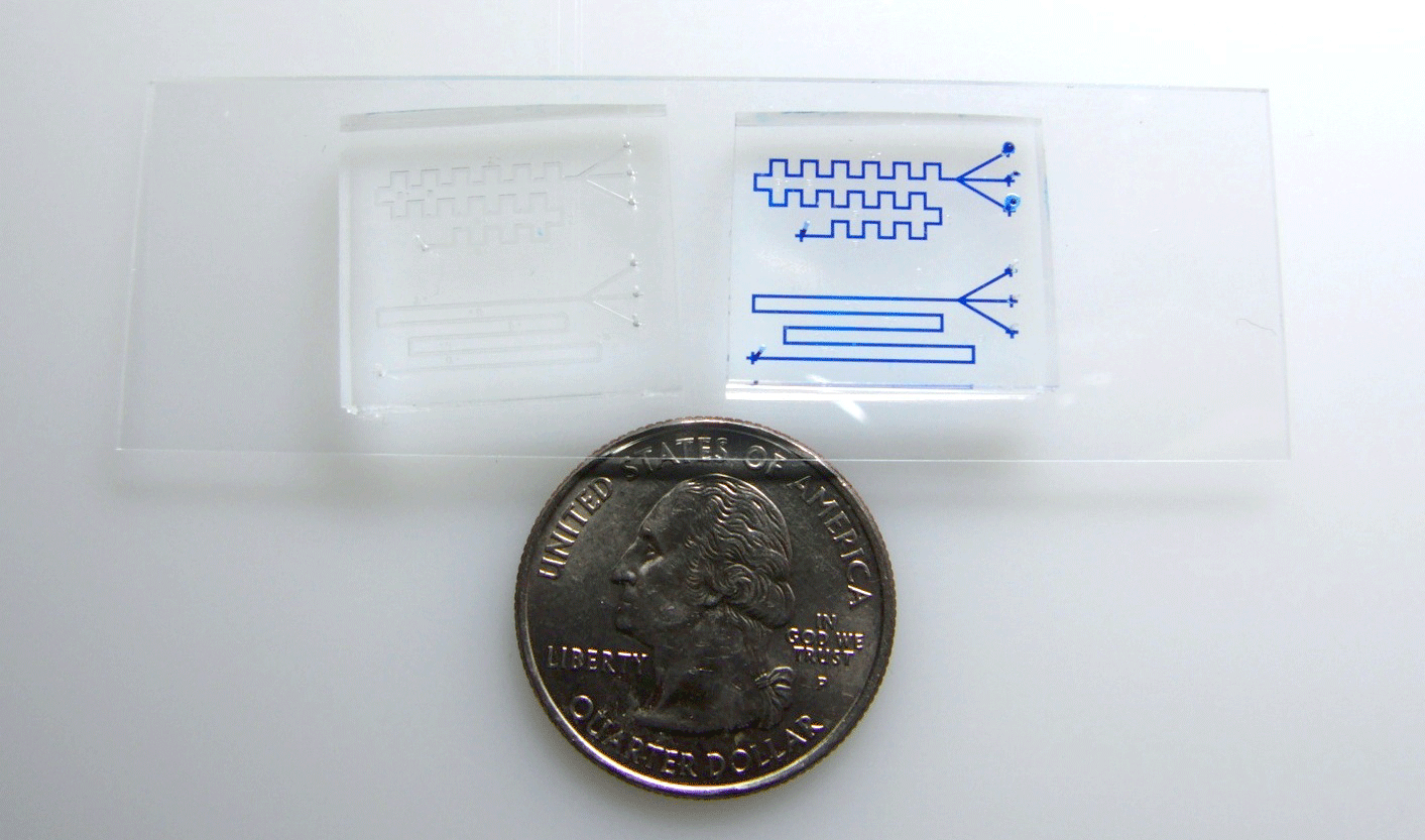
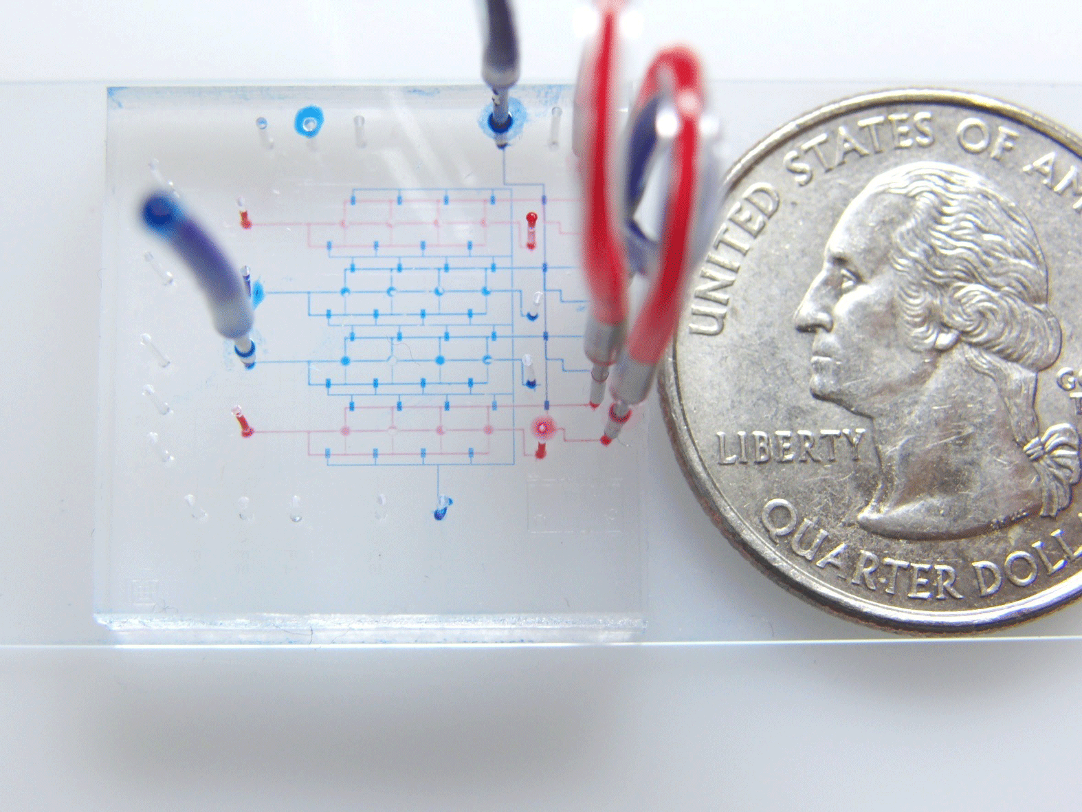
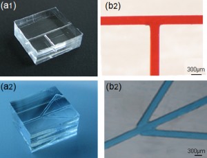
  7. Devices are ready for display. Notice the enhanced contrast of the colour filled channels vs the empty channels for the same device in Figure 2. While channels are visible only from some directions when they reflect light, colour-PDMS devices can be observed from every direction. Additionally, different device layers or areas can be specified by colour. In the figure control layers are blue and flow layers are red

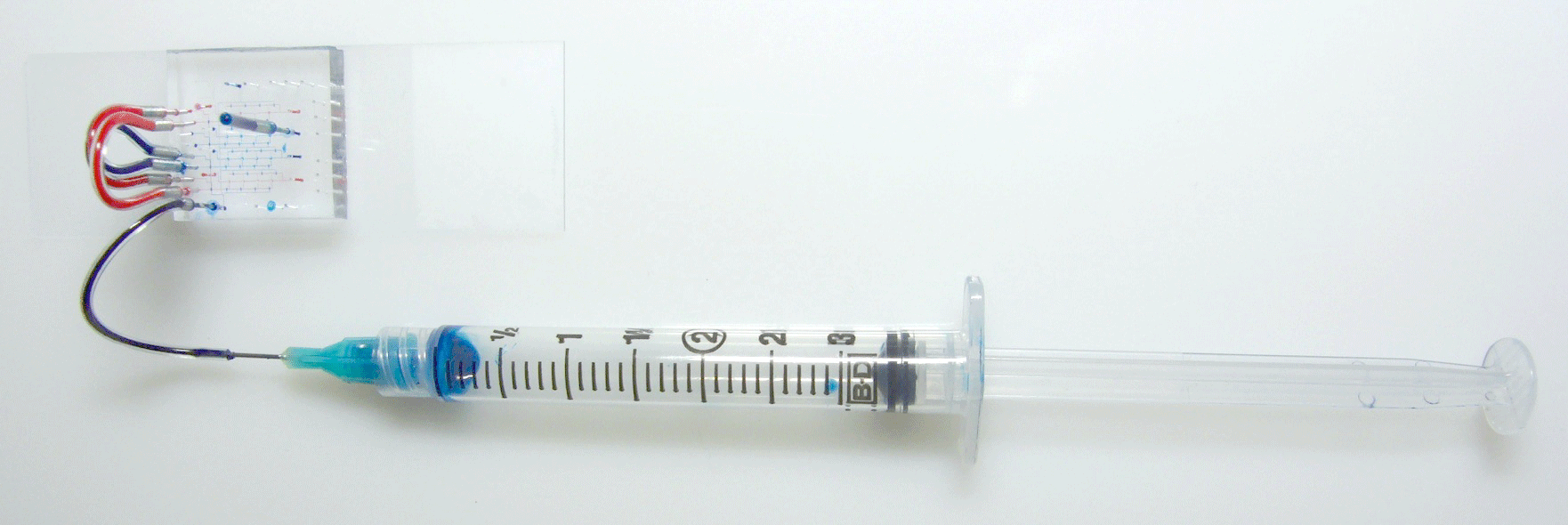
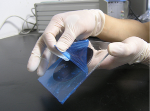
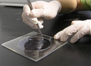
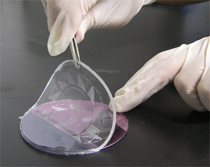
Fig. 1 Injection of PDMS through the channels. Air trapped inside the syringe provides a way to regulate the pressure applied to the device to minimize de-bonding. Compressing the air to 1/3 of original volume should provide enough pressure to drive the PDMS through.


Fig. 2 Enhanced channel contrast after injection, devices on the left side have empty channels and devices on the right have color PDMS inside.

Fig. 3 Different device zones can be identified by color. Here control layer is blue and flow layer is red. Secondary regulation channels are practically invisible when not filled.

References


[1] http://www.upchurch.com
[2] http://www.smooth-on.com

Digg This
Reddit This
Stumble Now!
Share on Facebook
Bookmark this on Delicious
Share on LinkedIn
Bookmark this on Technorati
Post on Twitter
Google Buzz (aka. Google Reader)

Drilling inlet and outlet ports in brittle substrates

R.J. Shilton, L. Y. Yeo, and J. R. Friend
MicroNanophysics Research Laboratory, Department of Mechanical and Aerospace Engineering, Monash University, Clayton, Victoria, 3800, Australia

Purpose


It is often necessary to form millimeter order holes in glass (and similar) substrates to form inlet or outlet ports in microfluidic devices. The easiest way to do this is to simply drill a hole in the required position, however owing to the brittle nature of most materials used in these devices, this can often lead to a high failure rate where the devices crack during the drilling process. Outlined here is a simple procedure for drilling ports in microfluidic devices, which has been tested with glass, silicon, and lithium niobate, with a very high success rate.

Materials


  • ~ 1mm diamond drill bit (UKAM Industrial Superhard Tools, Valencia, CA)
  • Drill press
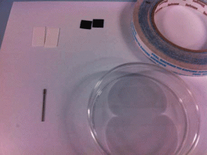
  • Double sided tape
  • Disposable plastic petri dish
  • Small piece of alumina (or other hard flat material)
  • Substrate to drill ports into

Procedure


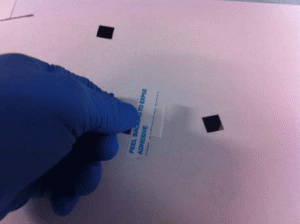
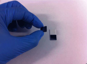
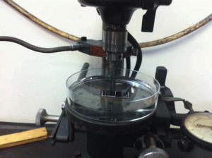
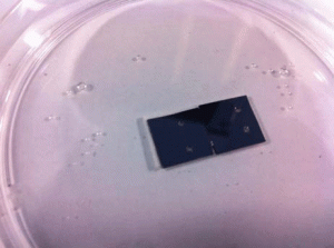
1. Attach microfluidic device to alumina with double sided tape. We have used this method reliably to drill ports in glass, silicon, and lithium niobate, however in principle it should work on a range of similar substrates. Silicon is shown in all images.

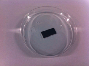
2.  Stick alumina to bottom of petri dish with a small amount of double sided tape.

3. Fill petri dish with a generous amount of water to cool down the drill site, and to remove particles into the fluid while drilling the hole.

4. Attach diamond drill bit to drill press. We successfully used drill bits of diameter 0.75 and 1 mm, however others should work equally as well.

5. Drill at a high speed (~10,000 RPM). Quite a bit of force can be applied without cracking the substrate, as it is stuck to a rigid backing. Drilling through a 0.5 mm thick substrate should take about ten to fifteen seconds, if it is attached firmly.


What else should I know?


  • Release the downward force a little near the hole exit, to avoid a rough hole on the other side.
  • Keep device immersed in water after drilling until it is ready to be cleaned to avoid particles becoming stuck in device channels etc…
  • Change water regularly to remove particle build up

MicroNanophysics Research Laboratory,

Department of Mechanical and Aerospace Engineering,

Monash University, Clayton, Victoria, 3800, Australia

Digg This
Reddit This
Stumble Now!
Share on Facebook
Bookmark this on Delicious
Share on LinkedIn
Bookmark this on Technorati
Post on Twitter
Google Buzz (aka. Google Reader)

A novel technique for aligning multiple microfluidic devices

Tiama Hamkins-Indik, Sandra Lam, Megan E. Dueck and Luke P. Lee
Department of Bioengineering, Berkeley Sensor and Actuator Center, Biomolecular Nanotechnology Center, University of California, Berkeley, CA 94720, US

Why is this useful?


Currently, there is no simple method for aligning multiple layers of PDMS microfluidic devices onto a glass slide.  We report a method for alignment that is easy, inexpensive, and has many relevant applications including printing proteins adjacently , flowing cells over previously printed proteins, and aligning two PDMS devices on top of each other for complex 3D geometries .  Currently, glass etching can be used to permanently mark glass, but this process is labor intensive and costly .  As a proof-of-concept, a 3-layered, 3 mm wide replica of Van Gogh’s Starry Night was created (Figure 5).

What do I need?


1.    Permanent marker (e.g. Sharpie™ marker)
2.    Syringe (1mL – 10mL) with needle tip (any gauge)
3.    Vacuum chamber
4.    Light microscope with 4X and 10X objectives
5.    Scotch™ Tape
6.    Alignment marker

The Alignment Marker:
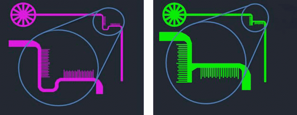
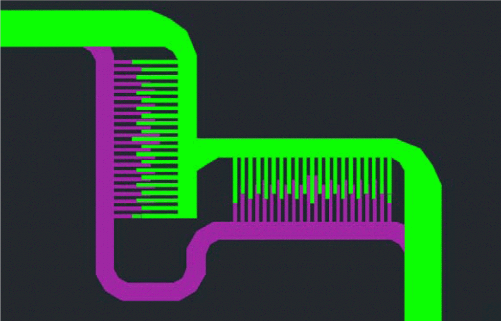
While any alignment marker can be used between layers, we suggest using the one shown in Figure 1.  The teeth act as Vernier scales in the x and y directions, thus, the degree of misalignment can be measured.  This alignment marker was designed with 10 µm wide teeth.  On the first layer (Figure 1, left), there are 22 posts which are 10 µm apart.  On the second layer, there are 20 posts each 11µm apart (Figure 1, right), making both markers 430µm wide.  The interlocking geometry of the markers is shown in Figure 2.  An inlet must be placed on the first layer’s alignment marker so that ink may flow through and stain the glass.

How do I do it?


1. Incorporate alignment marker

  • Add the alignment markers into the device design. On the silicon master that will be used to cast PDMS molds, The the channel height of the alignment marker on the silicon wafer should be in the 5 – 200 µm range.  The PDMS channel height only needs to be tall enough for Sharpie™ ink to flow through.

Figure 1. Alignment markers. The figure on the left is the alignment marker of the first level and the figure on the right is the alignment marker for the second layer.
Figure 2. Alignment markers interlocked.

2. Sharpie™ Ink Extraction

  • Put a needle onto a 1 mL – 10 mL syringe.
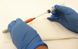
  • Insert the needle into the felt tip of a Sharpie™ marker, and slowly pull the plunger.  Slow extraction is necessary to allow air to diffuse into the marker as the ink escapes into the syringe.  Repeat as necessary.  (Figure 3)
  • Dispense the Sharpie™ ink into a microcentrifuge tube.  Sharpie™ extraction should result in 0.5 – 1 mL of Sharpie™ ink.
  • Dilute Sharpie™ ink 3X in 100% ethanol.

Figure 3. Sharpie™ extraction technique, pierce tip of Sharpie™ maker with syringe needle and slowly pull plunger.

3. First Layer

  • Cut and punch desired PDMS device.
  • Clean a glass slide and the device with Scotch™ tape.
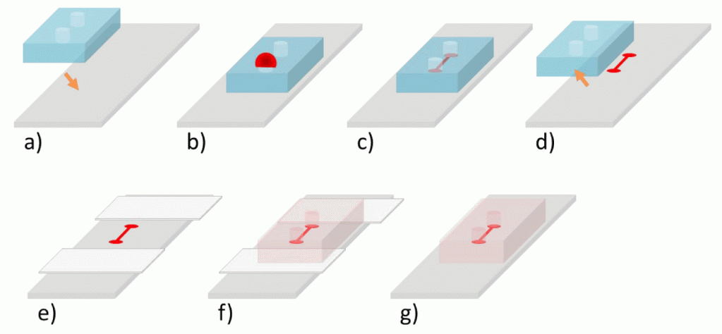
  • Reversibly bond the PDMS device onto a glass slide, by simply placing the cleaned PDMS onto the glass slide. (Figure 4a)
  • Load the Sharpie™ ink into the alignment marker channel.  This can be done by placing the device into a vacuum chamber for 5-10 minutes, removing the device from the vacuum, and placing a ~5 µL drop of Sharpie™ ink over the punch hole.  Only punch one entry hole for this method. (Figure 4b)
  • Allow the Sharpie™ ink to dry for 2 hours. (Figure 4c)
  • Remove PDMS. (Figure 4d)

Figure 4. Alignment technique schematic. a) place clean PDMS device on glass slide, b) load Sharpie™ ink, c) allow Sharpie™ ink to dry, d) remove PDMS device, e) place two pieces of scotch tape surrounding design, f) align second device to Sharpie™ ink alignment marker, g) remove scotch tape.

4. Second Layer

  • Cut and punch desired PDMS device.
  • Clean device with Scotch™ tape.
  • Place Scotch™ tape onto glass slide a few millimeters away from previous design.  (Figure 4e)
  • Under 4X or 10X magnification, bring the Sharpie™ alignment marker into the center of view and focus slightly above it.
  • Carefully place the cleaned second layer onto the scotch tape, but do not press down on the device so that the PDMS device is not in contact with the glass slide.
  • Still under the microscope, align the second layer with the Sharpie™ alignment marker by gently pushing the device along the Scotch™ tape.  The Scotch™ tape prevents the device from bonding with the glass slide.  (Figure 4f)
  • Attach the second layer by reversibly binding it to the glass slide by pressing down on the device.
  • Remove the scotch tape by holding the middle of the device down and pulling the scotch tape out from the edges of the PDMS device. (Figure 4g)
  • If additional layers are necessary, repeat second layer procedure.

What else should I know?


When using this technique by hand, the accuracy of the alignment between two layers can be down to 5 µm.  If a more precise alignment is necessary, a six axis alignment machine can be used.  As a proof-of-concept, we have reproduced Van Gogh’s Starry Night (Figure 5).  This design has two layers, and each layer was filled with Sharpie™ ink using vacuum loading.

Figure 5. 3 mm wide reproduction of Starry Night by Van Gough.

References


  1. Kane et al., Patterning proteins and cells using soft lithogrpahy, Biomaterials, 1999, 20, 2363-2376.
  2. Natarajan et al., Continuous-flow microfluidic printing of proteins for array-based applications including surface plasmon resonance imaging, Anal. Biochem., 2008, 373 (1), 141-146.
  3. Chiu et al., Patterned deposition of cells and proteins onto surfaces by using three-dimensional microfluidic systems, Proc. Natl. Acad. Sci. U. S. A., 2000, 97 (6), 2408 -2413.
  4. 3-Dimensional Molding for Making Microfluidic Devices, MicroDysis – Instrumentation Company with Micro- and Nano-fabrication, and Lab Automation, http://www.microdysis.com/TechMicrofab.aspx, 2010, accessed 16 April 2011.
Digg This
Reddit This
Stumble Now!
Share on Facebook
Bookmark this on Delicious
Share on LinkedIn
Bookmark this on Technorati
Post on Twitter
Google Buzz (aka. Google Reader)

The fabrication of PDMS interconnecting interface assisted by tubing fixation

Pengfei Li, Wei Xue, Jie Xu*

Mechanical Engineering, School of Engineering and Computer Science, Washington State University, Vancouver, WA 98686, USA.  E-mail: jie.xu[at]wsu.edu

Why is this useful?


Even though PDMS microfluidic devices have been widely applied in various research areas, it is still challenging to create the precise macro-to-micro interconnecting interface. Here we describe a method to connect a micro PDMS channel to the peripheral systems. With this method, we can avoid attaching exterior tubing using glue which is difficult to work with at the micro scale. In an effort to increase the connecting precision, a pre-curing step on a hot plate is adopted to prevent the tubing sliding away from the desired locations.

What do I need?


  • BD Vacutainer winged blood collection sets. (Model: 23G⨉3/4” ⨉12”, Fisher Scientific, Pittsburgh, PA, U.S.A.)
  • Dow Corning Silastic laboratory tubing. (11-189-15 Series, Fisher Scientific, Pittsburgh, PA, U.S.A.)
  • Sylgard 184 silicone elastomer kit. (Dow Corning, Midland, MI, U.S.A.)
  • SU-8 3050 permanent epoxy negative photoresist. (MicroChem, Newton, MA, U.S.A.)
  • 4-inch silicon wafer, pipette, aluminum foil, hot plate, and oven.

How do I do it?


1.    Prepare the SU-8 mold on a 4-inch silicon wafer with photolithography techniques. Based on the processes from the SU-8 data sheet supplied by MicroChem, SU-8 microstructures with approximately 50 µm in thickness are fabricated for this demonstration.

2.    Mix the elastomer base and the curing agent (mass ratio 10:1) to form PDMS, remove the air bubbles thoroughly using a desiccator or a centrifuge [1].

3.    Place the silicon wafer with the SU-8 channel molds on a hot plate. The temperate is set as 60oC under which the PDMS is able to cure in 10~20 minutes.

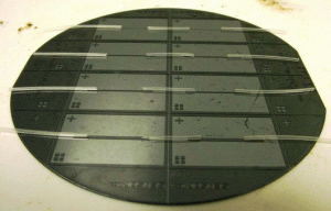
4.    Cut the Silastic tubing into short pieces with proper length. Align the tubing section with the channel molds.

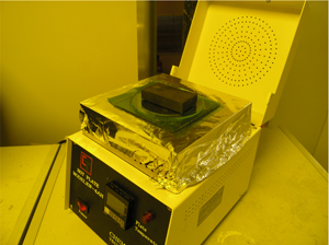
5.    Apply a small amount of PDMS to fix the tubing sections. Due to the relative high temperature of 60oC, the PDMS is nearly cured in 10~20 minutes. This step ensures that the tubing sections stay in the intended places where they are well aligned with channel molds. The tubing is still filled with air, though both ends of the tubing are sealed by PDMS.

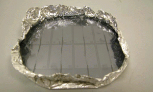
6.    Use aluminum foil to wrap the silicon wafer and create a container for storing PDMS. Extra PDMS is then quickly poured into the container and later cured in an oven [2].

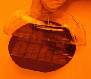
7.    Peel the cured PDMS off the channel molds and cut the PDMS into channel devices.

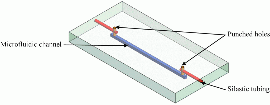
8.    Punch a small hole on each end of the channel. The tubing sections can be connected to the channels through the holes.

9.    Bond the PDMS channel to a glass substrate. Then bond another layer of PDMS as the top cover to seal the holes.

10.    Insert needles into the tubing sections. This creates the connection for the PDMS microchannel.

References


[1]  C. N. LaFratta, Degas PDMS in two minutes, Chips & Tips (Lab on a Chip), 17 August 2010.
[2] A. O’Neill, J. Soo Hoo and G. Walker, Rapid curing of PDMS for microfluidic applications, Chips and Tips , 23 October 2006.

Digg This
Reddit This
Stumble Now!
Share on Facebook
Bookmark this on Delicious
Share on LinkedIn
Bookmark this on Technorati
Post on Twitter
Google Buzz (aka. Google Reader)

Rapid, inexpensive and stress free drilling in glass substrates or thermally bonded glass chips using electrochemical spark erosion method

Arun Arora
KIST Europe, Korea Institute of Science and Technology, Campus E7.1, 66123 Saarbrücken, Germany

Background


An electrochemical spark erosion method is presented to drill holes in glass substrates or thermally bonded glass chips. Shohi and co-workers [1] reported this method in 1990. We have modified the method by using  a Pt wire 0.5 mm diameter anode instead of a steel needle cathode as a drilling needle and 50 V (instead of 35 V) was applied from a variable unregulated dc power supply to produce a constant spark in 8 M sodium hydroxide solution. When a steel needle cathode was used as reported in reference Shohi’s method, a large amount of brown precipitate was formed, which entered into the channel and created blocks.
Figure 1 shows a schematic diagram of the complete process and Figure 2 is a photo of the setup showing the process in operation.

Figure 1: Electro chemical spark erosion method of drilling holes in a thermally bonded glass device
Figure 2: Photo of the electro chemical spark erosion method for drilling holes in a thermally bonded glass device

The conventional methods to drill holes in glass include laser ablation or powder blasting. Both methods require expensive setup and trained staff therefore these methods are expensive.  The dentist drill also used to drill holes in bonded glass devices which can cause stress to the glass or can damage the channel walls. Sometimes glass particles can also clog the channel. A simple method for drilling access hole in a glass substrate or a thermally bonded glass chip is presented.

What do I need?


  • Pt wire (0.5 mm diameter)
  • Sodium Hydroxide solution (8M) (Caution)
  • Variable DC Power supply (100 V)
  • Petri dish

What do I do?


  1. Fixed a pt wire cathode on the side of the Petri dish as shown in schematic diagram in figure 1 or the photo in figure 2. Make sure that Pt wire reaches to the bottom of the petridish so it can make contact to the sodium hydroxide solution.
  2. Wear gloves and safety goggles and then place the Petri dish on a dry table. Fill it with sodium hydroxide (8M) solution (Caution: sodium hydroxide solution can cause severe burn if it comes in contact with skin. If any part of the body comes in contact, immediately wash it off with plenty of water.)
  3. Mark the position for the hole using a glass marker pencil on other side of the glass substrate where you want to drill the hole. Place it in the Petri dish facing the mark downward and immersed it approximately 5 mm deep into the sodium hydroxide solution.
  4. Connect the PVC covered anode and Pt wire cathode to the DC power supply.
  5. Hold the PVC covered Pt electrode precisely over the marked position on the glass substrate. Switch the power supply on and increase the voltage until the orange spark become visible. After 5 to 10 seconds of constant spark check the progress of drilling by taking out the glass substrate and viewing it with a hand held magnifying lens or a microscope (Figure 3).

Figure 3
Figure 3: Photo of the hole drilled by electrochemical spark erosion; a) Hole across a 60 µm wide channel. b) a 500 µm diameter hole across a 60 µm wide channel in a pre-bonded capillary electrophoresis glass device

References


[1] Shohi, S.; Esashi, M. Photoetching and Electrochemical Discharge Drilling of Pyrex Glass, Technical Digest of the 9th Sensor Symposium, Japan, 1990, 27.

Digg This
Reddit This
Stumble Now!
Share on Facebook
Bookmark this on Delicious
Share on LinkedIn
Bookmark this on Technorati
Post on Twitter
Google Buzz (aka. Google Reader)

Optimal protocol for moulding PDMS with a PDMS master

Jiaxing Wang1, Mingxin Zheng2, Wei Wang3,4*, and Zhihong Li3,4

1 Basic Medicine School, Peking University Health Science Center, Beijing, 100191, China
2 YuanPei College, Peking University, Beijing, 100871, China
3 Institute of Microelectronics, Peking University, Beijing, 100871, China
4 National Key Laboratory of Science and Technology on Micro/Nano Fabrication, Beijing, 100871, China
* E-mail: w.wang[at]pku.edu.cn

Why is this useful?


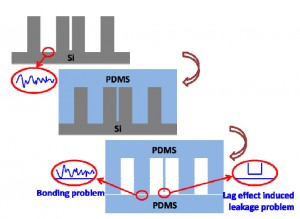
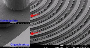
Nowadays, microfabricated silicon has been widely used as a master in PDMS-based soft lithography. It carries the merits of the microelectromechanical system (MEMS) technique, such as being able to construct complex 3D or high aspect ratio micro/nanostructures. However, the silicon master is easy to break during the PDMS moulding process. Due to the lag effects existed in deep reactive ion etching (DRIE) for silicon microfabrication, there will be a high risk of leakage between these two channels after PDMS moulding and bonding, if two parallel microchannels were designed too close, as shown in Fig. 1a. More seriously, the bonding surface of the PDMS replica will follow the morphology of the etched surface (Fig. 1a). Usually the etched silicon surface has high roughness, especially after a deep etching of silicon by DRIE (Fig. 1b), which makes the subsequent PDMS bonding being difficult, sometimes even impossible. To overcome the fragileness of the silicon master, it has been reported that moulding epoxy with PDMS replica, which is prepared by using the original microfabricated silicon as the master, to achieve a durable master in the PDMS moulding process. [1] However, this approach still suffers the aforementioned lag-induced leakage and bonding problems.

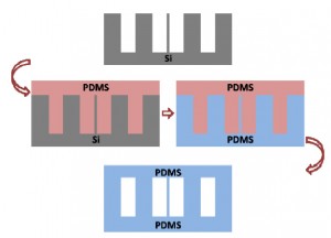
Moulding PDMS with a PDMS master replicated from the microfabricated silicon directly can overcome the aforementioned problems, as illustrated in Fig. 1c. It has been developed by silanizing the PDMS master with tridecafluoro- 1,1,2,2- tetrahydrooctyl- 1- trichlorosilane (in vacuo, 8h[2] or overnight[3]). In this Tip, we optimized the PDMS moulding process with the PDMS replica as the master. The optimal protocol is simple and easy to establish in any labs.
a

b

c

Fig. 1 Moulding PDMS with microfabricated silicon as master. (a) Schematic illustration of the lag effect induced leakage and bonding problems existed in the traditional soft-lithography when the PDMS is replicated from the microfabricated silicon master directly. (b) SEM photo of the microfabricated silicon master used in the present work (with the tilting angle of 45o). Inserted SEM photos give the morphology comparison between the etched silicon surface and the original one. (c) Schematic illustration of the moulding PDMS process with a PDMS master.

What do I need?


Microfabricated silicon, aluminum foil (Select HORECATM, Jinweiyuan Hotel Supplies Co. ltd.), mould release reagent (Micro90®, International Products Corporation, USA), PDMS (Sylgard 184; Dow Corning Co., MI), detergent (commercial available detergent, White CATTM, White Cat co. ltd.), ethanol (MOS grade, Beijing Chemical Reagent Institute).

What else should I know?


Process optimization

The present protocol contains two PDMS moulding processes: The first one is to construct the PDMS master from the microfabricated silicon while the second to make the final PDMS replica directly from the so-prepared PDMS master.

Considering the Corona-triggered PDMS-PDMS bonding technique [4], which will be adopted in the subsequent PDMS-microfluidic device construction, curing at 60oC for an hour of the PDMS pre-polymer prepared following the manufacture guideline was selected as the second PDMS moulding recipe. In this chips and tips, the first PDMS moulding process and the surface treatment of the PDMS master were optimized.

1) Optimization of the first PDMS moulding process

Table 1 Optimization of the first PDMS molding process

The first PDMS moulding process was optimized for the PDMS master preparation as shown in Table 1. Based on the above experiments, we found the optimal recipe for the first PDMS moulding process was curing the PDMS pre-polymer at 120oC for an hour with the mass ratio (Base to Curing agent) of 5:1. (Condition No.4)

2) Optimization of the surface treatment of the PDMS master

Table 2 Optimization of the surface treatment of the PDMS master

Surface treatment is another important factor for the PDMS moulding and was optimized as listed in Table 2. The results indicated that detergent dissolved in 75% ethanol (about 10 w.t.%) was the optimal recipe for the second PDMS moulding process. (Condition No.3) Herein, we name it the modified mold release agent. In later work we also use it for pre-treatment of the microfabricated silicon master.


How do I do it?


1. Make a holder with aluminium foil, according to the shape of the microfabricated silicon master. [5]
2. Mix the PDMS according to the manufacturer’s procedure, but with a mass ratio of the Base to the Curing agent of 5:1.
3. Pre-treat the microfabricated silicon master with the modified mold release agent.
4. Dry the microfabricated silicon master with nitrogen and put it into the holder.
5. Pour the PDMS pre-polymer into the holder, covering the whole microfarbicated silicon master, and then place the holder in an oven at 120oC for an hour.
6. Gently peel the PDMS replica (the PDMS master in the second PDMS molding process) off from the microfabricated silicon master. (Fig.2)
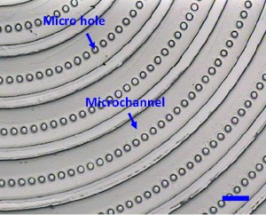
Fig. 2 The PDMS master, the PDMS replica from the microfabricated silicon. (The scale bar is 100μm.)

7. Treat the PDMS master with the modified mould release agent. For the deep holes (e.g. some holes with the depth of 100?m in Fig.2), treat the master with the modified mould release agent in the vacuum.
8. Place this PDMS master in a Petri dish, with the moulded-side upwards.
9. Mix PDMS prepolymer again according to the standard manufacturer’s procedure with a mass ratio of the base to the curing agent of 10:1.
10. Pour the PDMS mixture into the Petri dish, covering the whole PDMS master, and then cure at 60oC for an hour.
11. Carefully peel the PDMS replica (Fig.3) off from the PDMS master for the subsequent experiments.
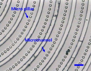
Fig. 3 The final PDMS replica from the PDMS master. (The scale bar is 100μm).

Acknowledgements


This work was financially supported by the 973 Program (Grant No. 2009CB320300).

References


[1] A. Estévez-Torres, A. Yamada and L. Wang,  An inexpensive and durable epoxy mould for PDMS, Chips & Tips (Lab on a Chip), 22 April 2009.
[2] J. R. Anderson, D. T. Chiu, R. J. Jackman, O. Cherniavskaya, J. C. McDonald, H. Wu, S. H. Whitesides and G. M. Whitesides, Fabrication of Topologically Complex Three-dimensional Microfluidic Systems in PDMS by Rapid Prototyping, Anal. Chem., 2000, 72, 3158-3164.
[3] J. L. Tan, J. Tien, D. M. Pirone, D. S. Gray, K. Bhadriraju, and C. S. Chen, Cells lying on a bed of microneedles: An approach to isolate mechanical force, Proc. Natl. Acad. Sci. U. S. A., 2003, 100(4), 1484-1489.
[4] C. Yang, W. Wang, and Z. Li, Optimization of Corona-triggered PDMS-PDMS Bonding Method, in Proceedings of the 4th IEEE International Conference on Nano/Micro Engineered and Molecular Systems, 319-322.
[5] A. O’Neill, J. Soo Hoo and G. Walker, Rapid curing of PDMS for microfluidic applications, Chips & Tips (Lab on a Chip), 23 October 2006.

Digg This
Reddit This
Stumble Now!
Share on Facebook
Bookmark this on Delicious
Share on LinkedIn
Bookmark this on Technorati
Post on Twitter
Google Buzz (aka. Google Reader)

Simple fabrication of microfluidic devices by replicating Scotch-tape masters

Anil B. Shrirao1 and Raquel Perez-Castillejos1,2
1 Department of Electrical and Computer Engineering, 2 Department of Biomedical Engineering, New Jersey Institute of Technology, Newark NJ, USA

Why is this useful?


We present a method for fabricating PDMS microfluidic devices based on replicating a master made of Scotch tape.  Often the fabrication of microfluidic devices by soft lithography is restricted to those who have access to a cleanroom that allows the fabrication of a master with micrometric features.  Here we demonstrate that patterned Scotch tape can be used (without the need of any chemical treatment) as a master for soft lithography yielding microfluidic devices with a uniform height of ~ 60 µm.  As a difference to previous Chips & Tips on rapid and easy prototyping techniques [1, 2], in this method the Scotch tape does not remain as part of the microfluidic device after fabrication.  Here we used a handheld cutting tool (scalpel) to pattern the Scotch tape.  A laser cutting machine could be used, instead, for masters requiring higher precision.

What do I need?


  • Glass slides, pre-cleaned from Manufacturer (Fisher Scientific, 75mm x 50mm x 1mm, Cat. No. 12-550-C)
  • Scotch tape (3M Scotch® Transparent Tape 600[6])
  • Stainless steel Scalpel or surgical blade with (Feather Safety Razor Co., LTD, Cat. No. 2976#11)
  • Polystyrene Petri dish (Fisher Scientific, 100mm x 15mm, Cat. No. 08-757-12)
  • Tweezers
  • Oven or hot plate (to work at 65°C)
  • Gloves (do not use latex gloves)
  • PDMS silicone elastomer base and curing agent (Sylgard 184, Dow Corning)

What do I do?


1. Attach a strip of Scotch tape to the glass slide.  The thickness of the Scotch tape will determine the height of the microchannel.  (To increase the height of the channel, attach additional strips of Scotch tape.)
2. Print the layout of the microchannel on regular paper.  Place the printout on a flat surface.
3. Place the glass slide on the printout, with the Scotch tape facing up.  Align the glass slide to the microchannel layout.  Fix the glass slide to the printout with a piece of Scotch tape on the corner of the slide.
4. Use the scalpel to cut the tape on the glass slide according to the layout.  (For cutting, we used another glass slide as a ruler)
5. Remove the Scotch tape from all regions of the glass slide except those in the layout of the microchannel.
6. Place the glass slide with patterned Scotch tape in a heating oven at 65°C for 2-3min; this improves the adhesion of the edges of patterned Scotch tape to the glass substrate.  At this point, the Scotch-tape pattern does not need any further treatment in order to be used as a master for soft lithography [1].
7. Mix the base and curing components of PDMS as recommended by the manufacturer [2].  Place the glass slide in a Petri dish, with the patterned Scotch tape facing up.  Pour the PDMS mixture in the Petri dish until the master (i.e., glass slide and Scotch tape) is covered completely.  Degas PDMS in vacuum (if needed) and allow it to cure for 1 hour at 65°C.
8. Use the scalpel to cut the slab of PDMS containing the microchannel.  Peel off the PDMS replica.  (The Scotch-tape master can be used again by repeating step 7.)
9. Punch holes in the PDMS replica for the inlets and outlets of the microfluidic device.
10. Seal the PDMS replica to a substrate, either (i) conformally-by bringing the PDMS replica in contact with a smooth, clean substrate-or (ii) irreversibly-by bringing the PDMS replica in contact with the substrate after oxidizing both parts in an oxygen plasma [3]).


Acknowledgements


R. P.-C. acknowledges the support of the New Jersey institute of Technology through starting faculty funds.

References


[1] R. J. Holmes and N. J, Goddard, Rapid prototyping of microfluidics, Chips & Tips (Lab on a Chip), 15 February 2007.
[2] R, Kumar, R. L. Smith, and M. G. Pappas, A method for rapid fabrication of microfluidic devices, Chips & Tips (Lab on a Chip), 30 June 2009.
[3] Y. Xia and G. M. Whitesides, Annu. Rev. Sci., 1998, 28, 153-184.
[4] Dow Corning Product Information, “Information about Dow Corning® brand Silicone Encapsulants”.
[5] M. K. Chaudhury and G. M. Whitesides, Langmuir, 1991, 7, 1013-1025.
[6] Scotch tape

Digg This
Reddit This
Stumble Now!
Share on Facebook
Bookmark this on Delicious
Share on LinkedIn
Bookmark this on Technorati
Post on Twitter
Google Buzz (aka. Google Reader)

Fast-iteration prototyping and bonding of complex plastic microfluidic devices

Jonathan Siegrist, Mary Amasia, and Marc Madou
Departments of Biomedical, Chemical, and Mechanical Engineering, University of California, Irvine, CA, 92697, USA

Why is this useful?


The fabrication and bonding of rapid-prototyped polymer-based microfluidic devices is of great interest. The emphasis is not only on the ability to produce large numbers of devices rapidly, but to perform fast design and test iterations. Here, we present a method for rapidly cutting thin plastic films into complex shapes, such that sophisticated 2.5-D microfluidic chips can be created. The plastic layers are bonded together using traditional, low-pressure thermal bonding to minimize channel deformation while achieving high bond strengths capable of withstanding high-pressure & temperature operations, such as polymerase chain reaction (PCR). The authors have found this method to be particularly amenable to fast design iterations, as one can easily go from a design on the computer screen to testing of a real part within 3 hours.

The method presented here of cutting plastic films using a commercially-available knife-based cutter/plotter avoids the use of traditional CNC milling, which can leave behind burrs and rough edges. While hot-embossing and laser machining can be superior alternatives to CNC milling, they are considerably more complex and expensive than the method presented here. Also, by using a computer-controlled cutter, obvious advantages are gained in terms of possible design complexity and reproducibility as compared to previous Tips [1, 2]. Finally, this cutting method can be used for many types of plastic films (< 400 um thick, depending on the machine being used) without the need for extensive optimization of cutting speeds or feed rates.

The use of thermal bonding allows for much higher bond strengths as compared to the use of pressure-sensitive adhesives or double-sided tapes, and also provides a large dynamic range in terms of the types of plastics that can be used. For example, thin films of either high glass-transition temperature (Tg) materials such as polycarbonate or low-Tg cyclic olefin copolymers can be used with this method, as compared to lamination methods that are limited to low Tg materials only [3].

What do I need?


  • Hydraulic Thermal Press, ideally with a max. temperature of at least 200ºC and a max. pressure of at least 1 MPa (the device used here was an MTP-8 from Tetrahedron Associates, Inc., CA, USA)
  • Computer-Controlled Cutter/Plotter (the device used here was a CE-2000 from Graphtec America, Inc., CA, USA)
  • CAD software
  • Bare Si Wafers, at least single-side polish
  • Plastic Films (the films used here were polycarbonate from McMaster-Carr, CA, USA)
  • Double-sided Tape (3M Scotch brand was used here)
  • Aluminium Foil
  • Tweezers
  • Alignment Pins (optional)
  • Oxygen Plasma System (optional)

What do I do?


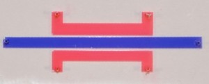
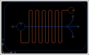
1. Design your microfluidic device using common CAD software (Fig. 1). Ensure the drawing for each layer is in a “polyline” format (i.e., each continuous line is defined as a single object) to facilitate smooth cutting. Import the CAD file into the software that controls the cutter/plotter.

A proof-of-concept microfluidic device laid out using CAD software, with different layers defined for each plastic part to be used.

2. Attach your plastic film (using double-sided tape) to a plastic sheet that will act as your support/sacrificial layer (e.g., 0.75 mm thick). Place this structure in the cutter/plotter, and cut the film (Fig. 2). Cutting force and number of cuts may need to be optimized depending on the film thickness. We found that 2x low-force cuts worked well for 127 µm-thick polycarbonate films, and 4x medium-force cuts worked well for 254 µm-thick films.

The plastic film to be cut is mounted on a support base using double-sided tape, and then placed in the cutter/plotter.

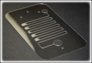
3. After all cuts are complete, carefully remove the part from the sacrificial layer and use tweezers to remove and discard the cut-out plastic features (Fig. 3). Repeat this for all layers, and then clean the plastic layers using isopropanol and water.

A single layer of polycarbonate cut out using the cutter/plotter, after removal of the cut features (for reference, the small loading and venting holes are 1 mm diameter).

4. Align all of the layers, and hold together using a clip. You may include alignment pin features on your layers, and use pins to assist in alignment. For example, you could use 1 mm-diameter pins (McMaster-Carr, CA, USA – #91585A001).
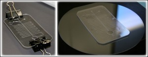
5. Place the assembled microfluidic device layers on the mirror-finished side of a Si wafer, and remove the clip(s) (Fig. 4). Then place the second wafer mirror-finished side down on top of the microfluidic device, being careful not to disturb the alignment.

All layers of the microfluidic device are aligned and held together using clips (left) and the part is then carefully placed on a Si wafer in preparation for thermal bonding (right).

6.  Place the assembly in the thermal press sandwiched between two pieces of foil, and thermally bond. Approximate parameters we found to work well for our proof-of-concept, multi-layer, polycarbonate device (1-top layer with loading/venting holes: 254 µm thick, 2-film with serpentine channel: 127 µm thick, 3-film with access holes and reservoirs: 127 µm thick, 4-film with linear channel: 127 µm thick, and 5-bottom layer: 254 µm thick) are shown in Fig. 5.

The thermal bonding parameters used for the proof-of-concept microfluidic device.

7.  Remove the microfluidic device from the thermal press, and test (Fig. 6).

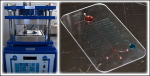
The thermal press used (left) and the completed microfluidic device (right) loaded with contrast agents for visualization.

What else should I know?


Thermal bonding parameters, such as bonding temperature, pressure, dwell times, and ramping rates, will need to be optimized to ensure complete bonding of the plastic layers. The main disadvantage of thermal bonding is possible microchannel deformation, which can affect fluidic function. Thus, the Tg of your material, the number of layers, and the required bond strength will all need to be considered. The previous Tip on prevention of chamber-sagging has obvious uses here [4].

The limitations on feature size using this method should also be noted. The thickness (z-axis) of your channels/chambers is determined by the thickness of the films. We found the feature size (x-y plane) limitations when cutting films to be ~ 200 µm, as limited by the cutter/plotter being used. Newer machines list resolutions on the order of 10s of µm.

Finally, while these plastic parts are inherently hydrophobic, they can be made hydrophilic via oxygen-plasma treatment. Common plasma-treatment parameters for our devices are 200 W for 2 mins at 200 mTorr O2-pressure to provide hydrophilicity for many weeks. This can facilitate liquid loading and also serve as a sterilization step.

Acknowledgements


We would like to thank the DARPA-MF3 center for funding, and Dr. Albert Yee of UC Irvine for generously allowing us to use his thermal press.

References


[1] R. J. Holmes and N. J, Goddard, Rapid prototyping of microfluidics, Chips & Tips, (Lab on a Chip), 15 February 2007.
[2] R, Kumar, R. L. Smith, and M. G. Pappas, A method for rapid fabrication of microfluidic devices, Chips & Tips, (Lab on a Chip), 30 June 2009.
[3] D. Olivero and Z. Fan, Lamination of plastic microfluidic devices, Chips & Tips, (Lab on a Chip), 30 July 2008.
[4] J. Xu and D. Attinger, How to prevent sagging during the bonding or lamination of chips with large aspect ratio chambers, Chips & Tips, (Lab on a Chip), 24 July 2009.

Digg This
Reddit This
Stumble Now!
Share on Facebook
Bookmark this on Delicious
Share on LinkedIn
Bookmark this on Technorati
Post on Twitter
Google Buzz (aka. Google Reader)

Rapid prototyping of branched microfluidics in PDMS using capillaries

S. Ghorbanian, M. A. Qasaimeh and D. Juncker
Biomedical Engineering Department, McGill University and Genome Quebec Innovation Centre, McGill University, 740 Dr. Penfield Avenue, Montreal, QC, H3A 1A4 Canada

Why is this useful?


Polydimethylsiloxane (PDMS) is widely used for the fabrication of microfluidic systems because it can readily be molded into the desired shape, is easy to seal onto substrates, and is transparent thus permitting visualization of the sample [1]. However, the fabrication of PDMS microfluidic devices depends on a microfabricated mould that needs to be made in a clean room using photolithography and microfabrication methods, all of which are costly and time consuming and beyond the reach of many researchers. Rapid prototyping techniques that circumvent the requirement for a clean room have been proposed, such as the use of double sided scotch tapes, but lack precision and control [2,3].

Here we present a method for rapid prototyping of branched microfluidics in PDMS with control over the architecture, channel width and depth. We propose using capillaries as the mould.  They are cut to size, then arranged on a flat PDMS according to the desired architecture and covered with PDMS which is then cured. The size of each channel can be adjusted by selecting a capillary with the desired diameter, and different branch architectures can readily be produced. The capillaries are then simply removed from the PDMS replica and leave behind a network of channels.

A key challenge is the gaps formed at the intersections between abutting capillaries. We found that a small flake of paraffin can be used which is then melted to fill up this gap. The use of capillaries with a square cross-section further facilitates the moulding, and allows for making complex networks with ease and good yield. This protocol only requires materials which are commercially available and comparatively inexpensive and takes less than one hour of hands-on time, followed by three hours of curing.

What do I need?


1. A layer of flat cured PDMS, Figure 1d
2. Uncured PDMS
3. Petri dishes
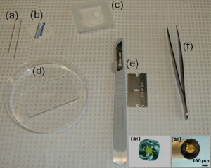
4. Square capillaries (preferably) or circular capillaries [4], Figure 1a
5. Ceramic cutting stone [4], Figure 1b
6. Paraffin [5], Figure 1c
7. Oven or hot plate
8. Sharp tweezers and razor blade, Figure 1(e,f)
9. Double-sided tape [6]

Figure 1. Required materials: (a) Glass capillaries, (a1) Cross section of the square glass capillary and (a2) the round glass capillary [4]. (b) Capillary cutting stone [4]. (c) Paraffin flakes [5]. (d) Flat cured PDMS piece in a Petri dish. (e) Razor blade. (f) Tweezers


What do I do?


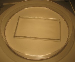
1. Cure a layer of PDMS in a Petri dish. Remove the PDMS, and cut it to the desired size. Flip this piece to obtain the flat surface on top and place it inside another Petri dish as shown in Figure 2.

Figure 2. Flat piece of a cured PDMS

2. Cut two pieces of capillary, Figure 1a (or as many needed to make the branched channels) using a ceramic cutting stone. The capillaries should be cut to a length that facilitates handling, typically three or more times the length of the final channels of interest and shorter than the diameter of the Petri dish. Also see “What else should I know I”. After cutting the capillaries, dip their tip at each side in melted paraffin or liquid glue and let it solidify. This helps prevent air trapped in the capillaries from exiting while degassing the PDMS, which can lead to bubbles and displacement of the capillaries.

3. Under the stereomicroscope grind the tip of the capillaries (which will be making the connections between capillaries) to remove the bumps seen in Figure 3a using a polishing stone, such as the side surface of a cutting stone, with horizontal movements, Figure 3b and flatten the tip completely, Figure 3c, which can be done at angles other than 90 degress as well, Figure 3d.

Figure 3. A square capillary before (a) and after grinding with the ceramic stone (b) and flattening its tip into a right angle (c) and other angles (d)

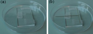
4. Stick double-sided tape at the outer extremities (preferably not at the connection sites) where the capillaries will be placed on the flat PDMS in the Petri dish, Figure 4a. Under a stereomicroscope place the capillaries according to the desired architecture. Figure 4b illustrates the placement of capillaries for fabricating T-shaped microchannels.

Figure 4. Placing the capillaries on the flat PDMS held by double side tape

5. Examine the gaps and connections between the capillaries to ensure good contact. A T-shaped connection and 45 degree connections are shown in Figure 5 below.

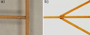
Figure 5. Different connections between capillaries: (a) Capillary connection in T-shape with a right angle and (b) connections with acute angles

6. Carefully place a small piece of paraffin using sharp tweezers on each of the capillary connections, Figure 6a. Heat the tip of the tweezers on a hot plate for a minute, and then carefully approach the tip to melt the paraffin, which fills the gaps between the capillaries due to capillary effects and joins them to one another, Figure 6b. The excess melted paraffin can be removed carefully wiht the tip of a razor or sharp tweezers.

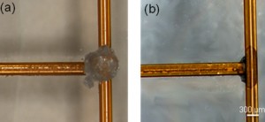
Figure 6. Filling the interconnection gaps with melted paraffin. (a) Piece of paraffin placed on the connection site. (b) The connection site with melted paraffin after cleaning

7. Pour a layer of uncured PDMS over the connected capillaries as shown in Figure 7 and degas the PDMS by placing the Petri dish in a vacuum desiccator to remove all air bubbles, for alternative methods see “What else should I know II”. Then, place the Petri dish in the oven at 65°C for 3 hours or more. For shorter curing time see “What else should I know III”.

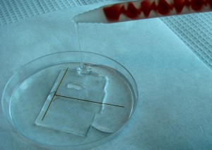
Figure 7. Pouring uncured PDMS over the capillaries network

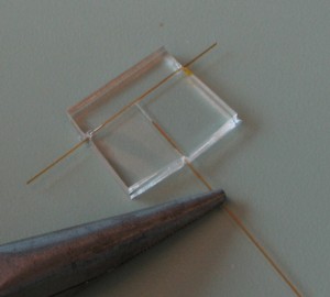
8. Remove the whole piece of cured PDMS from the Petri dish. Cut the sides of the cured PDMS using a razor blade, leaving a significant amount of the capillary exposed outside as shown below. Then carefully pull out the capillaries from the sides of the PDMS using pliers as shown in Figure 8. To make this process easier, the network can be immersed into or washed with acetone which will swell the PDMS and expand the channels prior to pulling out the capillaries.

Figure 8. Pulling off the capillaries after the PDMS is fully cured

If residues of paraffin are left inside the microchannels these can be dissolved and washed by flushing the microchannels with acetone.

9. Trim the microfluidic device to the desired shape using a razor blade or a cutter, Figure 9.

Figure 9. Fabricated branched microfluidics (a). Microchannels filled with red and blue dye (b).

What else should I know?


Several alternative fabrication tips are listed below:

I) To fabricate an open channel microfluidic network replace the flat cured PDMS layer (in step 1) with a clean glass slide. Once the PDMS is cured it can be separated from the glass slide. In this process smaller capillaries can be used to make the channels which can be removed using tweezers after detaching the PDMS from the substrate. This open network can also be bonded to another PDMS layer after the capillaries are removed.
II) If no vacuum desiccator is available to degas PDMS, the sample can be left to be degassed and cured at room temperature overnight followed by post-curing in an oven at 65°.
III) To increase the speed of PDMS curing in step 8, the Petri dish may be replaced by an aluminum foil or a glass plate and allow to use much higher temperatures inside an oven or on a hot plate to cure the PDMS within a few minutes.
IV) It is preferable to use square capillaries because there are no gaps formed at the connection sites due to the square shapes of the capillaries as opposed to the round capillaries which will have a small gap formed at the connection sites due to the rounded shape of the capillary walls, Figure 1(a1). These gaps can, however, get filled with melted paraffin.

References


[1] D. Duffy, J. McDonald, O. Schueller, G. Whitesides, Rapid prototyping of microfluidic systems in poly (dimethylsiloxane), Anal. Chem., 1998, 70, 4974-4984.
[2] R. J. Holmes and N. J, Goddard, Rapid prototyping of microfluidics, Chips & Tips, (Lab on a Chip), 15 February 2007.
[3] R, Kumar, R. L. Smith, and M. G. Pappas, A method for rapid fabrication of microfluidic devices, Chips & Tips, (Lab on a Chip), 30 June 2009.
[4] Polymicro technologies
[5] Fisher Scientific
[6] Scotch Tape

Digg This
Reddit This
Stumble Now!
Share on Facebook
Bookmark this on Delicious
Share on LinkedIn
Bookmark this on Technorati
Post on Twitter
Google Buzz (aka. Google Reader)

A simple and economical holder for casting PDMS chips

Gang Li*, Qiang Chen and Jianlong Zhao

Shanghai Institute of Microsystem and Information Technology, Chinese academic of Sciences, Shanghai, China

Why is this useful?


To fabricate PDMS chips, one usually pours an un-cured PDMS mixture into a Petri dish or an aluminum foil tray containing the patterned master, and then cures for 2 h at 85°C. However, these casting dishes or trays present some cost problems. Often a relatively high consumption of PDMS is demanded for these casting holders because quite a part of PDMS will seep into the gap between the wafer and trays due to capillary forces during PDMS pouring. On the other hand, the seepage of PDMS under the master wafer may tilt the master causing the thickness of the PDMS mold to vary from one side to the other. Furthermore these casting dishes or trays are generally throw-away holders for casting PDMS chips, which also increases the fabrication cost of PDMS chips.

This tip presents a new casting holder, made of a hollow plate whose bottom is enveloped by an adhesive tape. This casting holder can simplify the operation of releasing the cured PDMS casting, and minimize the consumption of PDMS. In addition, this holder can be used to fabricate double-side flat PDMS chips by placing a flat substrate on its top.

What do I need?


  • Circular hollow plate (home-made of PMMA)
  • Adhesive tape (Nitto Denko Corp., SPV-224)
  • PDMS (Dow Corning Sylgard 184)
  • Hotplate (Chemat Technilogy Inc., KW-4AH hotplate)
  • PET (Polyethylene terephthalate) film and flat glass slide (for optional double-side flat PDMS chips)  

What do I do?


1. Seal the bottom opening of the hollow plate with adhesive tape to form a casting holder (Figure 1), whose diameter is a little bigger than that of master wafer.

Figure 1

2. Place the master in the holder, and carefully apply a pressure to attach it to the tape, preventing any gaps from forming at the interface (Figure 2).

Figure 2

3. Mix the PDMS according to the manufacturer’s procedure [1].

4. Pour the PDMS mixture in the casting holder, and then place the holder on a hotplate at 85ºC for 2 hours.

5. Once the PDMS is cured, remove the holder from the hotplate and allow it to cool to room temperature.

6. Gently peel off tape from the bottom of holder (Figure 3).

Figure 3

7. Carefully cut around the edge of the PDMS mold using a sharp scalpel, and separate the PDMS from the holder (Figure 4).

Figure 4

8. Finally, peel the PDMS mold from the master wafer (Figure 5).

Figure 5

9. (Optional, for double-side flat PDMS chips) Prepare the casting holder as described above,  pour the PDMS mixture in the casting holder until the level of PDMS is a little higher than the top of holder, and then carefully drop a PET film onto the prepolymer mixture (Figure 6), which provides an easy way to remove the cover plates from the PDMS molds after curing.

Figure 6

10. Apply a pressure on the top of the film with a stack of glass slides and steel block to planarize the surface of the PDMS chips, and then cure the PDMS by placing the holder on a hotplate (Figure 7).

Figure 7

11. After curing, remove the glass slide and steel block from the top of holder, and then gently peel off the tape and PET film from the PDMS block (Figure 8).

Figure 8

12. Finally, separate the PDMS block from the holder and peel the PDMS mold from the master.

Acknowledgements


This material is based upon work supported by the Major State Basic Research Development Program of China (No. 2005CB724305), and the National High Technology Research and Development Program of China (No.2006AA02Z136).

References


[1] Dow Corning Product Information, “Information about Dow Corning® brand Silicone Encapsulants,” 2005.

Digg This
Reddit This
Stumble Now!
Share on Facebook
Bookmark this on Delicious
Share on LinkedIn
Bookmark this on Technorati
Post on Twitter
Google Buzz (aka. Google Reader)