Archive for the ‘Cells’ Category

Development of a cell culture microdevice with a detachable channel for clear observation

Eriko Kamata, Momoko Maeda, Kanako Yanagisawa, Kae Sato*

Department of Chemical and Biological Sciences, Faculty of Science, Japan Women’s University, Bunkyo, Tokyo 112-8681, Japan

*E-mail: satouk@fc.jwu.ac.jp

Why is this useful?

There have been many reports on microfluidic devices for cell culture having upper and lower microchannels separated by a thin PDMS membrane. In these devices, the lower channel often interferes with the microscopic observation of cells cultured in the upper channel. To avoid interference, a microdevice with a detachable lower channel was developed.

What do I need?

Materials:

PDMS (SILPOT 184w/c, Dow Corning Toray)

Hexane

PMMA sheet (56 × 76 × 2 mm)

Glass slides (52 × 76 mm and 26 × 76 mm)

Cover slip (24 × 60 mm)

1 kg weight

PTFE tubing (1 × 2 mm and 0.46 × 0.92 mm)

Tygon tubing (1.59 × 3.18 mm)

Biopsy Punches (1 mm and 2 mm, Kai corporation)

Equipment:

Vacuum desiccator

Oven (65 ˚C and 100˚C)

Spin coater

Plasma generator

Vacuum pump

What do I do?

  1. PDMS molding

Mix the elastomer and curing agent at a 10:1 mass ratio. De-gas the mixture under vacuum until no bubbles remain (20 min). Pour the degassed PDMS mixture onto a master, which has an upper channel (1 × 1 × 10 mm) or a lower channel (0.5 × 2 × 15 mm) structure, and then place it in an oven at 65˚C for 1 h. Peel off the PDMS replica from the master and adhere it to a glass slide (26 × 76 mm). Place it in an oven at 100˚C for 1 h (Fig.1).

  1. Preparation of a thin PDMS membrane

Spin coat 600 µL of the PDMS prepolymer (at a 10:1 mass ratio) on a PMMA sheet at 500 rpm for 20 s followed by 2,400 rpm for 600 s. Bake it at 65˚C for 1.5 h.

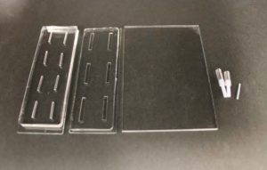
Fig.1 PDMS sheet with the upper channel, that with the lower channel, PDMS membrane, and tubing.

  1. Permanent bonding of the PDMS membrane and the sheet with the upper channel

Punch the inlet and outlet holes at both the ends of the upper channel with a 2-mm biopsy punch. Expose both the bonding surfaces of the PDMS membrane and the PDMS sheet with the upper channel (upper sheet) to plasma at 100 W, 35 s (Fig.2a and 2b). Laminate and bake them at 65˚C for 1 h (Fig.2c). Remove the PMMA sheet, and punch a hole to connect the sheet with the lower channel by using a 1-mm biopsy punch from the membrane side.
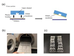
 

Fig.2 (a) Schematic diagram of bonding the PDMS membrane and the upper sheet. (b) Plasma treatment. (c) Bonded PDMS membrane and sheet.

  1. Detachable bonding of the PDMS membrane and the PDMS sheet with the lower channel

The PDMS sheet with the lower channel (lower sheet) is bonded to the PDMS membrane by using a PDMS prepolymer diluted with hexane (a dilution ratio of 1:3) as a glue1 (Fig.3a). Spin coat the diluted PDMS prepolymer (600 µL) at 2,000 rpm for 30 s on the surface of a glass slide (52 × 76 mm) to cover the slide with a thin layer of the glue and incubate it for 10 min to dry the solvent (Fig.3b). Place the lower sheet on the coated glass slide (Fig.3c). Apply glue at the four corners of the PDMS membrane bonded with the upper sheet (Fig.3d). Peel off the lower sheet from the glass slide and place the glue-coated surface of the sheet on the PDMS membrane (Fig.3e). After 30 min of incubation, bond the lower sheet to a cover slip by plasma bonding. Place a 1-kg weight and a glass slide on the device and bake it at 100˚C for 1 h (Fig.3f).
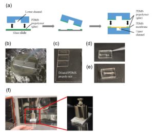
Fig.3 (a) Schematic diagram of bonding of the PDMS membrane and lower sheet. (b) Spin coat the diluted PDMS prepolymer (glue). (c) Lower sheet is placed on the thin film of the diluted PDMS prepolymer. (d) The diluted PDMS prepolymer is applied on the four corners of the PDMS membrane. (e)The glue-coated surface of the lower sheet is placed on the PDMS membrane. (f) A weight and a glass slide are placed on the device to bake it at 100˚C.

  1. Tubing

Connect polytetrafluoroethylene (PTFE) tubes (1 × 2 × 100 mm) with Tygon tubes (1.59 × 3.18 × 10 mm) to the holes present at both ends of the upper microchannel. Connect a PTFE tube (46 × 0.92 × 150 mm) to the hole of the lower channel. Apply PDMS prepolymer at the root of the tubes, and then bake it at 100˚C for 1 h for firm connection (Fig.4a and b).

  1. Cell culture

Introduce a cell suspension into the upper microchannel, which is manually precoated with 0.1 mg/mL of fibronectin. Incubate the device at 37˚C with 5% CO2 for 16 h to allow cells to adhere to the bottom of the upper channel (surface of the PDMS membrane).

  1. Detachment of the lower sheet for cell observation

Remove the lower sheet from the device carefully (Fig.4c and d). Place the rest of the device on a cover slip for observation with an inverted microscope (Fig.4 f and h).
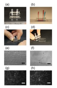
Fig.4 (a) The complete microdevice. (b) Side view of the microdevice. The cell culture channel (upper) is filled with water containing a red food color, while the lower channel is filled with water containing a blue food color. (c) and (d) The lower sheet is peeled off from the microdevice carefully. Phase contrast images of cells (e) before and (f) after detachment of the lower sheet. Fluorescent images of cells stained with CellTracker Red CMTPX (g) before and (h) after detachment.

Conclusion

We developed a microfluidic device with a detachable lower microchannel. It is important that different bonding techniques be used for each side of the PDMS membrane. If the lower channel is filled with air and the device is incubated in a CO2 incubator, dew condensation is often observed in the lower channel when the device is taken out from the incubator. The condensation in the lower channel makes observation difficult (Fig.4e and g). This problem was solved with the detachable device.

Acknowledgment

This work was supported in part by the Japan Society for the Promotion of Science (JSPS) KAKENHI Grant Number JP16H04170.

Reference

  1. Chueh, B.H., Huh, D., Kyrtsos, C.R., Houssin, T., Futai, N., Takayama, S. (2007). Leakage-free bonding of porous membranes into layered microfluidic array systems. Analytical Chemistry 79 (9), 3504–3508.
Digg This
Reddit This
Stumble Now!
Share on Facebook
Bookmark this on Delicious
Share on LinkedIn
Bookmark this on Technorati
Post on Twitter
Google Buzz (aka. Google Reader)

Rapid Inoculation and Recovery of Microbes in a Microfluidic Device

Greiner, A.1,2, Tekwa, E.W.1,3, Gonzalez, A.1, Nguyen, D.4

1Department of Biology, McGill University, 1205 Dr. Penfield, Montreal, QC, H3A 1B1, Canada.
2Department of Ecology and Evolution, University of Toronto, 25 Willcocks Street, Toronto, ON, M5S 3B2, Canada
3Department of Ecology, Evolution, and Natural Resources, Rutgers University, 14 College Farm Road, New Brunswick, NJ, 08901, USA.
4Meakins Christie Laboratories, Research Institute of the McGill University Health Centre, and Department of Medicine, McGill University, 1001 Decarie Blvd, Montreal, QC, H4A 3J1, Canada.


Why is this useful?

Microfluidic devices are used for many different types of experiments across the medical, ecological and evolutionary disciplines (Park et al., 2003; Keymer et al., 2008; Connell et al., 2013; Hol & Dekker, 2014). For example, microfluidic devices for microbial experiments require inoculation into smaller chambers that simulate natural microbial environments such as porous soils (Or et al., 2007) and biological hosts (Folkesson et al., 2012). These devices often involve complicated pump setups and irreversible seals. We developed a technique that requires only common lab equipment and makes the device reusable while also allowing the microbes to grow undisturbed (based on Tekwa et al., 2015; Tekwa et al., in review). Here, we provide a detailed guide for the assembly and the previously undocumented non-destructive disassembly of polydimethylsiloxane (PDMS) experimental devices to recover microbes in situ, which can then be plated for relative counts and further molecular analyses of population changes. This is complemented by videos for each step.

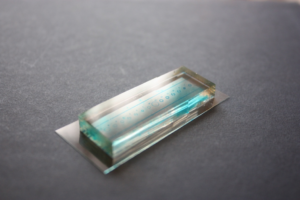
Figure 1: Microfluidic device containing 14 habitats on an elastomer (PDMS) layer pressed onto a 60mm x 24mm glass cover slip. The habitats are 10 or 20µm in depth, range from 1400 µm to 2670 µm in diameter and take the shape of a ring or network of patches. This device is used to test the effects of habitat patchiness on microbe dynamics. Habitats were dyed blue for visualization. For more information see Tekwa et al. (2015).


What do I need?

  • Single-layer PDMS devices with habitats on one side
  • Pipette + sterile pipette tips
  • Sterile petri dishes (1/device)
  • Sterile tweezers
  • Inoculum
  • Filtered water
  • Kimwipes
  • Autoclavable plastic container
  • Ethanol
  • Tinfoil
  • Sterile 1μL inoculating loop (1/habitat)
  • Sterile eppendorfs
  • Phosphate-buffered saline (PBS)
  • Biological safety cabinet (BSC)

How do I do it?

  1. Clean the PDMS devices: The PDMS device should be pre-treated once with 0.01N HCl for one hour and plasma-treated to keep it hydrophilic and amenable to bonding to glass or plastic substrates (Cho et al., 2007; Tekwa et al., 2015). Fill autoclavable plastic container 1/3rd full of 70% ethanol and PDMS devices and then cover with tinfoil, let them sit in this for >30 minutes before carefully disposing of the ethanol down the sink. Fill and empty the container with water 10 times in order to rinse the devices. Lastly, fill the container with filtered water, seal with tin foil and autoclave in order to sterilize the devices.
  2. Inoculating the devices (perform in a Biological Safety Cabinet, BSC): Set the devices to dry in the BSC for 30 minutes. Place the devices with features facing up on the lid of a petri dish and place a small amount (i.e. 0.7 µL) of inoculum onto each (of 14 in sample device Fig. 1) habitat. The amount of liquid must be sufficient to fill the habitats, but not too much as to prevent bonding between PDMS and the cover glass/petri dish (Fig. 2). Using sterile tweezers, pick up the device and place face down into the centre of a petri dish or cover glass, sealing it to the surface by pushing on the back with gloved fingers repeatedly, using a kimwipe to wick excess liquid away from the side. Then surround, but not touch, the device with kimwipes soaked in filtered water (Fig. 3) to ensure that the device does not dry out in the incubator, before closing the petri dish. Place upright in incubator for the desired amount of time. The experiment can now proceed untouched for up to 24 hours (see Supplementary video).

Figure 2. Device with bacteria droplets, 1 droplet per habitat.

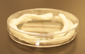
Figure 3. An ‘upright’ petri dish + kimwipes + device + coverslip ready to be incubated.


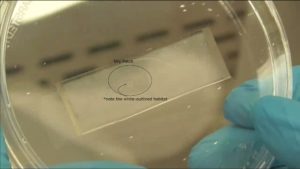
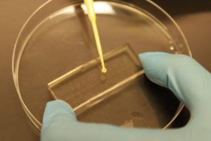
  1. Recovering from the devices (perform in a BSC): Open petri dish, carefully remove and discard kimwipes and then use sterile tweezers to gently unseal the device and place face up in the lid of the petri dish. By around 12 hours, the spaces between the habitats will be void of liquid from PDMS absorption, preventing microbes from being mixed across chambers during disassembly. Habitats that have dried out will appear white (Fig. 4) and cannot be used.  Dip sterile inoculating loop into an eppendorf with PBS, then use that loop to scrape one of the habitats (Fig. 5).  Dip that inoculating loop back into the eppendorf media again, which can then be grown overnight for further analyses such as plating for relative cell count (if there are different strains) and other molecular analyses. Repeat for the rest of the habitats that you are interested in, using new inoculating loops. The PDMS device can now be cleaned as in Step 1 and reused again.

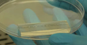
Figure 4. View of an inoculated and incubated device, looking through the bottom of a petri dish.


Figure 5. Recovering bacteria from a habitat in the disassembled PDMS device.


What else should I know?

The recovery technique can be used to estimate relative proportions of different types of microbes (e.g. morph frequencies) which is useful when performing competition assays and evolutionary experiments. Unlike in Tekwa et al. (2015), this technique forgoes the use of a confocal microscope; assessment of the contents of the device is instead performed through direct microbe recovery and standard plating procedures.


Links to Videos

These videos go through the specific procedure that we used to perform experiments on competition and cooperation in Pseudomonas aeruginosa and may be useful in determining specific amounts of media, growth times, etc. that may be utilized for experiments with similar PDMS microfluidic devices.

Part 1 – Intro + Washing the Devices https://youtu.be/Bne3ZN3wU4Q

Part 2 – Inoculating the Devices https://youtu.be/p-UYIRreYyM

Part 3 – Recovering from the Devices https://youtu.be/YLnmGxqMYgE

Supplementary – Fluorescent Bacteria Experiment https://youtu.be/lgMtryS62pA


Acknowledgements

AGr was supported by an NSERC Undergraduate Student Research Award and by an NSERC Discovery Grant. EWT was supported by the Fonds Québécois de la Recherche sur la Nature et les Technologies and the Québec Centre for Biodiversity Science. AGo was supported by the Canada Research Chair program and NSERC Discovery grants. DN was supported by a CFI Leaders Opportunity Fund (25636), a Burroughs Wellcome Fund CAMS award (1006827.01) and a CIHR salary award.


References

Cho, H., Jönsson, H., Campbell, K., Melke, P., Williams, J. W., Jedynak, B., … & Levchenko, A. (2007). Self-organization in high-density bacterial colonies: efficient crowd control. PLoS biology, 5(11), e302.

Connell, J. L., Ritschdorff, E. T., Whiteley, M., & Shear, J. B. (2013). 3D printing of microscopic bacterial communities. Proceedings of the National Academy of Sciences, 110(46), 18380-18385.

Folkesson, A., Jelsbak, L., Yang, L., Johansen, H. K., Ciofu, O., Høiby, N., & Molin, S. (2012). Adaptation of Pseudomonas aeruginosa to the cystic fibrosis airway: an evolutionary perspective. Nature reviews. Microbiology, 10(12), 841.

Hol, F. J., & Dekker, C. (2014). Zooming in to see the bigger picture: Microfluidic and nanofabrication tools to study bacteria. Science, 346(6208), 1251821.

Keymer, J. E., Galajda, P., Lambert, G., Liao, D., & Austin, R. H. (2008). Computation of mutual fitness by competing bacteria. Proceedings of the National Academy of Sciences, 105(51), 20269-20273.

Or, D., Smets, B. F., Wraith, J. M., Dechesne, A., Friedman, S. P. (2007). Physical constraints affecting bacterial habitats and activity in unsaturated porous media – a review. Advances in Water Resources, 30(6), 1505-1527.

Park, S., Wolanin, P. M., Yuzbashyan, E. A., Silberzan, P., Stock, J. B., & Austin, R. H. (2003). Motion to form a quorum. Science, 301(5630), 188-188.

Tekwa, E. W., Nguyen, D., Juncker, D., Loreau, M., & Gonzalez, A. (2015). Patchiness in a microhabitat chip affects evolutionary dynamics of bacterial cooperation. Lab on a Chip, 15(18), 3723-3729.

Tekwa, E.W., Nguyen, D., Loreau, M., Gonzalez, A. Defector clustering is linked to cooperation in a pathogenic bacterium. In review.

Digg This
Reddit This
Stumble Now!
Share on Facebook
Bookmark this on Delicious
Share on LinkedIn
Bookmark this on Technorati
Post on Twitter
Google Buzz (aka. Google Reader)

Rapid and easy fabrication of glass-bottom culture dishes for long-term live cell imaging

Ayako Yamada123, Jean-Louis Viovy123, Catherine Villard123 and Stéphanie Descroix123

1 Laboratoire Physico Chimie Curie, Institut Curie, PSL Research University, CNRS UMR168, Paris, France.

2 Sorbonne Universités, UMPC Univ. Paris 06, Paris, France

3 Institut Pierre-Gilles de Gennes, Paris, France

Email: ayako.yamada@curie.fr


Why is this useful?

Glass is a versatile surface for chemical treatments, and it still is by far the most used substrate for surface engineering (e.g. micropatterning, surface chemistry) or plasma-bonding of PDMS microfluidic devices. For cell culture on such substrates, glass-bottom culture dishes are desired to keep over the cells well defined medium volumes, and to protect the cells from contamination and medium evaporation. Moreover, they are optically better suited for microscopy observation than polystyrene dishes routinely used for cell culture. Although glass-bottom culture dishes are commercially available (e.g. Fluorodish from WPI), the presence of plastic walls limits the treatments that can be performed onto the glass bottom surface and those are more expensive ( 5 € per dish; φ 50 mm) than polystyrene dishes (e.g. φ 40 mm dish from TPP, 0.5 € per dish). In this Tip, we describe an easier way than a previous Tip1 to transform a polystyrene culture dish into a glass-bottom one, while preserving the possibility to apply to the glass any treatment before its assembly into a dish. Note that in this method, the body of the culture dish will be upside down and the lid is thus no longer lifted above the dish opening by lid stoppers. However, the gas exchange through the gap between the body and its lid seems to be enough to culture cells healthily in this dish. In summary, this Tip provides a low-cost and rapid solution for cell culture in a microfluidic device or on an engineered surface directly in a culture dish, suited for a long-term live cell imaging.


What do I need?      

  • φ 40 mm polystyrene tissue culture dish (e.g. TPP #93040)
  • φ 40 mm cover slide (e.g. Thermo Fisher Scientific #11757065, ∼0.2 € per slide)
  • Large screw driver (or a similar tool)
  • Uncured mixture of PDMS base and curing agent (10:1 w/w)
  • Oven or hotplate
  • Ferromagnetic metal plate (e.g. lid of a PDMS container, optional)
  • Cylindrical magnets (optional)


What do I do?

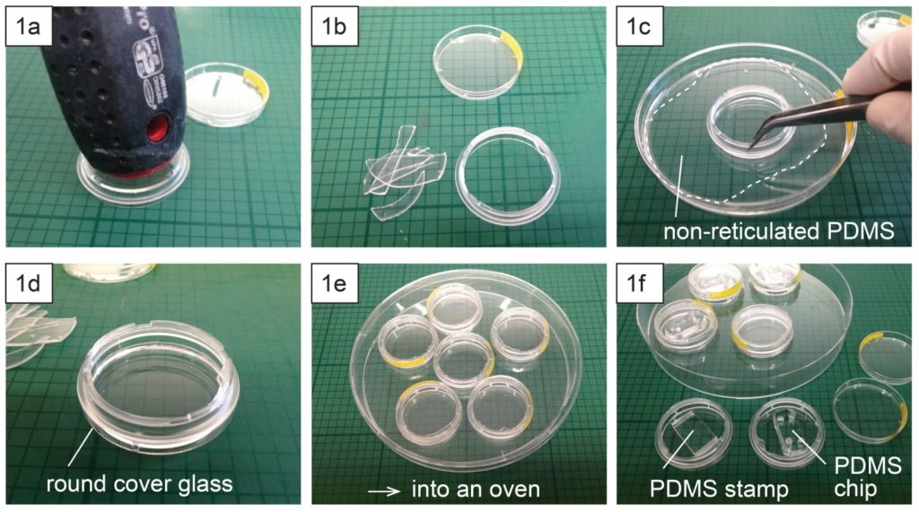
  1. Place a polystyrene culture dish upside down on a surface and hit a few times the center of the dish bottom with the grip of a large screw driver (Fig. 1a) until the dish bottom falls apart from the dish wall (Fig. 1b). The bottom should fall easily with the success rate around 9 over 10. Avoid breaking the dish wall by hitting the bottom too strongly.
  2. Spread uncured PDMS mixture on a flat substrate (e.g. a larger plastic Petri dish) and coat the edge of the dish (broken part up) with PDMS (Fig. 1c).
  3. Place the dish (broken part up) on a cover glass slide (Fig. 1d) and cure PDMS in an oven or on a hotplate e.g. at 80 °C for 10 min (Fig. 1e).
  4. Surface treatment (e.g. micro-contact printing) or plasma-bonding of a PDMS chip to the glass surface can be performed after or before the dish assembly (Fig. 1f).

  1. To keep humidity for on-chip cell culture, the dish can be filled with e.g. phosphate buffered saline (Fig. 2a). Dishes with chips or micropatterns loaded with cells can be placed in a CO2 incubator with or without further protection (Fig. 2b).
  2. Long-term live cell imaging can be performed using a stage top incubator (Fig. 2c).


What else should I know?

  1. Depending on the support type of microscopes, it might be necessary to well align the contours of the dish and the glass slide. This can be done using cylindrical magnets (3 per dish) and a ferromagnetic metal plate (Fig. 3a) during PDMS curing in an oven or on a hotplate (Fig. 3b).


Acknowledgement

This work is supported by the French National Research Agency (ANR) as part of the “Investissements d’Avenir” program (reference: ANR 10-NANO 0207) and ERC Advanced Grant CellO (FP7-IDEAS-ERC-321107).


Reference

[1] Caballero D, Samitier J, Different strategies for the fabrication of cell culture chambers for live-cell imaging studies. Chips and Tips, 02 Dec 2014 (https://blogs.rsc.org/chipsandtips/2014/12/02/different-strategies-for-the-fabrication-of-cell-culture-chambers-for-live-cell-imaging-studies/)


Digg This
Reddit This
Stumble Now!
Share on Facebook
Bookmark this on Delicious
Share on LinkedIn
Bookmark this on Technorati
Post on Twitter
Google Buzz (aka. Google Reader)

A simple, bubble-free cell loading technique for culturing mammalian cells on lab-on-a-chip devices

Sahl Sadeghi1­* and Meltem Elitas1

1 Faculty of Engineering and Natural Sciences, Sabanci University, 34956, Istanbul, Turkey

* Sahl Sadeghi wrote the paper.


Purpose

Lab-on-a-chip (LOC) devices significantly contribute different disciplines of science. Polydimethylsiloxane (PDMS) is one of the main materials, which is widely used for the fabrication of biological LOCs, due to its biocompatibility and ease of use. However, PDMS and some other polymeric materials are intrinsically water repellant (or hydrophobic), which results in difficulties in loading and operating LOCs. The eminent consequence of hydrophobicity in LOCs for biological systems is the entrapment of air bubbles in microfluidic channels. Although the oxygen plasma treatment of PDMS reduces the surface hydrophobicity for a certain period of time, the hydrophilic property of PDMS vanishes over time1. The persistent problem of bubbles in the microfluidics led to several studies conducted to overcome it. Some of these solutions suggested implementing bubble traps2,3, surface treatment of LOCs through hydrophilic coatings4, and using actively controlled bubble removal systems 5,6.

Although the aforementioned design complexities are introduced to LOCs in order to reduce the clogging problem caused by the bubbles, these modifications also result in higher production cost, complex operation, and long device preparation time. In many single-cell experiments without losing or damaging the rare cells, these cells needs to be introduce into the LOCs. Here, we present a simple method that enables loading a small number of cells without introducing bubbles in the microfluidics channels.


Materials

·         PDMS (Dow Corning Sylgard 184 Silicon Elastomer Kit)
·         Pipette tips (20-200 ul, Eppendorf, # 3120000917)
·         Pipetman (Gilson, P200, #69989-5)
·         Aqueous ethanol 70% (ZAG Chemistry)
·         Cell culture medium (DMEM, PAN Biotech, #P04-01548)
·         Mammalian cells (MCF7, ATCC-HTB-22)
·         Sterile syringe (BD 10 ml Syringe, Luer-Lok Tip, #300912)
·         Sterile Hamilton syringe (Hamilton, 100 ul SYR, #84884)


Procedure

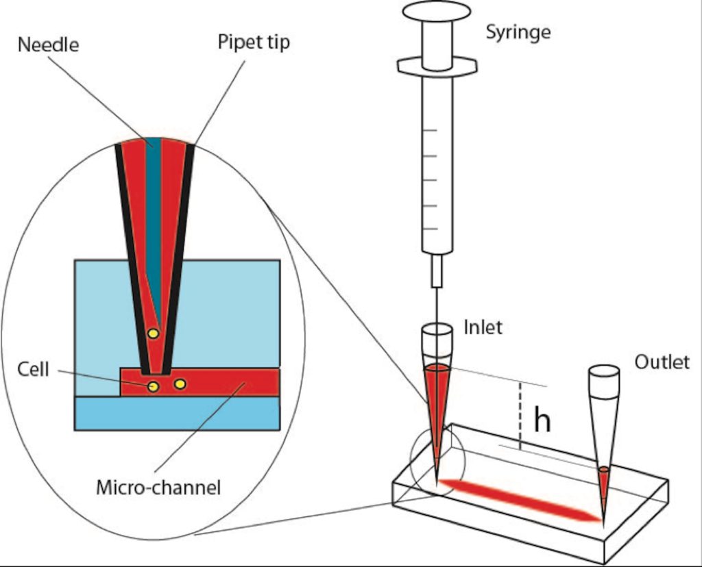
Step 1: Insert two 200-ul pipet tips at the inlet and outlet ports of the PDMS device as illustrated in Figure 1.

Step 2: Introduce a 70% aqueous ethanol into the inlet-pipet tip using a pipetman. Thus, the inner surface of microfluidic channels will be disinfected and the fluid flow will be tested within the micro channels as it is applied in many other protocols for LOCs7,8.

Step 3: Gently apply pressure pressing the pipetman to force ethanol solution flow through the micro channels and cavities of the PDMS device. Take care to avoid applying negative pressure from the outlet-pipet tip, which might create air leakage through the pipet connections. Besides, applying a negative pressure will directly affect the amount of a gas dissolved in the liquid according to Henry’s law9 that might contribute formation of more bubbles inside the micro-channels. The positive pressure will facilitate removal of the air bubbles via dissolving them.

Step 4: After flushing the chip with ethanol solution, inspect the chip to ensure bubble removal. In case of air bubbles, repeat the steps 2 and 3.

Step 5: Fill the syringe (10 ml) with medium or phosphate buffered saline (PBS). Take care to remove the air bubbles inside the syringe, mount and lock the needle on the syringe. Then, flow medium through the needle too make sure the needle is full of medium without any bubble. Insert the needle in the inlet-pipet tip; gently apply positive pressure to replace the ethanol with medium. Next, collect the excess medium from the outlet-pipet tip.

Step 6: Fill the inlet pipet with fresh medium in such a way that due to certain height (h) between the levels of the medium in the inlet and outlet pipet tips, very slow medium flow will be established inside the micro-channels.

Step 7: Load your cells into the Hamilton syringe and take care to ensure that there is no air bubble inside its needle and syringe. Insert the needle of the Hamilton syringe into the inlet-pipet tip as explained in Step 5, Figure 1. Introduce the cells via applying gentle positive pressure to the syringe. Established flow streams in the PDMS chip will deliver the released cells to the desired positions in the chip. Flow rate can be arranged adjusting the applied positive pressure and amount of medium collected in the inlet and outlet pipet tips. The excess supernatant from the outlet-pipet tip can be collected, and fresh medium can be supplied through the inlet-pipet tip during the experiment.


References

  1. Tan, S.H., N.T. Nguyen, Y.C. Chua, and T.G. Kang,. Biomicrofluidics, 2010. 4(3).
  2. Zheng, W.F., Z. Wang, W. Zhang, and X.Y. Jiang,. Lab on a Chip, 2010. 10(21): p. 2906-2910.
  3. Wang, Y., D. Lee, L.S. Zhang, H. Jeon, J.E. Mendoza-Elias, T.A. Harvat, S.Z. Hassan, A. Zhou, D.T. Eddington, and J. Oberholzer, . Biomedical Microdevices, 2012. 14(2): p. 419-426.
  4. Wang, Y.L., C.E. Sims, and N.L. Allbritton,. Lab on a Chip, 2012. 12(17): p. 3036-3039.
  5. Karlsson, J.M., M. Gazin, S. Laakso, T. Haraldsson, S. Malhotra-Kumar, M. Maki, H. Goossens, and W. van der Wijngaart,. Lab on a Chip, 2013. 13(22): p. 4366-4373.
  6. Cortes, D.F., T.-X. Tang, D.G.S. Capelluto, and I.M. Lazar,. Sensors and Actuators B: Chemical, 2017. 243: p. 650-657.
  7. Benavente-Babace, A., D. Gallego-Perez, D.J. Hansford, S. Arana, E. Perez-Lorenzo, and M. Mujika,. Biosensors & Bioelectronics, 2014. 61: p. 298-305.
  8. Yesilkoy, F., R. Ueno, B.X.E. Desbiolles, M. Grisi, Y. Sakai, B.J. Kim, and J. Brugger,. Biomicrofluidics, 2016. 10(1).
  9. Henry, W., Phil. Trans. R. Soc. Lond., 1803. 93: 29–274.

Figure 1 – Schematic of cell loading procedure in a microfluidic PDMS device.


Digg This
Reddit This
Stumble Now!
Share on Facebook
Bookmark this on Delicious
Share on LinkedIn
Bookmark this on Technorati
Post on Twitter
Google Buzz (aka. Google Reader)

A novel low cost method to prepare a cross-linked gelatin membrane for potential biological applications

Gabriele Pitingolo1 and Valerie Taly1

 1 INSERM UMRS1147, CNRS SNC 5014, Université Paris Descartes, Equipe labellisée Ligue Nationale contre le cancer 2016. Paris, France.

email: gabriele.pitingolo@parisdescartes.fr


Why is this useful?

In recent years, several gelatin types (i.e. GelMA, A or B) have been used in pharmaceutical formulation and tissue engineering due to their excellent biocompatibility, propensity to cell differentiation and availability at low cost.1 The use of gelatin as biomaterial is also advantageous for the possibility to tune the mechanical properties of the substrate, changing the concentration in water and the degree of cross-linking. However, Transwell® is the most used permeable support with microporous membranes and is a standard method for culturing cells.2 This commercial type of support has been widely used to study the molecular secretion by different cell types and also to reproduce several in vitro physiological barriers (i.e. blood brain barrier).3 Transwell® cell culture inserts are convenient, because they are sterile and easy-to-use, but they are very expensive (~ 300 $ for a 12 pack) and possess a limited range of biomaterial properties because they are made from polyester or polycarbonate. Furthermore, as shown by Falanga and colleagues, these porous membranes are often integrated onto microfluidic chip for permeability studies.4

Recently, Yong X. Chen et al. proposed an alternative method to prepare a suspended hydrogel membrane platform for cell culture, designing a complicated protocol to synthesize the GelMA and to fabricate an open-grid structure made of polylactic acid (PLA) polymer using a commercial printer.5 Our tip shows a novel low-cost method for preparing a cross-liked gelatin membrane as a permeable support useful for potential biological applications. In addition, the proposed protocol doesn’t require the use of sophisticated fabrication technologies or expensive materials. It uses gelatin from porcine skin and the formaldehyde vapor technique to cross-link the gelatin membrane. As proof of concept, we integrate the gelatin membrane into a microfluidic chip, to show the possibility to develop a platform for comparable studies in static and dynamic conditions.

Furthermore, to demonstrate the resistance to culture temperature (around 37° C) of the prepared gelatin membrane, we tested the mechanical properties (Young’s modulus) before and after the incubation time (2 days). Finally, we observed the preservation of the mechanical properties and structural integrity that makes the membrane usable for studies with cell culture.


What do I need?

  • Transwell insert
  • Porcine gelatin type A
  • Formaldehyde solution
  • Scalpel
  • PMMA milled chamber or similar


What do I do?

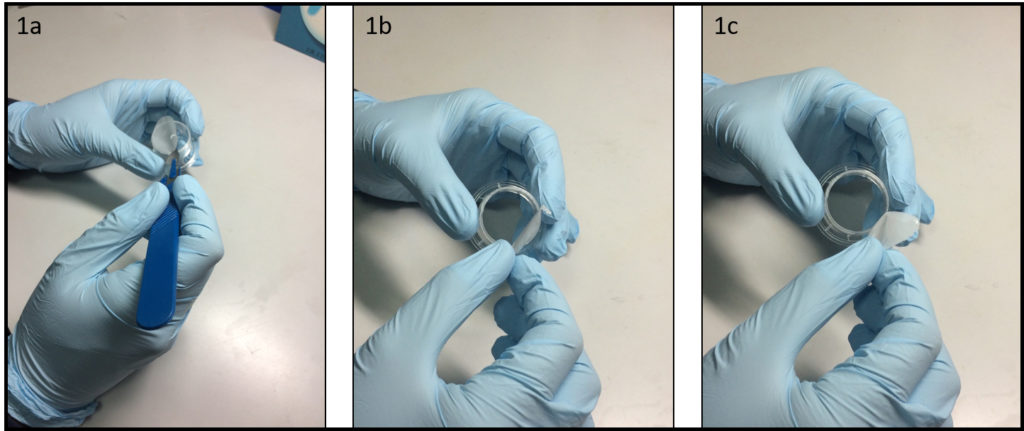
  1. Remove the porous membrane from the Transwell® insert (Fig. 1a-1c) or alternatively use a similar homemade support. To facilitate this step is convenient to use a scalpel to incise the membrane along the entire diameter.


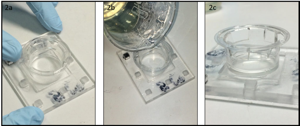
  1. After the removal of the membrane, the Transwell® support is ready to use. Position the structure at the center of the PMMA chamber (depth 2 mm at least) (Fig 2a) and pour liquid 10% w/v gelatin, without bubbles, onto the PMMA chamber and inside the Transwell® support (Fig 2b). After 2 min of stabilization, put the system in the fridge, for at least 10 minutes.


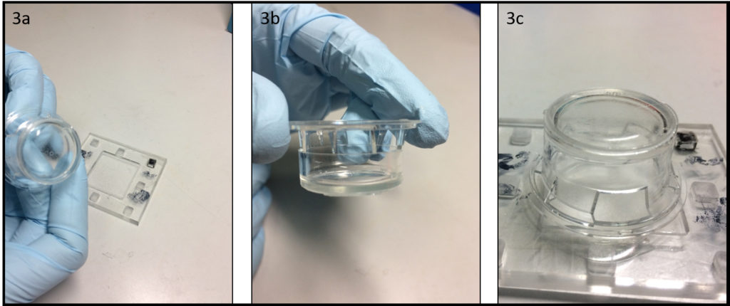
  1. After the gelation time (10 minutes at 4°C) it is possible to remove the formed gelatin membrane from the PMMA chamber, with the aid of a scalpel to facilitate the detachment (Fig 3a-3b). As shown in Figure 3c the gelatin membrane it appears very flat, an ideal characteristic for cell cultivation (Fig 3c). To guarantee the preservation of the mechanical properties during the cell culture step, cross-link the gelatin membrane using the classical protocol “cells-biocompatible”, such as glyceraldehyde6, formaldehyde7 and glutaraldehyde8 methods or natural products such as genipin.9


  1. In this case, we used the vapor formaldehyde method to cross-link the prepared gelatin membrane and to obtain a system with lower aqueous solubility, higher mechanical strength and stability against enzymatic degradation. We exposed the gelatin membrane to formaldehyde vapors for 1 day. In Figure 4a we show the difference, after 48 h of culture conditions, between a sample cross-linked (left) and not (right). The final depth of the cross-linked gelatin membrane is around 1 mm, however, it is possible to change the depth tuning the liquid gelatin amount. Finally, we calculated, before and after the incubation time, the young’s modulus of the cross-linked gelatin membrane, observing a similar value of 40 kPa (compression test by using hydraulic testing system Instron DX).


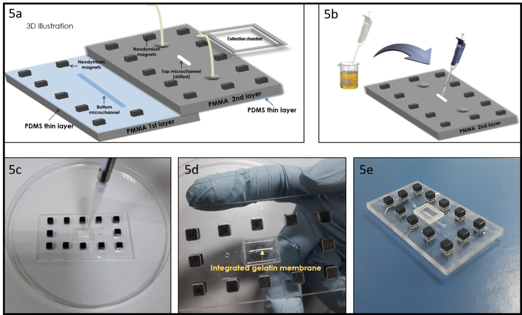
  1. Integration of the gelatin membrane into a microchip. In this section, we detail the integration of the gelatin membrane into a microfluidic chip. As example, we used the same geometry proposed in our previous work4, to make a device for a permeability experiment (Figure 5a). As shown in Figure 5b, just pour the liquid gelatin into the smaller drilled microchannel, using a pipette to form a thin uniform layer of gelatin (Figure 5c). After gelation at 4° C, cross-link the formed gelatin membrane using the previously described method, the result is shown in Figure 5d. To bond the different PMMA-PDMS substrates we propose here a magnetic approach recently developed by our group10, to preserve the gelatin membrane by physical-chemical stress as in the case of solvent evaporation and plasma bonding. Figure 5e shows the final chip.


Conclusions: In this tip, a novel biocompatible gelatin permeable support was obtained by using a simple and low cost fabrication method. Vapor formaldehyde method or other chemical crosslinking approach can be applied to crosslink the integrated gelatin membrane for the use as potential scaffolds for cell culture. Furthermore, the Young’s modulus and the thickness of the permeable membrane can be adjusted by changing the initial concentration of the gelatin, the degree of cross linking and the amount of liquid gelatin. Finally, we showed the integration of the gelatin membrane into a modular microchip. Therefore, we propose an easy and low cost method to prepare a permeable gelatin membrane for cell biology and for other applications.


Acknowledgements

This work was carried out with the support of the Pierre-Gilles de Gennes Institute equipment (“Investissements d’Avenir” program, reference: ANR 10-NANO 0207).


References

  1. Geckil, Hikmet, et al. “Engineering hydrogels as extracellular matrix mimics.” Nanomedicine 5.3 (2010): 469-484.
  2. https://www.corning.com/worldwide/en/products/life-sciences/products/permeable-supports/transwell-guidelines.html
  3. Guarnieri, Daniela, et al. “Shuttle‐Mediated Nanoparticle Delivery to the Blood–Brain Barrier.” Small 9.6 (2013): 853-862.
  4. Falanga, A. P., Pitingolo, G., Celentano, M., Cosentino, A., Melone, P., Vecchione, R. & Netti, P. A. (2016). Shuttle‐mediated nanoparticle transport across an in vitro brain endothelium under flow conditions. Biotechnology and Bioengineering.
  5. Chen, Yong X., et al. “A Novel Suspended Hydrogel Membrane Platform for Cell Culture.” Journal of Nanotechnology in Engineering and Medicine 6.2 (2015): 021002.
  6. Sisson, Kristin, et al. “Evaluation of cross-linking methods for electrospun gelatin on cell growth and viability.” Biomacromolecules 10.7 (2009): 1675-1680.
  7. Usta, M., et al. “Behavior and properties of neat and filled gelatins.” Biomaterials 24.1 (2003): 165-172.
  8. Talebian, A., et al. “The effect of glutaraldehyde on the properties of gelatin films.” Kemija u industriji 56.11 (2007): 537-541.
  9. Bigi, A., et al. “Stabilization of gelatin films by crosslinking with genipin.” Biomaterials 23.24 (2002): 4827-4832.
  10. Pitingolo Gabriele, et al. “Fabrication of a modular hybrid chip to mimic endothelial-lined microvessels in flow conditions.” Journal of Micromechanics and Microengineering” (Accepted manuscript)
Digg This
Reddit This
Stumble Now!
Share on Facebook
Bookmark this on Delicious
Share on LinkedIn
Bookmark this on Technorati
Post on Twitter
Google Buzz (aka. Google Reader)

Different strategies for the fabrication of cell culture chambers for live-cell imaging studies

David Caballero1,2, Josep Samitier1,2

1 Nanobioengineering group, Institute for Bioengineering of Catalonia (IBEC), Barcelona, Spain

2 Centro de Investigación Biomédica en Red de Bioingeniería, Biomateriales y Nanomedicina (CIBER-BBN), Zaragoza, Spain.

Why is this useful?


Long-term imaging of cells is typically performed using standard Petri dishes. Frequently, these ´chambers´ are not convenient when sample manipulation and treatment (e.g. functionalization, immunostaining…) is needed, both before and after the experiment1. To overcome this ´problem´, glass coverslips are used. They can be easily manipulated when following multistep protocols prior to cell deposition. For live-cell microscope imaging, the customized coverslips are secured into holders (chambers) containing the adequate cell culture medium2, 3. These holders are either supplied by the microscope manufacturers or fabricated in a mechanical workshop; this implies time for the design and money.

In this work we show two different strategies for the simple, fast and cheap fabrication of chambers for live-cell imaging, using materials and simple tools typically available in a bioengineering laboratory. These materials include Petri dishes, polydymethylsiloxane (PDMS), Falcon tube cap and glass coverslips. In the first strategy (A) we drill a hole in a Petri dish where a customized glass coverslip is adhered at the bottom using wax. In the second strategy (B), a PDMS frame is used to hold the coverslip inside a Petri dish. Depending on the user final application one strategy is recommended over the other (see below). All the material is biocompatible and simple to obtain. These two methods provide several advantages: (i) they are easy and cheap; (ii) the chambers can be fabricated in a short-time period; (iii) this approach avoids the purchase of commercially-available holders or ordering the fabrication to a mechanical workshop.

What do I need?


    Figure 1

Figure 1. Material needed for the fabrication of the home-made chambers.

(1) PDMS
(2) Glass coverslip #1, 25 mm in diameter
(3) Cap of a 15 mL Falcon (any brand)
(4) Syringe 5mL
(5) Polishing paper
(6) Wax
(7) P35 plastic Petri dish (any brand)
(8) Sharp Tweezers
(9)Glass Pasteur pipette

You will also need:

  • Hot plate
  • Bunsen burner (optional)
  • Soldering iron (or drill)
  • EtOH 70%
  • Oven
  • SYLGARD 184 PDMS and crosslinker agent (Dow Corning)


What do I do?


Figure 1 (above) shows all the material needed for the fabrication of the home-made chambers using both strategies A and B. The steps describing both strategies are detailed next.

STRATEGY A:

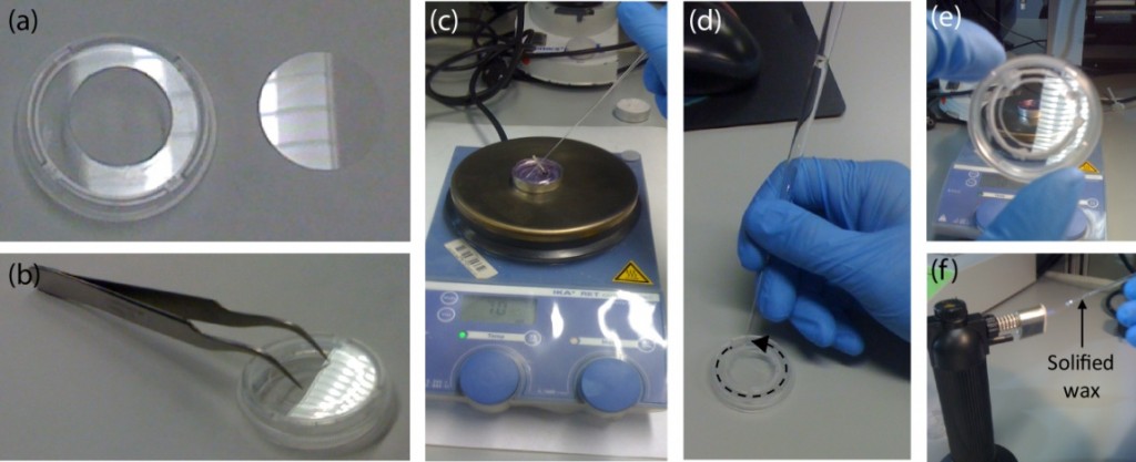
1. Make a circular hole of around 2 cm in diameter in the middle of the lower part of a P35 Petri dish using a pre-heated, sharp-tip soldering iron. Alternatively, a drill can be used. Ensure a perfect circular hole by using a 15 mL Falcon cap as a template (see Fig.2a-c).

Figure 2. Fabrication of the cell culture chamber using Strategy A. Drilling a hole on the lower part of a Petri. (a) First, draw a circumference of about 2 cm in diameter in the center of the lower part of a Petri dish. Use a 15 mL Falcon cap as template. (b-c) Next, a soldering iron is used to drill a hole. (d) Finally, the edge of the hole is polished using thin polishing paper.

2. Polish the hole using polishing paper (see Fig.2d). Check that the Petri is free of debris. Rinse the sample with etOH 70%. (Optional: Sonicate).

3. Use the tweezers to place and secure the glass coverslip on the back side of the holey dish with its customized side (e.g. functionalized) facing the inner part of the Petri (see Fig.3a-b).

Figure 3. Adhering the glass coverslip to the lower part of the drilled Petri dish using wax. (a) The drilled Petri dish and glass coverslip #1, 25 mm in diameter are (b) placed together and secured using the tweezers. (c) Next, the wax is melted using a heat plate. A small volume is absorbed by capillarity using a glass Pasteur pipette. (d) Following the edge defined by the coverslip and the dish, the chamber is sealed. This step is critical to ensure a good sealing. (e) Check that no open remaining points are left. (f) A Bunsen burner or equivalent can be used to melt the solidified wax inside the pipette.

4. Melt the wax using the hot plate and fill the Pasteur pipette with a small volume (see Fig.3c). NOTE: Capillarity will make the liquid wax to flow inside the pipette.

5. Gently, put in contact the tip of the pipette (filled with wax) with the coverslip. Follow the edge formed by the coverslip and the Petri (see Fig.3d). Cover it completely with wax until the entire contour is sealed (see Fig.3e). Refill the pipette if necessary. NOTE 1: The wax may solidify inside the pipette quickly. If so, melt it again using the Bunsen burner (or equivalent) (see Fig.3f). NOTE 2: Ensure that no empty spaces are left; cell medium will flow through them.

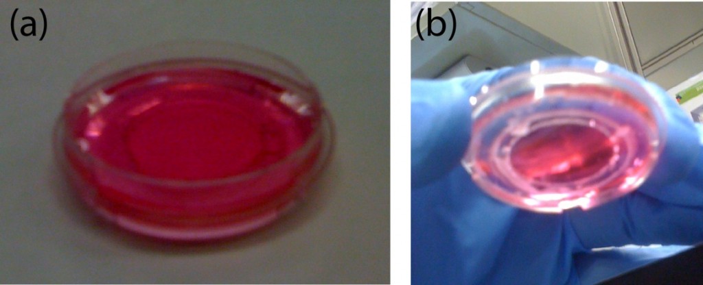
6. Culture the cells of interest (see Fig.4). Place the chamber inside the microscope and start the live-cell imaging experiment. NOTE: Manipulate gently the sample.

Figure 4. Finished chamber for live-cell imaging experiments. (a) Front view of the chamber filled with cell culture medium. (b) Back side view showing the wax-sealed region. No leakage is observed. The coverslip can be easily recovered after the experiment by pushing it down gently with the tweezers.

7. At the end of the experiment, the medium can be removed and the coverslip detached by pushing it down gently with the tweezers. Treat the sample as desired (e.g. immunostaining).

STRATEGY B:

1. Insert upside down the cap of a 15 mL Falcon in the middle of a P35 Petri dish (see Fig.5a).

Figure 5. Fabrication of the cell culture chamber using Strategy B. (a) A 15 mL Falcon cap is placed in the center of a P35 Petri dish. The empty region is filled with PDMS using a syringe. (b) The sample is degassed and cured. (c) The cap is removed and the PDMS frame released.

2. Fill the empty space with a syringe (or equivalent) with PDMS in a 10:1 ratio (pre-polymer:cross-linker). Degass and cure it at 65ºC for 4h (see Fig. 5b). NOTE 1: Holding the cap with adhesive tape will ensure that it remains in the center during curing. In this case, curing must be performed at RT overnight. NOTE 2: A very thin PDMS layer may appear after the removal of the cap. Remove it manually to ensure a through-hole in the PDMS. Alternatively, a weight can be applied on top of the cap.

3. Remove the cap using the tweezers and release the PDMS frame (see Fig. 5c).

4. Sterilize the PDMS frame. Rinse it with etOH 70% and UV-irradiate for 15 min.

5. Working in the cell culture room, deposit a drop (~50 uL) of culture medium in the center of a new P35 Petri dish (see Fig. 6a).

Figure 6. Finished chamber for live-cell imaging experiments. (a) A small drop of cell culture medium is deposited in the center of a new P35 Petri dish. (b) The customized coverslip is placed on top of it. (c) The PDMS frame is introduced inside the Petri and pushed down to hold the coverslip forming the chamber. Finally, the chamber is filled with cells.

6. Place the (customized) glass coverslip facing-up on top of the drop (see Fig. 6b). NOTE: This will ensure that no air bubbles are formed. Hold it with the PDMS frame.

7. Culture the cells of interest (see Fig. 6c). Place the sample inside the microscope and start the experiment.

8. At the end of the experiment, the medium can be removed and the coverslip released by removing the PDMS frame with the tweezers. Treat the sample as desired (e.g. immunostaining).

Figure 7. Microfabricated coverslips for live-cell imaging studies. (a) Glass coverslip covered with PDMS microstructures. (b) The modified coverslip can be used for the fabrication of chambers using both strategies. (c) Zoomed image of the microstructures (parallel grooves). The dimensions are: 1 um x 1 um x 1 cm (HxWxL); the separation between grooves is 1 um. Scale bar: 10 um. (d) NIH3T3 fibroblasts aligned parallel to the grooves. The arrow shows the direction of the structures. Scale bar: 50 um.


What else should I know?


By using these two approaches, chambers can be easily fabricated in the lab. Most importantly, this approach allows the manipulation of coverslips before and after the experiment with cells. If the coverslips are (bio)chemically modified (e.g. micropatterned with proteins of the extracellular matrix), manipulation must be performed carefully and fast to avoid sample degradation. Similarly, for cell guidance assays, coverslips can be easily modified with microfabricated structures to orient cell growth and motility (see Fig. 7), following the same steps described in this protocol.

For Strategy A, users must be aware of the melting temperature of wax (around 45ºC). This implies that the sealing may be fragile when performing the experiments at 37ºC and manipulation must be performed gently to avoid liquid leakage. Other materials and shapes, besides circular glass coverslips, could be used. This will depend on the experimental requirements of the user. For Strategy B, the PDMS frame and the coverslip could be used without the Petri in some applications. However, for live-cell imaging, a perfect fit between the chamber and the microscope stage is needed and the use of the Petri is therefore strongly recommended.

Finally, the user must consider the magnification needed for the experiment and the thickness of the sample on each strategy. Strategy A is recommended for high magnification microscopy (40X – 100X) and Strategy B for low magnification (4X – 20X). If needed, thinner coverslips (#0) could be used.


Acknowledgements

Dr. Daniel Riveline, Dr. Jordi Comelles (Laboratory of Cell Physics ISIS/IGBMC, Strasbourg, France) and David Izquierdo (Nanobioengineering group – IBEC, Barcelona, Spain) are acknowledged for technical help and discussions.

References

1.      Zanella F, Lorens JB, Link W. High content screening: seeing is believing. Trends Biotech 2010; 28:237-45.

2.      Caballero D, Voituriez R, Riveline D. Protrusion Fluctuations Direct Cell Motion. Biophysical Journal 2014; 107:34-42.

3.      Riveline D, Buguin A. Devices and methods for observing the cell division WO/2010/092116, 2009.

Digg This
Reddit This
Stumble Now!
Share on Facebook
Bookmark this on Delicious
Share on LinkedIn
Bookmark this on Technorati
Post on Twitter
Google Buzz (aka. Google Reader)

A method for periodic sterile sample collection during continuous cell culture in microfluidic devices

Dmitry A. Markov,1,2 Elizabeth M. Lillie,1,2 Philip C. Samson,2,3 John P. Wikswo,1,2,3,4 Lisa J. McCawley2,5
1 Department of Biomedical Engineering, Vanderbilt University, Nashville, TN
2 Vanderbilt Institute for Integrative Biosystems Research and Education (VIIBRE), Vanderbilt University, Nashville, TN
3 Department of Physics and Astronomy, Vanderbilt University, Nashville, TN
4 Department of Molecular Physiology and Biophysics, Vanderbilt University Medical Center, Nashville, TN
5 Department of Cancer Biology, Vanderbilt University Medical Center, Nashville, TN

Why is this useful?


Allows for sterile sample collection during long-term slow perfusion experiments. Current developments in microfluidic devices and their applications in biology during long-term cell and tissue culture have introduced additional challenges associated with periodic sterile collections of effluent for storage and further analysis. Pumps (either mechanical or electroosmotic) and gravity-fed systems work well for delivering media at very slow flow rates (< 1 µL/min). However, sample collection at these rates becomes a non-trivial task, especially when one-, two-, or three-week-long experiments are required. For example, it would take 24 hours to collect 1 mL of effluent at a flow of 0.7µL/min. Two important parameters to consider are sample evaporation during the collection step and sterility maintenance during multiple periodic collections. These are especially critical for the gravity-fed systems where the output port of the cell culture device has to be opened to the atmospheric pressure. To overcome these challenges, we have tested use of Breathe-Easy gas permeable adhesive membranes from Electron Microscopy Sciences (cat. # 70536-10) as aseptic filters on top of sample collection vials. These membranes are inexpensive, easy to use, and gas permeable, allowing for unimpeded fluid flow into the sample collection reservoir, while maintaining sterility of the whole system during sample collection. During our three-week-long cell culture experiments, we were able to perform multiple 24-hour effluent collections from our bioreactor with a 5-day period while maintaining sterility of the whole system. Upchurch valves were used to switch the flow between the waste reservoir (4 days) and the sample collection vials (1 day). The evaporation was reduced by keeping the complete system at 100% humidity within a CO2 cell culture incubator.

What do I need?


  • 1.5 mL microcentrifuge tube
  • Butane torch or Bunsen burner
  • Small 25 gauge heated needle for puncturing
  • Short piece of small 23 gauge stainless steel tubing to be inserted into the microcentrifuge tube
  • 90 second epoxy (Araldite 2043 or similar)
  • Small bore Tygon tubing (OD = 1/16th “, ID=0.020”)
  • Drill bit #53
  • Breathe-Easy membrane (Electron Microscopy Sciences, cat. # 70536-10)

How do I do it?


Tube insertion using stainless steel interface connector:

  • Heat the puncturing needle with a cigarette lighter, Bunsen burner, or a small butane torch and carefully make a hole at the bottom of the microcentrifuge tube.
  • Use a 4-6 mm long piece of small gauge stainless steel interface tubing, and insert it into the punctured hole.  Outer diameter of this tubing should be larger by 1 or 2 gauges than the puncturing needle (23 ga. tubing has larger diameter than 25 ga. tubing). The fit should be snug.
  • Apply a small bead of 90 second curing epoxy to affix the interface tube to the microcentrifuge tube.
  • After the epoxy dries, feed tygon tubing onto the stainless steel interface tube.
  • In our case, we used a 25 ga needle to create a hole and a 23 ga stainless steel tubing (OD=0.022″) as an interface between a 1.5 ml microcentrifuge tube and flexible Tygon tubing (OD=1/16th, ID=0.020″).

Alternative procedure for direct flexible tube insertion:

  • Drill a small hole in the bottom of the microcentrifuge tube
  • Insert the Tygon tube and apply 90 second epoxy to glue the two parts together.
  • We used a #53 drill bit (Ø = 0.0595″) for 1/16th OD tygon tubing.

To apply the filter:
The following procedure should be performed wearing gloves and under aseptic conditions.  Biosafety cabinets or laminar flow hoods with proper filtration are adequate.  The Breathe-Easy membranes typically come sterile from the supplier and should be opened inside the biosafety hood and handled with sterile tweezers.  The modified microcentrifuge tube with attached Tygon tubing can be sterilized using common procedures involving autoclave, alcohol, or gamma radiation sterilization (gas sterilization was not tested). All mentioned approaches performed reasonably well and were quite adequate; however, we have found that multiple autoclave sterilization cycles often softened the epoxied connections and alcohol sterilization required excessive handling of the components.  In our opinion, gamma ray sterilization was found to be the easiest, simplest, and the most convenient approach.

  • Start with sterile membrane and microcentrifuge tube with proper tubing attached.
  • Cut off the snap lid of the microcentrifuge tube.
  • Peel one half of the protective film from one side of Breathe-Easy filter and attach exposed adhesive side of filter to the top rim of the tube.
  • Then, remove the second half of the protective film and fully attach filter (make certain the seal is good around the perimeter with firm pressure applied with flat edge of forceps).
  • When ready to use the assembly, carefully remove the top protective layer.

We used this method to periodically sample effluent from our bioreactor cartridges during long term cell culture.  Each cartridge contains 4 reactors that are connected to a single manifold shown in Figure 4D.

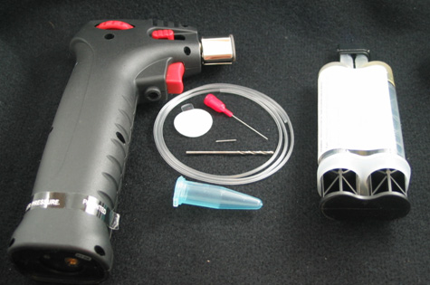
Figure 1. Tools and supplies needed

Figure 2. Preparing the microcentrifuge tube – Method 1. A) Hole punched with a hot needle. B) A piece of stainless steel needle inserted into the punctured hole. C) Inserted tube fixed in place with a 90 second epoxy. D) Flexible Tygon tubing attached to the interface tube.

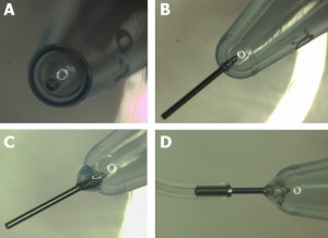
Figure 3. Preparing the microcentrifuge tube – Method 2. A) Hole being drilled at the bottom. B) View of the drilled hole. C) Insertion of the small diameter Tygon tubing. D) Application of the 90 second epoxy to fix the inserted tube in place.

Figure 4. Preparation of the sterile cover. A) Protective plastic is removed from the adhesive side of the filter. B) Filter membrane is attached to the tube rim. C) Use caution to remove top protective plastic from the membrane attached to the tube. D) View of the tube with the filter mounted in the sample holder and attached to the collection network.

Acknowledgements


We would like to acknowledge support provided by the following grants: NIH/NCI R21 CA126728-01A1 and DOD/BCRP W81XWH-09-1-0444.

Digg This
Reddit This
Stumble Now!
Share on Facebook
Bookmark this on Delicious
Share on LinkedIn
Bookmark this on Technorati
Post on Twitter
Google Buzz (aka. Google Reader)